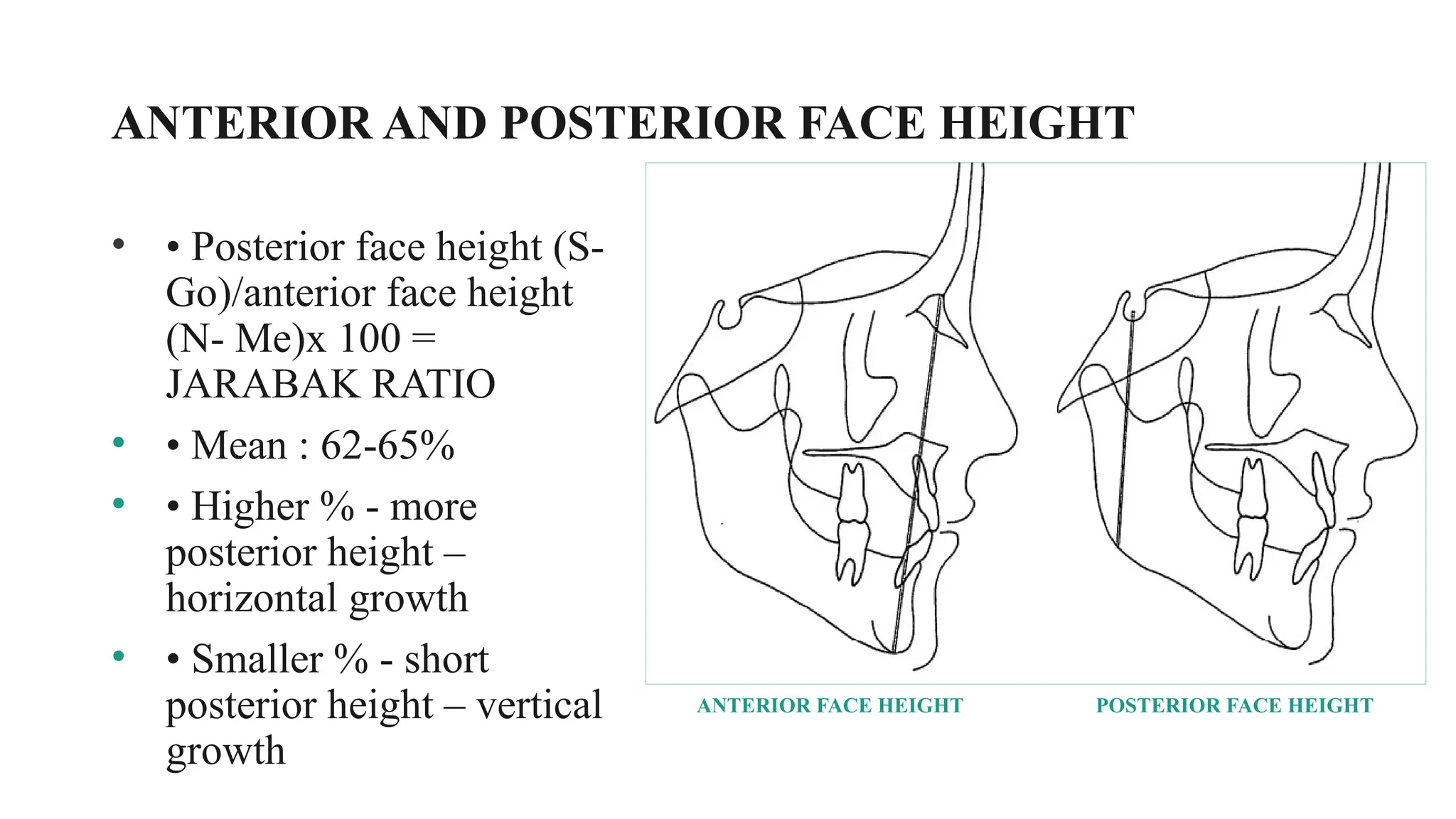
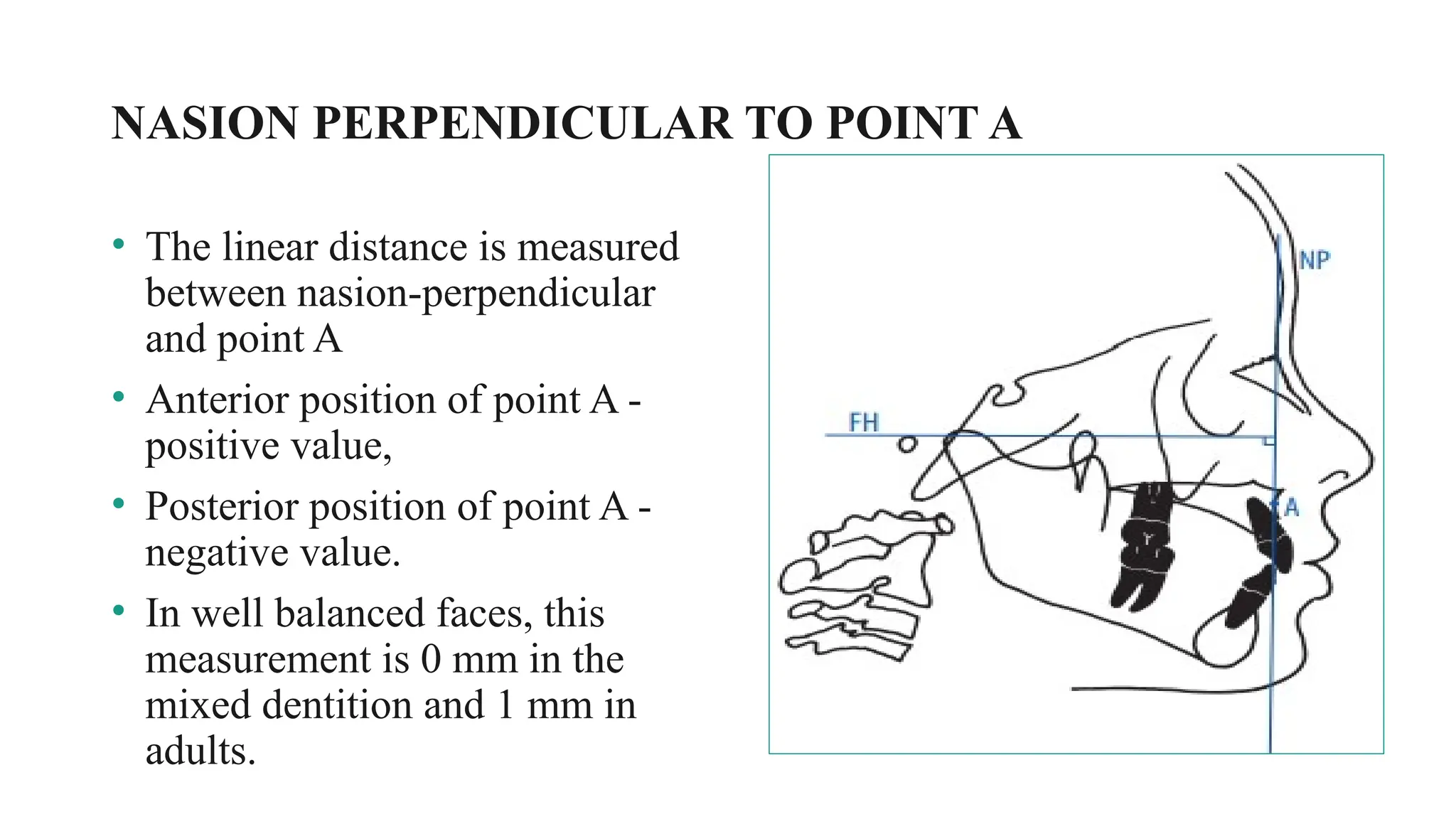
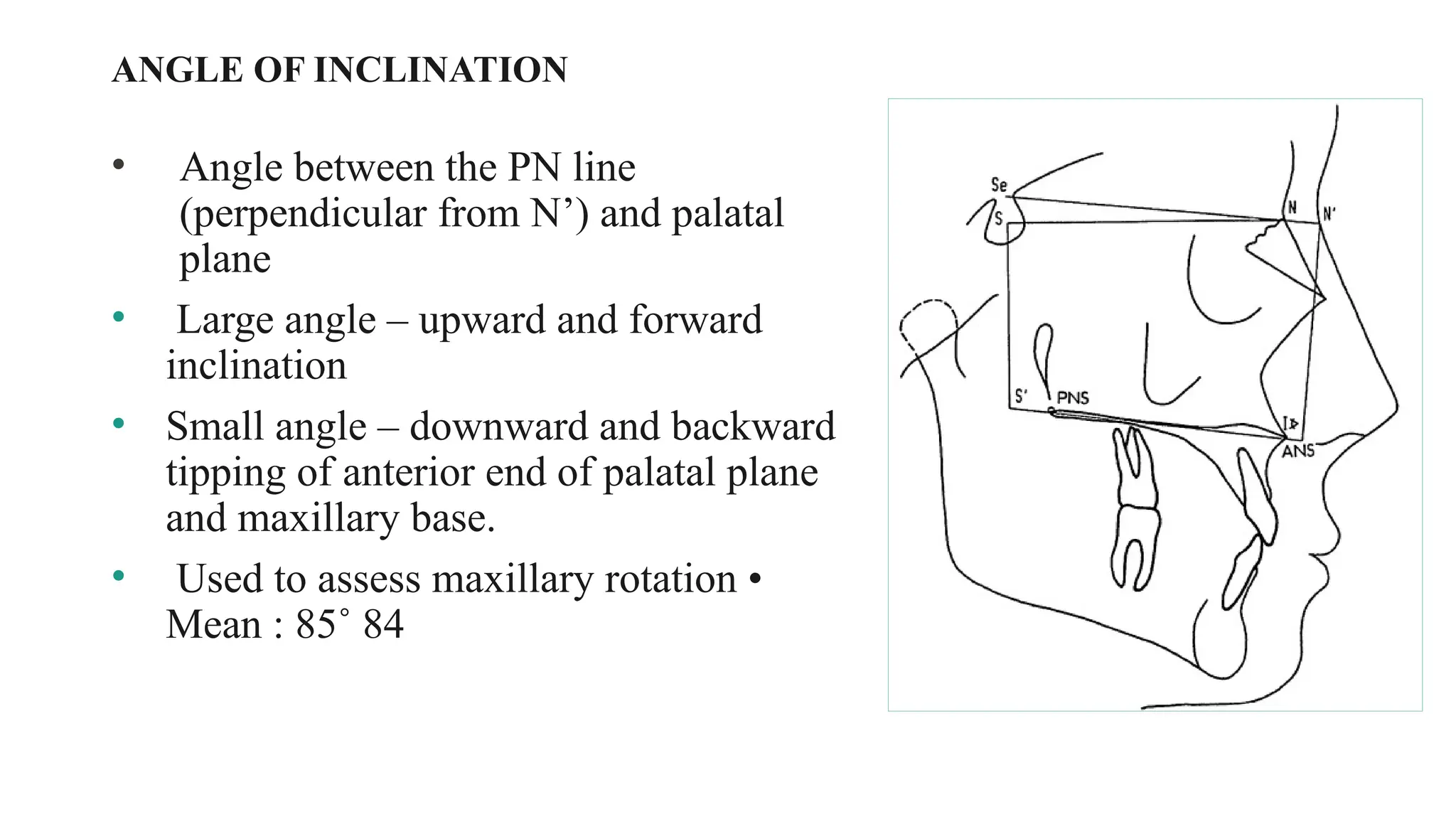
CEPHALOMETRIC ANALYSIS
• INTRODUCTION
• HISTORY OF CEPHALOMETRY
• CEPHALOMETRIC IMAGING SYSTEM
• TYPES OF CEPHALOGRAM
• LANDMARKS
• TRACING OF CEPHALOGRAMS
• CLASSIFICATION OF CEPHALOMETRIC ANALYSES
• HARD TISSUE ANALYSIS
• SHORTCOMINGS OF ANB ANGLE AND WITS
CONTENTS
INTRODUCTION
• Cephalometry developed from Craniometry.
• Craniometry in the Edinburgh Encyclopaedia of 1813 is defined as
“the art of measuring skulls of animals so as to discover their specific
differences”.
• For many years anatomists and anthropologists were confined to
measuring craniofacial dimensions using the skull of dead individuals.
•Cephalometry is concerned with measuring the head inclusive of soft
tissues, be it living or dead.
Craniometry – Measuring skull.
Cephalometry – Measuring head.
However Cephalometry had its limitations due to the inaccuracies that
resulted from varying thickness of soft tissues.
DEFINITIONS
•Radiographic technique for abstracting the human head into
geometric shape.
•Scientific measurement of the dimensions of the head.
•A scientific study of the measurements of head with relation to
specific reference points ; used for evaluation of facial growth
and development, including soft tissue profile.
History Of Cephalometric Radiography
• In 1895, Prof. Wilhelm Conrad Roentgen made a remarkable
contribution to science with the discovery of x-rays.
• Prof. Wilhem Koening & Dr. Otto Walkhoff simultaneously made the
first dental radiograph in 1896.
• Justus A.W. van Loon, was probably the first to introduce
cephalometrics into orthodontics.
• The first x- ray pictures of skull in the standard lateral view were
taken by A.J.Pacini & Carrera in 1922.
• Paccini identified the following landmarks : Gonion, Pogonion, Nasion, and
Anterior Nasal Spine. He also located the sella turcica & external auditory
meatus.
• In 1931 cephalometric radiography came to full function when Hofrath in
Germany and Broadbent in the United States provided both a research and a
clinical tool for the study of malocclusion and underlying skeletal
disproportions.
1. There was little mention of a frontal view
2. The path of the central ray was not fixed in relation to the head
3. No plan was suggested for superimposition of subsequent films
4. Considerable emphasis was placed on the recording of soft tissuehere w
CEPHALOMETRIC IMAGING SYSTEM
X-RAY APPARATUS
AN IMAGE
RECEPTOR
A CEPHALOSTAT
 Extra oral film - 8 x 10” or 10 x 12”
Sensitive to the fluorescent light radiated from the inten­
sifying screen.
 Intensifying screens -
Emit either blue or green light after being exposed to radiation.
 Cassette -
Light Tight Box
 A grid -
A device used to reduce the scatter radiation.
 A soft-tissue shield -
To act as a filter and reduce over penetration of x-rays into soft tissue.
Image receptor system consists of :
THE CEPHALOMETER
• “The Reserve craniostat” was developed by T. Wingate Todd.
• After Broadbent completed his training with Angle, he received an
appointment as research fellow in the Western Reserve Department of Anatomy
under Todd.
• Together they designed a roentgenographic craniostat that made possible
precise standardization of cranial x-rays of dry skull.
• The next step was to adapt the device to the heads of living subjects: The
Roentgenographic Cephalometer.
Broadbent cephalostat with head holder positioned
with cassette in place for a lateral cephalogram
• During 1920’s Broadbent refined the
craniostat in to craniometer by the
addition of metric scales.
• The patient’s head was centered in the
cephalostat with the superior borders of
the external auditory meatus resting on
the upper parts the two ear rods.
• The lowest point on the inferior bony
border of the left orbit, indicated by the
orbital marker, was at the level of the
upper parts of the ear rods.
•Nose clamp was fixed at the root of the nose to
support the upper face.
•The focus film distance was set at 5 feet (152.4
cm) and the subject film distance could be
measured to calculate image magnification.
•With the two X ray tubes at right angles to
each other in the same horizontal plane, two
images (lateral & PA) could be simultaneously
produced.
TYPES OF CEPHALOGRAMS
• Lateral Cephalogram
• Frontal Cephalogram
USES OF CEPHALOGRAM
a) In orthodontics diagnosis and treatment planning
b)In classification of skeletal and dental abnormalities.
c) In eastablishing facial types.
d)In evaluation of treatment results.
e) In predicting growth related changes and changes associated with
surgical treatment.
f) Valuable aid in research work involving the craniodentofacial region.
PRINCIPLE OF CEPHALOMETRIC ANALYSIS
• To compare the patient with a normal reference group, so that
difference between the patient’s actual dentofacial relationships and
those expected for his/her racial or ethnic group are revealed.
GOALS OF CEPHALOMETRICS
• To evaluate the relationship, both horizontally and vertically, of the
five major functional components of the face:
• The cranium and the cranial base.
• The skeletal maxilla.
• The skeletal mandible.
• The maxillary dentition and the alveolar process.
• The mandibular dentition and the alveolar process.
Tracing Of A Cephalogram
• Thorough familiarity with the gross anatomy
is required before the tracing.
• By convention the bilateral structures (eg,
the rami and inferior borders of the
mandible) are first traced independently. An
average is then drawn by visual
approximation, which is represented by a
broken line.
• A lateral cephalogram
• Acetate matte tracing paper (.003 inches thick, 8×10 inches)
• A sharp 3H drawing pencil or a very fine tipped pen
• Masking tape and a few sheets of cardboard (preferably black) and a
hollow cardboard tube.
• A protractor and tooth symbol tracing template for drawing the teeth. Also
templates for tracing the outlines of ear rods.
• Dental casts trimmed to maximum intercuspation of the teeth in occlusion.
• Viewbox (variable rheostat desirable but not essential).
Tracing Supplies And Equipments
Requirements
• Should be easily seen on the roentgenogram,
• Be uniform in out line, and easily reproducible.
• Should have significant relationship to the vectors of growth.
• Should permit valid quantitative measurements of lines and
angles projected from them.
• Measurements should be amenable to statistical analyses
Hard Tissue Landmarks
Ba
ANS
A
Cephalometric Planes
• Frankfurt Horizontal plane: This plane is drawn from
Porion to Orbital.
• Sella-Nasion plane: It represents the anterior cranial base.
• Basion-Nasion plane: This plane is used in the Rickett’s
analysis.
• Palatal plane: Plane passing through the ANS and the
PNS.
• Occlusal plane: It is the plane passing through the cusp
tips of the upper and lower first molars and a point
• Bolton - Nasion plane
• Mandibular plane: Different definitions are given in different
analysis :
1.Downs analysis - extends from Go to Me.
2.Steiner’s analysis - extends from Go to Gn.
3.Salzmann and Tweed - tangent to the lower border of the mandible as
the mandibular plane.
Cephalometric Analysis
• The major use of radiographic cephalometry is in characterizing the patient’s dental and
skeletal relationships.
• This led to the development of a number of cephalometric analyses to compare a patient
to the population standards.
• William. B. Downs in 1948 developed the first cephalometric analysis.
• Its significance was that it presented an objective method of portraying many factors
underlying malocclusion.
• Followed by other analyses by Steiner (1953), Tweed (1953), Ricketts (1958), Enlow
(1969), Jaraback (1970), Jacobson (1975) etc.
Cephalometric analysis can be classified in different ways:
1. Methodological classification
2. According to the area of analysis
3. Normative classification
Classification Of Analysis
Based on the basic units of the analysis they can be classified into:
Angular Analysis
The basic units are angle
in degrees.
Linear Analysis
The facial skeleton is
analyzed by determining
certain linear dimensions.
1. Dimensional Analysis
2. Proportional Analysis
3. Analysis to determine
Position
Methodological Classification
Classification According To Area Of Analysis
Dentoskeletal Analysis
Soft tissue Analysis
Functional Analysis.
Normative Classification
Analysis may be classified according to the concept on which
normal values have been based.
• Mononormative Analysis: Single norm is used.
• Multinormative Analysis: A series of norms including Age,
Sex are used.
• Correlative Analysis: Used to assess individual variations
of facial structure to establish their mutual relationships.
Analysis of Facial Skeleton
EXTENT OF ANTERIOR CRANIAL BASE
• Sella – nasion
• Distance is used to assess the proportional
lengths of maxillary and mandibular bases.
• Mean : 75mm
• 9 year old average length of anterior
cranial base is 68.8mm (horizontal) and
63.8mm (vertical).
• Acc. To holdaway , It increases by ¾ mm
annually.
• Increased : horizontal growth pattern
• Decreased : vertical growth pattern
EXTENT OF POSTERIOR CRANIAL BASE
• Sella- articulare
• Also k/a lateral cranial base length
• It relates to the position of fossa and
posterior facial height
• Short cranial base : vertical growth
pattern/ skeletal open bites
• Mean : 32-35mm
• A mean rate of increase of 8mm
between age 6 and 16.
ORIENTATION ANGLE
• It is the angular relation between the
Frankfurt Horizontal plane and Sella-Nasion
plane.
• Mean - 7 degrees
SADDLE ANGLE
• N-S-Ar angle between the
anterior and posterior cranial
base.
• Mean : 123 ˚ ± 5 ˚
• Increased angle – posterior
position of the fossa /
mandibular retrognathia
• Decreased angle – anterior
position of the fossa/mandibular
prognathism
ARTICULAR ANGLE
• S-Ar-Go angle - Constructed angle
between upper and lower contours of
facial skeleton
• Increased angle : mandibular
retrognathism
• Decreased angle : mandibular
prognathism –
• Mean : 143 ˚ ± 6 ˚
GONIALANGLE
• Ar-Go-Me angle : formed by the tangents to
the body of mandible and posterior border of
ramus
• expression for the form of the mandible, with
reference to the relation between body and
ramus.
• Mean : 128 ˚ ± 7 ˚
SUM OF POSTERIOR ANGLES (BJORK SUM)
• Sum of angles (saddle, articular and gonial) is
396˚ ± 6˚
• >396 ˚ – vertical growth / clockwise rotation
• <396 ˚ – horizontal growth / anticlockwise
rotation
• If the sum is less than 360˚ then it is favourable
for functional appliance therapy.
UPPER AND LOWER GONIALANGLE
• The gonial angle may be divided by a line drawn
from nasion to gonion. This gives a lower and upper
angle.
• The upper angle is formed by ascending ramus and
line joining nasion and gonion.
• Mean value : 50˚-55 ˚
• The lower angle is formed by line joining nasion
and gonion and lower border of mandible.
• Mean value : 72 ˚- 75 ˚
• Large upper angle – horizontal growth
• Small upper angle – caudal growth
• Large lower angle – vertical growth
• Small lower angle – sagittal growth
ANTERIOR AND POSTERIOR FACE HEIGHT
• • Posterior face height (S-
Go)/anterior face height
(N- Me)x 100 =
JARABAK RATIO
• • Mean : 62-65%
• • Higher % - more
posterior height –
horizontal growth
• • Smaller % - short
posterior height – vertical
growth
ANTERIOR FACE HEIGHT POSTERIOR FACE HEIGHT
ANALYSIS OF JAW BASES
Maxilla (SNA)
• Point A : The anterior limits of the
apical base of the maxilla.
• SNA: to determine whether maxilla
is positioned more anteriorly or
posteriorly to the cranial base.
• The mean SNA reading is 82°.
MAXILLA TO MANDIBLE
ANTEROPOSTERIOR RELATIONSHIP
• Effective length of maxilla - condylion to point A.
• Effective length of mandible - condylion to anatomic gnathion.
• Maxillomandibular differential = MIDFACIAL LENGTH –
MANDIBULAR LENGTH
EXTENT OF MAXILLARY BASE
• Distance from PNS to perpendicular
drawn from point A to palatal plane.
• Mean : 45.5 mm at age 8 years.
• Annual increase of 1.2 mm for boys
and 0.8 mm for girls.
NASION PERPENDICULAR TO POINT A
• The linear distance is measured
between nasion-perpendicular
and point A
• Anterior position of point A -
positive value,
• Posterior position of point A -
negative value.
• In well balanced faces, this
measurement is 0 mm in the
mixed dentition and 1 mm in
adults.
ANGLE OF INCLINATION
• Angle between the PN line
(perpendicular from N’) and palatal
plane
• Large angle – upward and forward
inclination
• Small angle – downward and backward
tipping of anterior end of palatal plane
and maxillary base.
• Used to assess maxillary rotation •
Mean : 85˚ 84
MANDIBLE (SNB)
• To assess whether the mandible
is protrusive or recessive
relative to the cranial base.
• the sella–nasion–point B
(SNB) angle is read.
• mean: 80°
• < 80°- recessive mandible
• >80° - prognathic mandible
FACIALANGLE:
• Used to measure the degree of retrusion
or protrusion of the mandible.
• This is the inferior inside angle in
which the facial line (nasion-pogonion
[N-Pog] intersects the FH.
• Mean reading : 87.8°.
• Range of 82 to 95°.
• > 87.8 = prominent chin
⁰
• < 87.8 = retrusive chin
⁰
EXTENT OF MANDIBULAR BASE
• Distance between gonion – pogonion
• Mean : 68 mm at 8 years of age
• Ideally , it should be 3mm more than N- Se
distance.
• Increased : horizontal growth pattern
• Decreased : vertical growth pattern
EXTENT OF ASCENDING RAMUS
• • Distance between gonion and condylon
• • Mean (at age 8) – 46mm - With an
annual of 2mm for boys and 1.2 mm for
girls upto age 16.
• Increased length : horizontal growth
pattern
• Decreased length : vertical growth pattern
ANB
• difference between SNA and
SNB.
• The ANB angle provides a
general idea of the
anteroposterior discrepancy of
the maxillary to the mandibular
apical bases.
• The mean reading : 2°
• >2°- Class II skeletal tendency.
• <2°- Class III skeletal
relationship
WITS APPRAISAL
• Linear measurement and not an analysis in itself.
• Measure to the extent to which the jaws are related to each other
• Used to identify instances in which the ANB reading does not accurately
reflect the extent of anteroposterior jaw dysplasia.
• Drawing perpendicular lines on a lateral cephalometric headfilm tracing
from points A and B on the maxilla and mandible, respectively, onto the
occlusal plane, which is drawn through the region of the overlapping cusps
of the first premolars and first molars.
• The points of contact on the occlusal plane from points A and B are labeled
AO and BO, respectively
• Distance between AO and BO gives
the AP relationship.
• Average jaw relationship is -1mm
for men and 0 mm for women.
• Positive reading : BO behind AO –
class II
• Negative reading : BO ahead of AO
- class III
• Greater the Wits reading deviation
from –1.0 mm in men and 0 mm in
women, the greater the horizontal or
anteroposterior jaw disharmony.
2. ANGLE OF CONVEXITY
• This angle measures the degree of the
maxillary basal arch at its anterior limit
(point A) relative to the total facial profile
(N-Pog).
• Mean value: 0°
• Range: 8.5 to 10°
• Positive angle: protrusive mandible
• Negative angle: retrusive mandible
ANALYSIS OF VERTICAL
SKELETAL RELATIONSHIP
MANIBULAR PLANE
• Drawn between gonion and
gnathion.
• Angle is formed by relating it to the
anterior cranial base (SN).
• Mean reading: 32°.
FMA- FRANKFORT MANDIBULAR PLANE ANGLE
• It is the angle formed by the
intersection of Frankfort
horizontal plane with the
mandibular plane.
• Indicates direction of lower
facial growth horizontally and
vertically
• Range : 22 ˚ to 28 ˚
• Mean : 25 ˚
5. Y- AXIS ( GROWTH AXIS)
• Measured as the acute angle formed by the
intersection of a line from the sella turcica
to gnathion with the FH.
• The range: 53° to 66 °
• Mean reading: 59.4°
• Increase angle- vertical growth pattern.
• Decrease angle- horizontal growth pattern
BASAL PLANE ANGLE
• Measures an angle to FH.
• On average, this angle is 26 degrees at
9 years of age and decreases
approximately 1 degree every 3 years.
• A high or steep mandibular plane angle
implies that an open bite may be caused
by the skeletal morphologic
characteristics of the mandible.
• A low mandibular plane suggests the
opposite (ie, a deep bite).
UPPER 1 TO SN
• • Long axis of the upper incisor is
extended to intersect the SN line and
posterior angle is measured.
• • Mean : 102˚ ± 2˚
• • 102˚ angulation is achieved only 1 or 2
years after eruption.
• • increased angle – maxillary incisor
protrusion
• • Decreased angle – lingually tipped
incisors
UPPER1 TO PALATAL PLANE
• Anterior angle between the long axis of the incisor and palatal plane is
measured.
• Mean : 70˚ ± 5˚
• Large angle : upright incisors
• Small angle : incisor protrusion
MAXILLARY INCISOR POSITION
• Angle formed by the intersection of
the long axis of the upper central
incisiors and the line joining nasion
to point A.
• U1 to NA {LINEAR} – shows
forward and backward position of
teeth
• U1 to NA {ANGLE} – relative
axial inclination of teeth
• Mean {LINEAR} – 4mm
{ANGLE} - 22˚
PROTRUSION OF MAXILLARY INCISORS
• Measured as the distance between the
incisal edge of the maxillary central
incisor to the line from point A to Pog.
• Range: –1 mm to 5 mm
• Mean: 2.7 mm
• Positive reading: maxillary dental
protrusion
• Negative reading: retruded position of
maxillary central incisior.
IMPA- INCISOR MANDIBULAR PLANE ANGLE
• It is the angle formed by the
intersection of the long axis
of the lower incisor with the
mandibular plane.
• • Used as a guide in
maintaining or positioning
lower incisors in relation to
the underlying basal bone.
• • Range : 85˚ - 90 ˚
• • Mean: 87˚
MANDIBULAR INCISOR POSITION
• Determined by relating the teeth to
the line from nasion to point B (NB).
• L1 to NB {LINEAR} – shows
forward and backward position of
teeth.
• L1 to NB {ANGLE} – relative axial
inclination of teeth.
• Mean {LINEAR} – 4mm
{ANGLE} - 25˚
LOWER INCISOR – CHIN RELATIONSHIP
• The degree of prominence of the chin
contributes to the determination of the
placement of the teeth in the arch.
• Ideally, according to Holdaway, 5 the
distance between the labial surface of
the lower incisor to the NB line should
be equal (ie, 4 mm).
• 2mm discrepancy is acceptable
• 3mm – less desirable
• > 4mm- corrective measures
INTERINCISALANGLE
• The interincisal angulation relates
the relative position of the
maxillary incisor to that of the
mandibular incisor.
• • Mean : 130˚
• • >130˚ / obtuse : requires
advancement anteriorly.
• • <130˚ / acute : both maxillary
and mandibular teeth requires
uprighting.
Bjork’s Polygon
• In this analysis a polygon is used to assess the anterior and
posterior facial height relationships and also to predict the
direction of growth change in the lower face.
The basis of this is the relationship of the 3 angles.
• Saddle angle (N.S Ar),
• Articulare angle (S-Ar-Go),
• Gonial Angle (Ar-Go_me)
>396°
Clockwise
<396°
Anticlockwise
396°
N
S
Ar
G
• Clockwise Change: Indicates that the anterior facial height is increasing
more rapidly than posterior facial height and it could be associated with
backward growth at the symphysis leading to anterior openbite tendency
• Counter Clockwise Change: Indicate that the posterior face height is
increasing more rapidly giving rise to forward growth of chin and anterior
deep bite tendency
BETAANGLE
• C Y BAIK - 2004
• To assess skeletal discrepancy between
maxilla and mandible in sagittal plane.
• Angle is formed by line joining pt. A and pt.
B , pt.B to pt. C (center of condyle) and a
perpendicular is dropped from pt.A to C-B
line.
• The angle between a perpendicular line and
pt. A pt. B line is ß angle.
• 27˚ - 35˚ - class I
• < 27˚ - class II
• > 35˚ - class III
W ANGLE
• W A BHAD et al – 2013
• Point S – sella turcica
• Point M – midpoint of premaxilla
• Point G – center of the largest circle
that is tangent to internal inferior,
anterior and posterior surfaces of
mandibular symphysis.
• Values:
• 51-56= class I skeletal pattern
• < 51˚ = class II skeletal pattern
• > 56˚ = class III skeletal pattern
SHORTCOMINGS OF ANB ANGLE
• Jacobson suggested that the antero-posterior and vertical position of
the nasion point and the rotational changes of the jaws influence the
reliability of the ANB angle and thus recommended the use of Wits
appraisal.
• It changes significantly with age whereas the Wits does not change
significantly with age.
• Bishara et al. suggested using both ANB and Wits together.
SHORTCOMINGS OF WITS APPRAISAL
• Change in degree of occlusal plane angle will have the effect on wits,
decrease in occlusal plane angle will increase the wits.
• Increase in vertical distance bw point A and B will also increse the
wits.
• Tracing error of occluasal plane.
• Wits appraisal is not related to the skull base and nasion point.
Use Of AXD angle
• Beatty in 1975, devised the AXD angle, where point X is located by
projecting point A on to a perpendicular to SN line.
• Point D is located in the bony symphysis as described by Stiener.
REFERENCES
• ORTHODONTIC CEPHALOMETRY- ATHANASOIS E.
ATHANASOIS
• ATLAS AND MANUAL OF CEPHALOMETRY RADIOGRAPHY-
THOMAS RAKOSI
• RADIOGRAPHY CEPHALOMETRY FROM BASICS TO
VIDEOIMAGING- ALEXANDER JACOBSON
• Beatty EJ. A modified technique for evaluating apical base relationships.
Am J Orthod. 1975 Sep;68(3):303-15.

Cephalometrics analysis in orthodontics.pptx

  • 1.
  • 2.
    • INTRODUCTION • HISTORYOF CEPHALOMETRY • CEPHALOMETRIC IMAGING SYSTEM • TYPES OF CEPHALOGRAM • LANDMARKS • TRACING OF CEPHALOGRAMS • CLASSIFICATION OF CEPHALOMETRIC ANALYSES • HARD TISSUE ANALYSIS • SHORTCOMINGS OF ANB ANGLE AND WITS CONTENTS
  • 3.
    INTRODUCTION • Cephalometry developedfrom Craniometry. • Craniometry in the Edinburgh Encyclopaedia of 1813 is defined as “the art of measuring skulls of animals so as to discover their specific differences”. • For many years anatomists and anthropologists were confined to measuring craniofacial dimensions using the skull of dead individuals.
  • 4.
    •Cephalometry is concernedwith measuring the head inclusive of soft tissues, be it living or dead. Craniometry – Measuring skull. Cephalometry – Measuring head. However Cephalometry had its limitations due to the inaccuracies that resulted from varying thickness of soft tissues.
  • 5.
    DEFINITIONS •Radiographic technique forabstracting the human head into geometric shape. •Scientific measurement of the dimensions of the head. •A scientific study of the measurements of head with relation to specific reference points ; used for evaluation of facial growth and development, including soft tissue profile.
  • 6.
    History Of CephalometricRadiography • In 1895, Prof. Wilhelm Conrad Roentgen made a remarkable contribution to science with the discovery of x-rays. • Prof. Wilhem Koening & Dr. Otto Walkhoff simultaneously made the first dental radiograph in 1896. • Justus A.W. van Loon, was probably the first to introduce cephalometrics into orthodontics. • The first x- ray pictures of skull in the standard lateral view were taken by A.J.Pacini & Carrera in 1922.
  • 7.
    • Paccini identifiedthe following landmarks : Gonion, Pogonion, Nasion, and Anterior Nasal Spine. He also located the sella turcica & external auditory meatus. • In 1931 cephalometric radiography came to full function when Hofrath in Germany and Broadbent in the United States provided both a research and a clinical tool for the study of malocclusion and underlying skeletal disproportions. 1. There was little mention of a frontal view 2. The path of the central ray was not fixed in relation to the head 3. No plan was suggested for superimposition of subsequent films 4. Considerable emphasis was placed on the recording of soft tissuehere w
  • 8.
    CEPHALOMETRIC IMAGING SYSTEM X-RAYAPPARATUS AN IMAGE RECEPTOR A CEPHALOSTAT
  • 10.
     Extra oralfilm - 8 x 10” or 10 x 12” Sensitive to the fluorescent light radiated from the inten­ sifying screen.  Intensifying screens - Emit either blue or green light after being exposed to radiation.  Cassette - Light Tight Box  A grid - A device used to reduce the scatter radiation.  A soft-tissue shield - To act as a filter and reduce over penetration of x-rays into soft tissue. Image receptor system consists of :
  • 11.
    THE CEPHALOMETER • “TheReserve craniostat” was developed by T. Wingate Todd. • After Broadbent completed his training with Angle, he received an appointment as research fellow in the Western Reserve Department of Anatomy under Todd. • Together they designed a roentgenographic craniostat that made possible precise standardization of cranial x-rays of dry skull. • The next step was to adapt the device to the heads of living subjects: The Roentgenographic Cephalometer.
  • 12.
    Broadbent cephalostat withhead holder positioned with cassette in place for a lateral cephalogram • During 1920’s Broadbent refined the craniostat in to craniometer by the addition of metric scales. • The patient’s head was centered in the cephalostat with the superior borders of the external auditory meatus resting on the upper parts the two ear rods. • The lowest point on the inferior bony border of the left orbit, indicated by the orbital marker, was at the level of the upper parts of the ear rods.
  • 13.
    •Nose clamp wasfixed at the root of the nose to support the upper face. •The focus film distance was set at 5 feet (152.4 cm) and the subject film distance could be measured to calculate image magnification. •With the two X ray tubes at right angles to each other in the same horizontal plane, two images (lateral & PA) could be simultaneously produced.
  • 14.
    TYPES OF CEPHALOGRAMS •Lateral Cephalogram • Frontal Cephalogram
  • 15.
    USES OF CEPHALOGRAM a)In orthodontics diagnosis and treatment planning b)In classification of skeletal and dental abnormalities. c) In eastablishing facial types. d)In evaluation of treatment results. e) In predicting growth related changes and changes associated with surgical treatment. f) Valuable aid in research work involving the craniodentofacial region.
  • 16.
    PRINCIPLE OF CEPHALOMETRICANALYSIS • To compare the patient with a normal reference group, so that difference between the patient’s actual dentofacial relationships and those expected for his/her racial or ethnic group are revealed.
  • 17.
    GOALS OF CEPHALOMETRICS •To evaluate the relationship, both horizontally and vertically, of the five major functional components of the face: • The cranium and the cranial base. • The skeletal maxilla. • The skeletal mandible. • The maxillary dentition and the alveolar process. • The mandibular dentition and the alveolar process.
  • 18.
    Tracing Of ACephalogram • Thorough familiarity with the gross anatomy is required before the tracing. • By convention the bilateral structures (eg, the rami and inferior borders of the mandible) are first traced independently. An average is then drawn by visual approximation, which is represented by a broken line.
  • 19.
    • A lateralcephalogram • Acetate matte tracing paper (.003 inches thick, 8×10 inches) • A sharp 3H drawing pencil or a very fine tipped pen • Masking tape and a few sheets of cardboard (preferably black) and a hollow cardboard tube. • A protractor and tooth symbol tracing template for drawing the teeth. Also templates for tracing the outlines of ear rods. • Dental casts trimmed to maximum intercuspation of the teeth in occlusion. • Viewbox (variable rheostat desirable but not essential). Tracing Supplies And Equipments
  • 20.
    Requirements • Should beeasily seen on the roentgenogram, • Be uniform in out line, and easily reproducible. • Should have significant relationship to the vectors of growth. • Should permit valid quantitative measurements of lines and angles projected from them. • Measurements should be amenable to statistical analyses
  • 21.
  • 22.
    Cephalometric Planes • FrankfurtHorizontal plane: This plane is drawn from Porion to Orbital. • Sella-Nasion plane: It represents the anterior cranial base. • Basion-Nasion plane: This plane is used in the Rickett’s analysis. • Palatal plane: Plane passing through the ANS and the PNS. • Occlusal plane: It is the plane passing through the cusp tips of the upper and lower first molars and a point
  • 23.
    • Bolton -Nasion plane • Mandibular plane: Different definitions are given in different analysis : 1.Downs analysis - extends from Go to Me. 2.Steiner’s analysis - extends from Go to Gn. 3.Salzmann and Tweed - tangent to the lower border of the mandible as the mandibular plane.
  • 24.
    Cephalometric Analysis • Themajor use of radiographic cephalometry is in characterizing the patient’s dental and skeletal relationships. • This led to the development of a number of cephalometric analyses to compare a patient to the population standards. • William. B. Downs in 1948 developed the first cephalometric analysis. • Its significance was that it presented an objective method of portraying many factors underlying malocclusion. • Followed by other analyses by Steiner (1953), Tweed (1953), Ricketts (1958), Enlow (1969), Jaraback (1970), Jacobson (1975) etc.
  • 25.
    Cephalometric analysis canbe classified in different ways: 1. Methodological classification 2. According to the area of analysis 3. Normative classification Classification Of Analysis
  • 26.
    Based on thebasic units of the analysis they can be classified into: Angular Analysis The basic units are angle in degrees. Linear Analysis The facial skeleton is analyzed by determining certain linear dimensions. 1. Dimensional Analysis 2. Proportional Analysis 3. Analysis to determine Position Methodological Classification
  • 27.
    Classification According ToArea Of Analysis Dentoskeletal Analysis Soft tissue Analysis Functional Analysis.
  • 28.
    Normative Classification Analysis maybe classified according to the concept on which normal values have been based. • Mononormative Analysis: Single norm is used. • Multinormative Analysis: A series of norms including Age, Sex are used. • Correlative Analysis: Used to assess individual variations of facial structure to establish their mutual relationships.
  • 29.
  • 30.
    EXTENT OF ANTERIORCRANIAL BASE • Sella – nasion • Distance is used to assess the proportional lengths of maxillary and mandibular bases. • Mean : 75mm • 9 year old average length of anterior cranial base is 68.8mm (horizontal) and 63.8mm (vertical). • Acc. To holdaway , It increases by ¾ mm annually. • Increased : horizontal growth pattern • Decreased : vertical growth pattern
  • 31.
    EXTENT OF POSTERIORCRANIAL BASE • Sella- articulare • Also k/a lateral cranial base length • It relates to the position of fossa and posterior facial height • Short cranial base : vertical growth pattern/ skeletal open bites • Mean : 32-35mm • A mean rate of increase of 8mm between age 6 and 16.
  • 32.
    ORIENTATION ANGLE • Itis the angular relation between the Frankfurt Horizontal plane and Sella-Nasion plane. • Mean - 7 degrees
  • 33.
    SADDLE ANGLE • N-S-Arangle between the anterior and posterior cranial base. • Mean : 123 ˚ ± 5 ˚ • Increased angle – posterior position of the fossa / mandibular retrognathia • Decreased angle – anterior position of the fossa/mandibular prognathism
  • 34.
    ARTICULAR ANGLE • S-Ar-Goangle - Constructed angle between upper and lower contours of facial skeleton • Increased angle : mandibular retrognathism • Decreased angle : mandibular prognathism – • Mean : 143 ˚ ± 6 ˚
  • 35.
    GONIALANGLE • Ar-Go-Me angle: formed by the tangents to the body of mandible and posterior border of ramus • expression for the form of the mandible, with reference to the relation between body and ramus. • Mean : 128 ˚ ± 7 ˚
  • 36.
    SUM OF POSTERIORANGLES (BJORK SUM) • Sum of angles (saddle, articular and gonial) is 396˚ ± 6˚ • >396 ˚ – vertical growth / clockwise rotation • <396 ˚ – horizontal growth / anticlockwise rotation • If the sum is less than 360˚ then it is favourable for functional appliance therapy.
  • 37.
    UPPER AND LOWERGONIALANGLE • The gonial angle may be divided by a line drawn from nasion to gonion. This gives a lower and upper angle. • The upper angle is formed by ascending ramus and line joining nasion and gonion. • Mean value : 50˚-55 ˚ • The lower angle is formed by line joining nasion and gonion and lower border of mandible. • Mean value : 72 ˚- 75 ˚ • Large upper angle – horizontal growth • Small upper angle – caudal growth • Large lower angle – vertical growth • Small lower angle – sagittal growth
  • 38.
    ANTERIOR AND POSTERIORFACE HEIGHT • • Posterior face height (S- Go)/anterior face height (N- Me)x 100 = JARABAK RATIO • • Mean : 62-65% • • Higher % - more posterior height – horizontal growth • • Smaller % - short posterior height – vertical growth ANTERIOR FACE HEIGHT POSTERIOR FACE HEIGHT
  • 39.
  • 40.
    Maxilla (SNA) • PointA : The anterior limits of the apical base of the maxilla. • SNA: to determine whether maxilla is positioned more anteriorly or posteriorly to the cranial base. • The mean SNA reading is 82°.
  • 41.
    MAXILLA TO MANDIBLE ANTEROPOSTERIORRELATIONSHIP • Effective length of maxilla - condylion to point A. • Effective length of mandible - condylion to anatomic gnathion. • Maxillomandibular differential = MIDFACIAL LENGTH – MANDIBULAR LENGTH
  • 43.
    EXTENT OF MAXILLARYBASE • Distance from PNS to perpendicular drawn from point A to palatal plane. • Mean : 45.5 mm at age 8 years. • Annual increase of 1.2 mm for boys and 0.8 mm for girls.
  • 44.
    NASION PERPENDICULAR TOPOINT A • The linear distance is measured between nasion-perpendicular and point A • Anterior position of point A - positive value, • Posterior position of point A - negative value. • In well balanced faces, this measurement is 0 mm in the mixed dentition and 1 mm in adults.
  • 45.
    ANGLE OF INCLINATION •Angle between the PN line (perpendicular from N’) and palatal plane • Large angle – upward and forward inclination • Small angle – downward and backward tipping of anterior end of palatal plane and maxillary base. • Used to assess maxillary rotation • Mean : 85˚ 84
  • 46.
    MANDIBLE (SNB) • Toassess whether the mandible is protrusive or recessive relative to the cranial base. • the sella–nasion–point B (SNB) angle is read. • mean: 80° • < 80°- recessive mandible • >80° - prognathic mandible
  • 47.
    FACIALANGLE: • Used tomeasure the degree of retrusion or protrusion of the mandible. • This is the inferior inside angle in which the facial line (nasion-pogonion [N-Pog] intersects the FH. • Mean reading : 87.8°. • Range of 82 to 95°. • > 87.8 = prominent chin ⁰ • < 87.8 = retrusive chin ⁰
  • 48.
    EXTENT OF MANDIBULARBASE • Distance between gonion – pogonion • Mean : 68 mm at 8 years of age • Ideally , it should be 3mm more than N- Se distance. • Increased : horizontal growth pattern • Decreased : vertical growth pattern
  • 49.
    EXTENT OF ASCENDINGRAMUS • • Distance between gonion and condylon • • Mean (at age 8) – 46mm - With an annual of 2mm for boys and 1.2 mm for girls upto age 16. • Increased length : horizontal growth pattern • Decreased length : vertical growth pattern
  • 50.
    ANB • difference betweenSNA and SNB. • The ANB angle provides a general idea of the anteroposterior discrepancy of the maxillary to the mandibular apical bases. • The mean reading : 2° • >2°- Class II skeletal tendency. • <2°- Class III skeletal relationship
  • 51.
    WITS APPRAISAL • Linearmeasurement and not an analysis in itself. • Measure to the extent to which the jaws are related to each other • Used to identify instances in which the ANB reading does not accurately reflect the extent of anteroposterior jaw dysplasia. • Drawing perpendicular lines on a lateral cephalometric headfilm tracing from points A and B on the maxilla and mandible, respectively, onto the occlusal plane, which is drawn through the region of the overlapping cusps of the first premolars and first molars. • The points of contact on the occlusal plane from points A and B are labeled AO and BO, respectively
  • 52.
    • Distance betweenAO and BO gives the AP relationship. • Average jaw relationship is -1mm for men and 0 mm for women. • Positive reading : BO behind AO – class II • Negative reading : BO ahead of AO - class III • Greater the Wits reading deviation from –1.0 mm in men and 0 mm in women, the greater the horizontal or anteroposterior jaw disharmony.
  • 53.
    2. ANGLE OFCONVEXITY • This angle measures the degree of the maxillary basal arch at its anterior limit (point A) relative to the total facial profile (N-Pog). • Mean value: 0° • Range: 8.5 to 10° • Positive angle: protrusive mandible • Negative angle: retrusive mandible
  • 54.
  • 55.
    MANIBULAR PLANE • Drawnbetween gonion and gnathion. • Angle is formed by relating it to the anterior cranial base (SN). • Mean reading: 32°.
  • 56.
    FMA- FRANKFORT MANDIBULARPLANE ANGLE • It is the angle formed by the intersection of Frankfort horizontal plane with the mandibular plane. • Indicates direction of lower facial growth horizontally and vertically • Range : 22 ˚ to 28 ˚ • Mean : 25 ˚
  • 57.
    5. Y- AXIS( GROWTH AXIS) • Measured as the acute angle formed by the intersection of a line from the sella turcica to gnathion with the FH. • The range: 53° to 66 ° • Mean reading: 59.4° • Increase angle- vertical growth pattern. • Decrease angle- horizontal growth pattern
  • 58.
    BASAL PLANE ANGLE •Measures an angle to FH. • On average, this angle is 26 degrees at 9 years of age and decreases approximately 1 degree every 3 years. • A high or steep mandibular plane angle implies that an open bite may be caused by the skeletal morphologic characteristics of the mandible. • A low mandibular plane suggests the opposite (ie, a deep bite).
  • 59.
    UPPER 1 TOSN • • Long axis of the upper incisor is extended to intersect the SN line and posterior angle is measured. • • Mean : 102˚ ± 2˚ • • 102˚ angulation is achieved only 1 or 2 years after eruption. • • increased angle – maxillary incisor protrusion • • Decreased angle – lingually tipped incisors
  • 60.
    UPPER1 TO PALATALPLANE • Anterior angle between the long axis of the incisor and palatal plane is measured. • Mean : 70˚ ± 5˚ • Large angle : upright incisors • Small angle : incisor protrusion
  • 61.
    MAXILLARY INCISOR POSITION •Angle formed by the intersection of the long axis of the upper central incisiors and the line joining nasion to point A. • U1 to NA {LINEAR} – shows forward and backward position of teeth • U1 to NA {ANGLE} – relative axial inclination of teeth • Mean {LINEAR} – 4mm {ANGLE} - 22˚
  • 62.
    PROTRUSION OF MAXILLARYINCISORS • Measured as the distance between the incisal edge of the maxillary central incisor to the line from point A to Pog. • Range: –1 mm to 5 mm • Mean: 2.7 mm • Positive reading: maxillary dental protrusion • Negative reading: retruded position of maxillary central incisior.
  • 63.
    IMPA- INCISOR MANDIBULARPLANE ANGLE • It is the angle formed by the intersection of the long axis of the lower incisor with the mandibular plane. • • Used as a guide in maintaining or positioning lower incisors in relation to the underlying basal bone. • • Range : 85˚ - 90 ˚ • • Mean: 87˚
  • 64.
    MANDIBULAR INCISOR POSITION •Determined by relating the teeth to the line from nasion to point B (NB). • L1 to NB {LINEAR} – shows forward and backward position of teeth. • L1 to NB {ANGLE} – relative axial inclination of teeth. • Mean {LINEAR} – 4mm {ANGLE} - 25˚
  • 65.
    LOWER INCISOR –CHIN RELATIONSHIP • The degree of prominence of the chin contributes to the determination of the placement of the teeth in the arch. • Ideally, according to Holdaway, 5 the distance between the labial surface of the lower incisor to the NB line should be equal (ie, 4 mm). • 2mm discrepancy is acceptable • 3mm – less desirable • > 4mm- corrective measures
  • 66.
    INTERINCISALANGLE • The interincisalangulation relates the relative position of the maxillary incisor to that of the mandibular incisor. • • Mean : 130˚ • • >130˚ / obtuse : requires advancement anteriorly. • • <130˚ / acute : both maxillary and mandibular teeth requires uprighting.
  • 67.
    Bjork’s Polygon • Inthis analysis a polygon is used to assess the anterior and posterior facial height relationships and also to predict the direction of growth change in the lower face. The basis of this is the relationship of the 3 angles. • Saddle angle (N.S Ar), • Articulare angle (S-Ar-Go), • Gonial Angle (Ar-Go_me) >396° Clockwise <396° Anticlockwise 396° N S Ar G
  • 68.
    • Clockwise Change:Indicates that the anterior facial height is increasing more rapidly than posterior facial height and it could be associated with backward growth at the symphysis leading to anterior openbite tendency • Counter Clockwise Change: Indicate that the posterior face height is increasing more rapidly giving rise to forward growth of chin and anterior deep bite tendency
  • 69.
    BETAANGLE • C YBAIK - 2004 • To assess skeletal discrepancy between maxilla and mandible in sagittal plane. • Angle is formed by line joining pt. A and pt. B , pt.B to pt. C (center of condyle) and a perpendicular is dropped from pt.A to C-B line. • The angle between a perpendicular line and pt. A pt. B line is ß angle. • 27˚ - 35˚ - class I • < 27˚ - class II • > 35˚ - class III
  • 70.
    W ANGLE • WA BHAD et al – 2013 • Point S – sella turcica • Point M – midpoint of premaxilla • Point G – center of the largest circle that is tangent to internal inferior, anterior and posterior surfaces of mandibular symphysis. • Values: • 51-56= class I skeletal pattern • < 51˚ = class II skeletal pattern • > 56˚ = class III skeletal pattern
  • 71.
    SHORTCOMINGS OF ANBANGLE • Jacobson suggested that the antero-posterior and vertical position of the nasion point and the rotational changes of the jaws influence the reliability of the ANB angle and thus recommended the use of Wits appraisal. • It changes significantly with age whereas the Wits does not change significantly with age. • Bishara et al. suggested using both ANB and Wits together.
  • 72.
    SHORTCOMINGS OF WITSAPPRAISAL • Change in degree of occlusal plane angle will have the effect on wits, decrease in occlusal plane angle will increase the wits. • Increase in vertical distance bw point A and B will also increse the wits. • Tracing error of occluasal plane. • Wits appraisal is not related to the skull base and nasion point.
  • 73.
  • 74.
    • Beatty in1975, devised the AXD angle, where point X is located by projecting point A on to a perpendicular to SN line. • Point D is located in the bony symphysis as described by Stiener.
  • 75.
    REFERENCES • ORTHODONTIC CEPHALOMETRY-ATHANASOIS E. ATHANASOIS • ATLAS AND MANUAL OF CEPHALOMETRY RADIOGRAPHY- THOMAS RAKOSI • RADIOGRAPHY CEPHALOMETRY FROM BASICS TO VIDEOIMAGING- ALEXANDER JACOBSON • Beatty EJ. A modified technique for evaluating apical base relationships. Am J Orthod. 1975 Sep;68(3):303-15.