![[object Object]](data:image/gif;base64,R0lGODlhAQABAIAAAAAAAP///yH5BAEAAAAALAAAAAABAAEAAAIBRAA7)
Recommended
More Related Content
What's hot
What's hot (20)
Viewers also liked
Viewers also liked (20)
Similar to Sex Determination
Similar to Sex Determination (20)
Recently uploaded
Recently uploaded (20)
Sex Determination
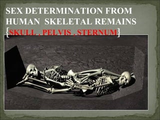
- 1. SEX DETERMINATION FROM HUMAN SKELETAL REMAINS { SKULL , PELVIS , STERNUM }
- 4. The human skeleton consists of both fused and individual bone. Fused bones include those of the pelvis and the cranium. At birth a newborn baby has approximately 300 bones, whereas on average an adult human has 206 bones . The difference comes from a number of small bones that fuse together during growth, such as the sacrum and coccyx of the vertebral column . Introduction Skeleton is an excellent material in living and non-living population for genetic, anthropological, odontologic and forensic investigations. Skull and bone features vary from male to female and differentiation is usually based on the male features that are typically more pronounced and marked than female features.
- 5. Krongman ranks accuracy of sex determination using the pelvis at 95% , the skull at 90%, the pelvis and skull at 98% and long bones at 80%. The determination of sex by examination of the skeleton is based upon the appearances of: 1. Pelvis (innominates + sacrum ) 2. Skull (cranium + mandible ) 3. Long bones (Humerus , Femur ) 4. Sternum 5. Scapula , metacarpal bones
- 6. SEXUAL DIMORPHISM Differences between men and women include all the features related to reproductive role, notably the endocrine (hormonal) systems and their physical, psychological and behavioral effects.
- 7. SEXUAL DIMORPHISM – BASIC PRINCIPLES The evaluation of sexual dimorphism in skeleton is generally based on two factors: 1. Size difference 2. Function related differences.
- 9. The skull is a bony structure which serves as the general framework for the head . The skull supports the structures of the face and protects the head against injury. The skull can be subdivided into two parts: the cranium and the mandible .
- 13. MALE FEMALE When compared, the female skull appears smaller and more gracile. The male skull is usually larger and more rugged.
- 14. This is the region directly above the orbit and nose, or the "brow ridge“. Less pronounced=female More pronounced=male Supraorbital ridges
- 15. ROUNDISH WITH SHARP MARGINS MALE FEMALE SQUARISH WITH ROUNDISH MARGINS ORBITS
- 16. The frontal bone (forehead) of males tends to be slanted back and on females it tends to be more rounded
- 17. GLABELLA WELL DEVELOPED ILL DEVELOPED MALE FEMALE
- 18. Zygomatic arches In females, the zygomatic arch is less pronounced, and tends to not extend posteriorly beyond the external auditory meatus. In males, the zygomatic arch is more pronounced or robust, and tends to extend posteriorly beyond the external auditory meatus.
- 20. MASTOID PROCESSES The mastoid processes are located on the inferior portion of the temporal bone, just posterior to the external auditory meatus.
- 21. EXTERNAL OCCIPITAL PROTUBERANCE
- 22. MALE FEMALE
- 23. The mandible together with the maxilla , the largest and strongest bone of the face . It forms the lower jaw and holds the lower teeth in place.
- 24. The male mandible tends to have a “square” shape. Mandible of female tends to have a pointed chin . Chin (anterior mandible)
- 25. Mandible in the male is closer to a right angle than the female. In the female, the ramus is an obtuse angle to the lower jaw bone, i.e., greater than 90 degrees. The ramus in the male is wider and larger. RAMUS (rear of the mandible) MALE FEMALE
- 26. LARGER,WIDER AND BROADER SMALLER AND NARROWER PALATE MALE FEMALE
- 27. The pelvis comprises the two innominates and the sacrum. PELVIS
- 28. The hip bone (or innominate bone ) is a large, flattened, irregularly shaped bone. Together with the sacrum and coccyx , it comprises the pelvis . Components It consists of three parts, the ilium , ischium , and pubis , which are distinct from each other in the young subject, but are fused in the adult. The best indicator of sex on the adult skeleton is the shape of the pubic bone of the pelvis. HIP BONE
- 29. Sex Determination from Pelvic Morphology The pelvic girdle is the most sexually dimorphic region of the skeleton, and it can be used to determine sex with a high degree of accuracy. The sexual dimorphism of the pelvis is primarily the result of reproductive mechanics, and is not readily apparent until adolescence.
- 32. Five features in innominate that indicate sex in pubic region are: (1) width of sciatic notch (inferior ilium) (2) subpubic angle (concavity) (3) ventral arc (on the pubis, near the symphysis, ventral) (4) ischio-pubic ramus (bone connecting pubis and ischium) PELVIS (5) acetabulum diameter (lateral innominate)
- 33. MALE FEMALE SMALL AND DEEP WIDER AND SHALLOWER SCIATIC NOTCH
- 34. SCIATIC NOTCH Generally, the sciatic notch tends to be wider in the female and narrower in the male.
- 36. The subpubic angle is much wider in females than in males, typically more that 90 degrees and less than 90 degrees, respectively. LESS AND “V” SHAPED WIDE AND TEND TO “U” SHAPED SUB-PUBIC ANGLE MALE FEMALE
- 38. It is the curved ridge of bone on anterior surface of the pubic bone. It is common in females and almost never seen in males.
- 41. The space in the middle of the pelvic bone (the pelvic inlet) is larger in women to facilitate birthing. The pelvic inlet is the space you see when both innominates and sacrum are articulated. PELVIC INLET
- 43. Sexual dimorphism The sacrum is noticeably sexually dimorphic . In the female the sacrum is shorter and wider than in the male. The bone is also directed more obliquely backward; this increases the size of the pelvic cavity. Straighter in males & curved in females. MALE FEMALE
- 45. <125 mm = female >155 mm = male These figures are from Bass