Downloaded 231 times


![MVO2[/min O2 in cardiac muscle] α CBF
Sales
• cardiac myocyte shortening
• contractility
• Relaxation
• myocardial wall tension
• heart rate
1st Qtr
2nd Qtr
3rd Qtr
4th Qtr](https://image.slidesharecdn.com/coronaryphysiology-140302125946-phpapp02/85/Coronary-physiology-11-320.jpg)
![Coronary collaterals imply
• Attenuate the degree of ischemia
• Degree of collaterals is variable[number,size and location]
• Chronic severe stenosis
• Less long term mortality](https://image.slidesharecdn.com/coronaryphysiology-140302125946-phpapp02/85/Coronary-physiology-12-320.jpg)


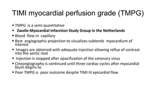
![The seat of coronary resistance
• Precapillary arterioles[R1] connect the epicardial arteries to the
myocardial capillaries and are the primary determinants of coronary
resistance and flow](https://image.slidesharecdn.com/coronaryphysiology-140302125946-phpapp02/85/Coronary-physiology-15-320.jpg)


![Multivessel disease and FFR
Reduces number of stents[FAME ]
No survival benefit revascularising FFR<0.75 of single stenosis
MACE are more with intervention
Revascularisation approach PCI vs. CABG is modified
Functional SYNTAX score improves the management style](https://image.slidesharecdn.com/coronaryphysiology-140302125946-phpapp02/85/Coronary-physiology-33-320.jpg)


The document discusses coronary hemodynamics, emphasizing the measurement of coronary function through various methods such as FFR, CFR, and IVUS. It highlights the complexities of coronary resistance, the role of auto-regulation in maintaining myocardial perfusion, and the significant impact of stenosis on myocardial blood flow. Additionally, it explores the relationship between hemodynamic assessments and interventions in the context of coronary artery disease.


