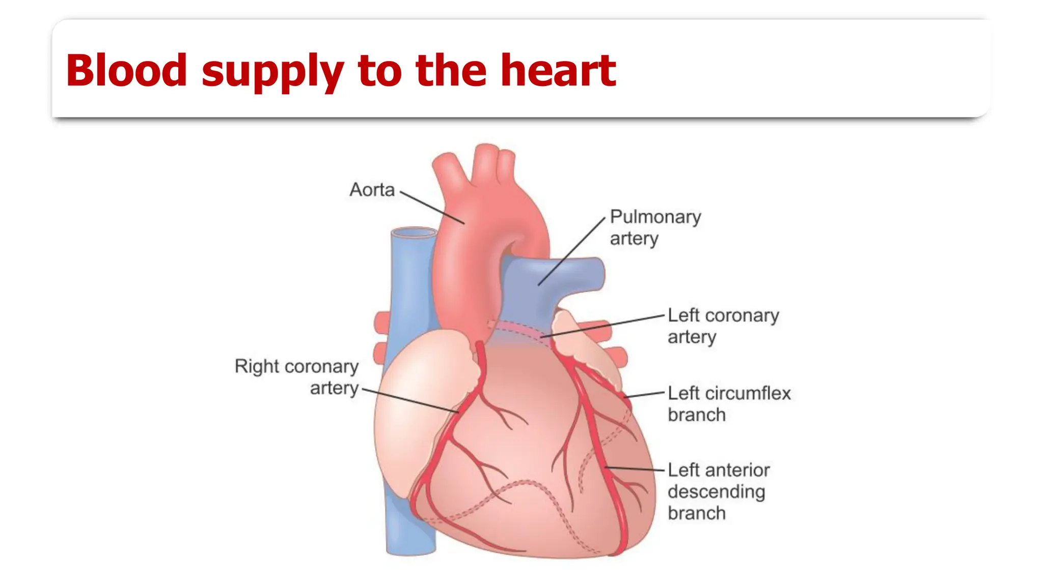
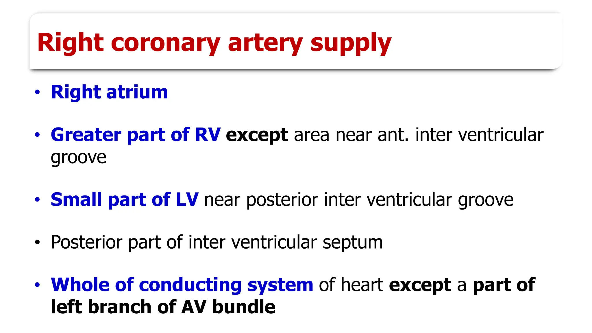
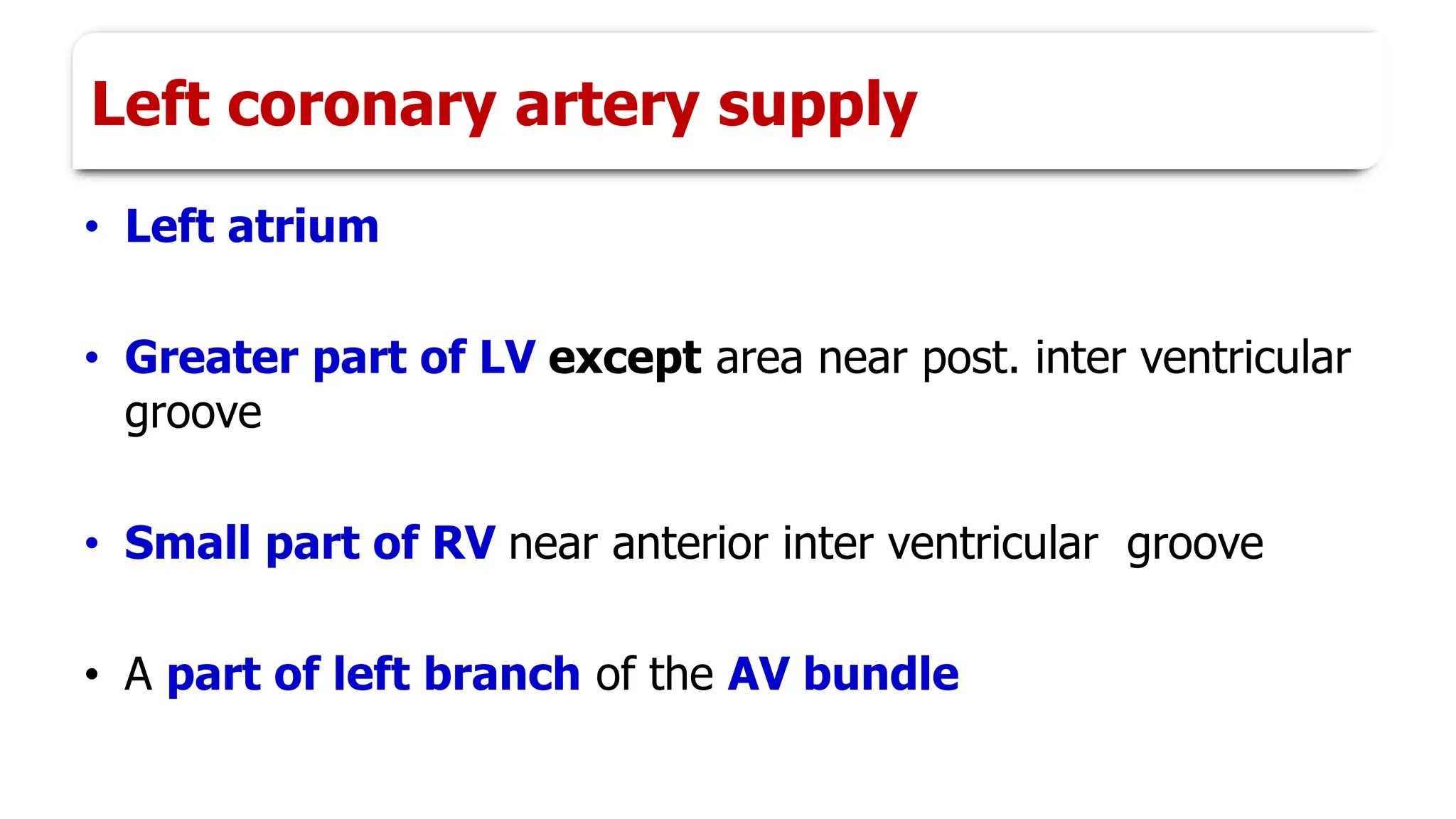
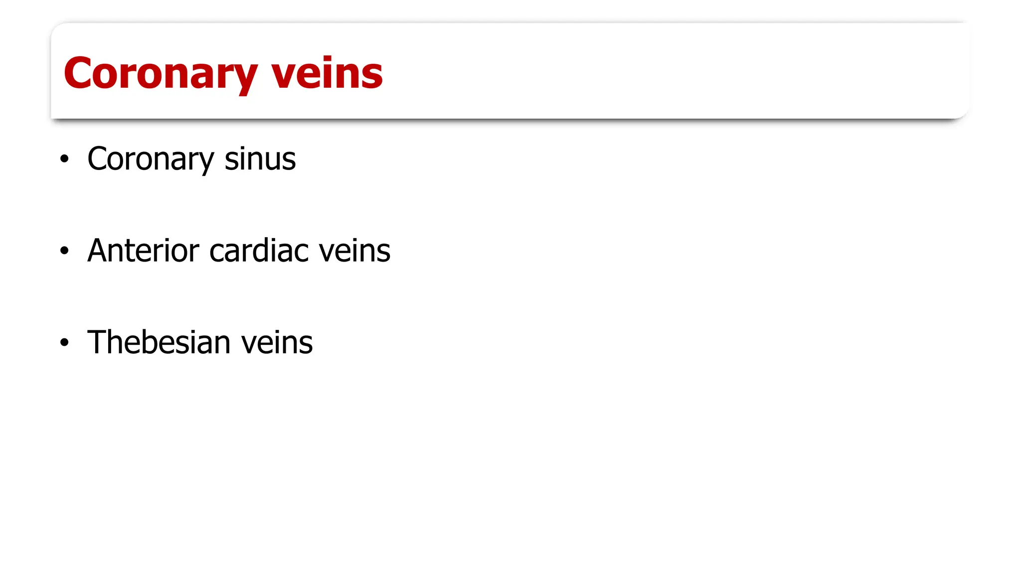
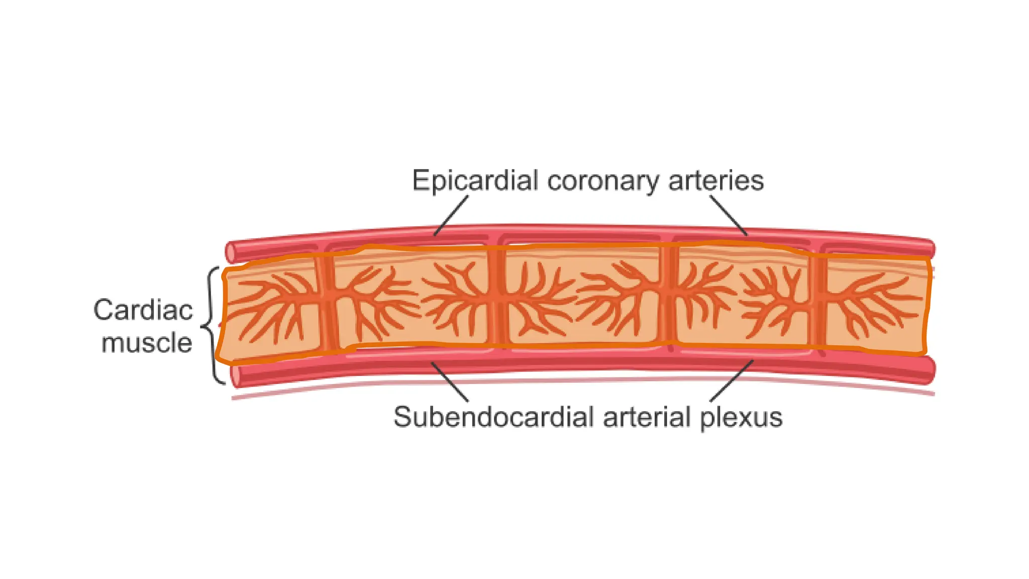
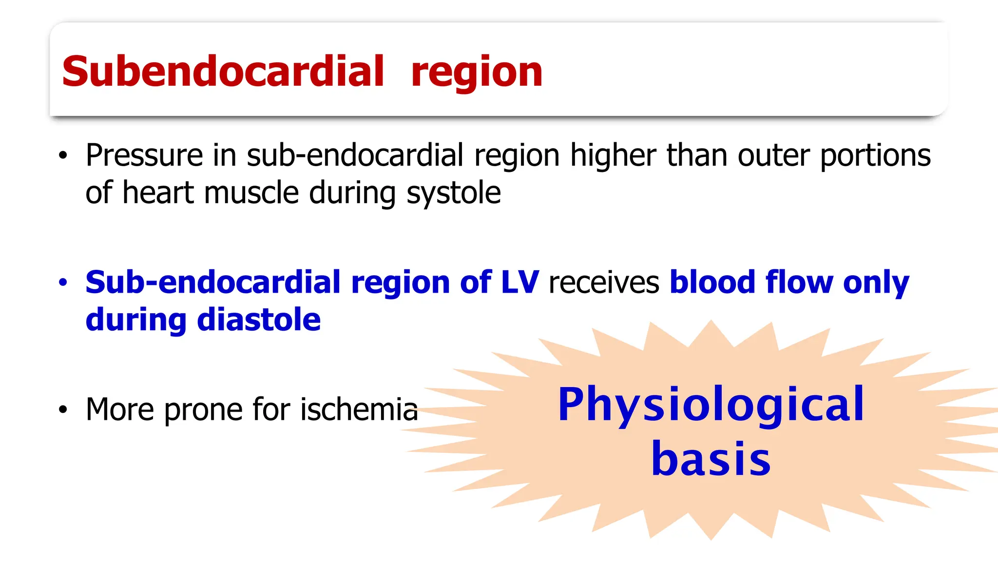
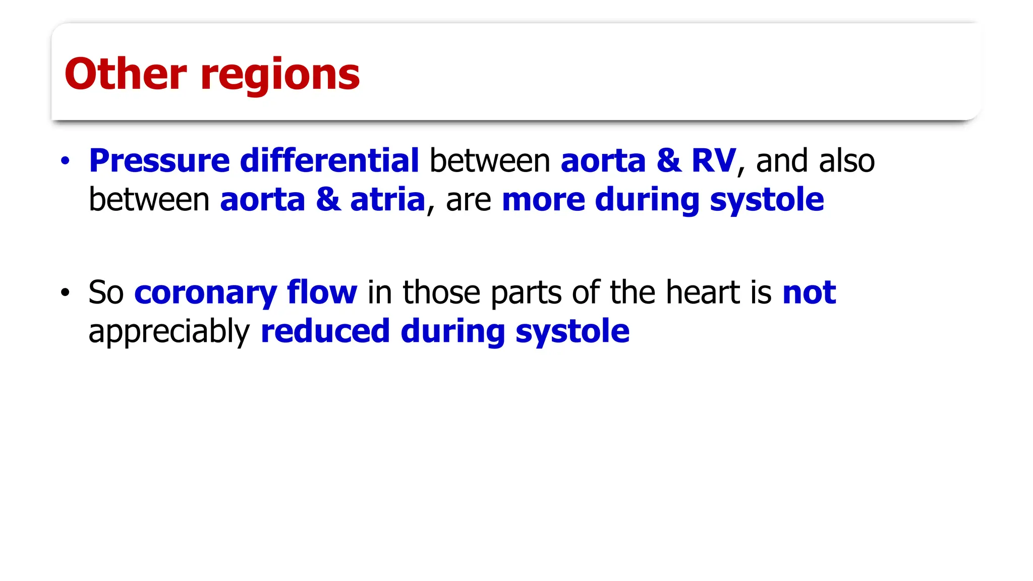
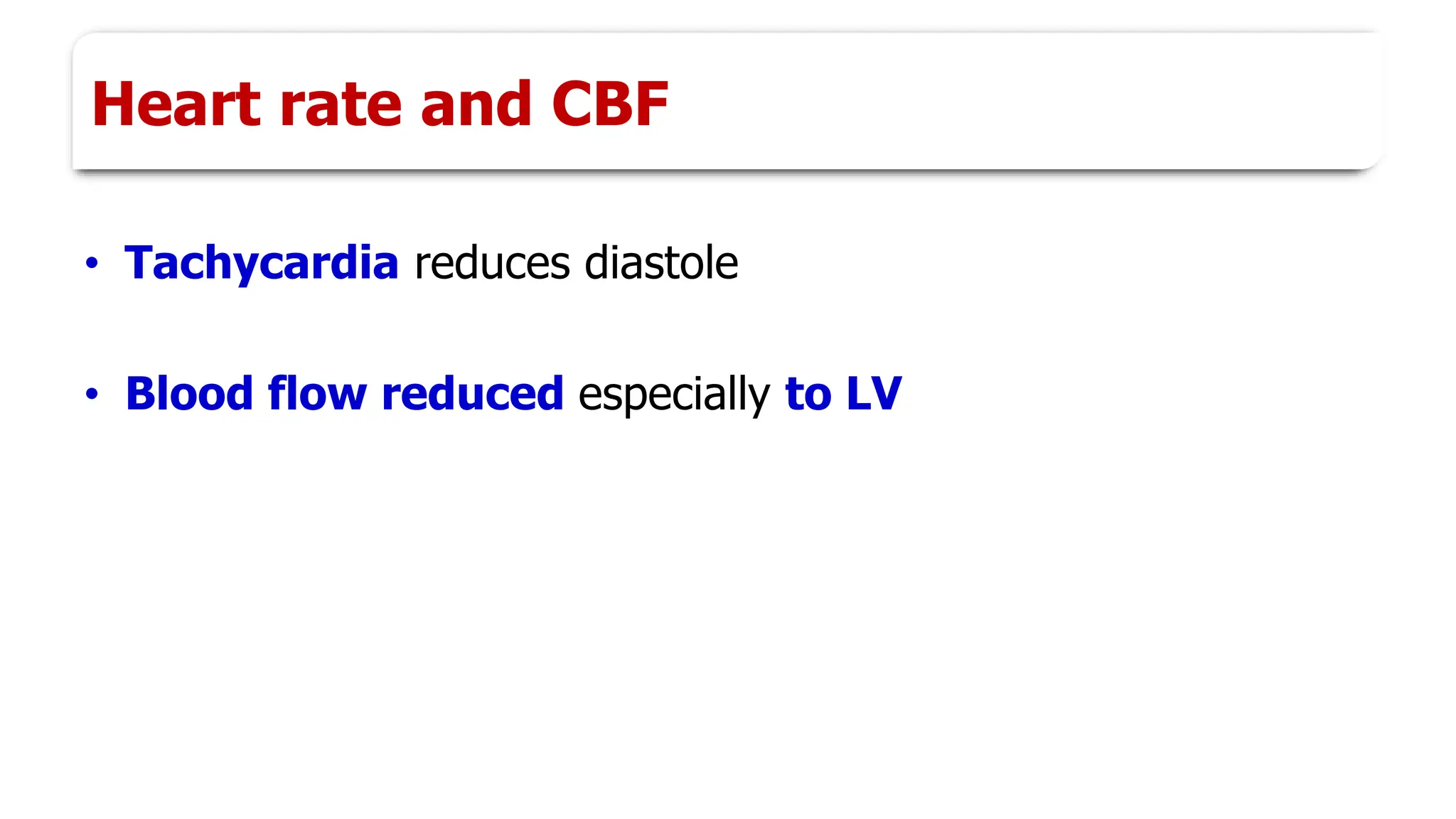
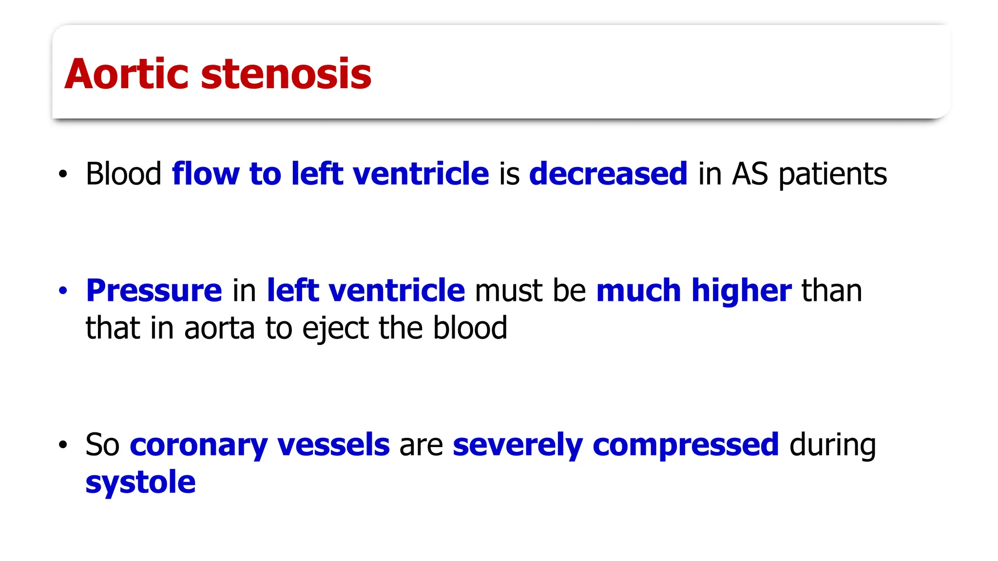
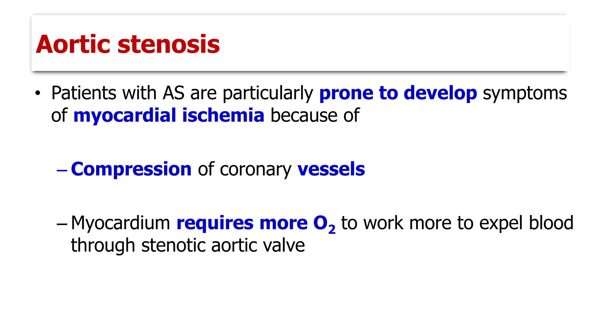
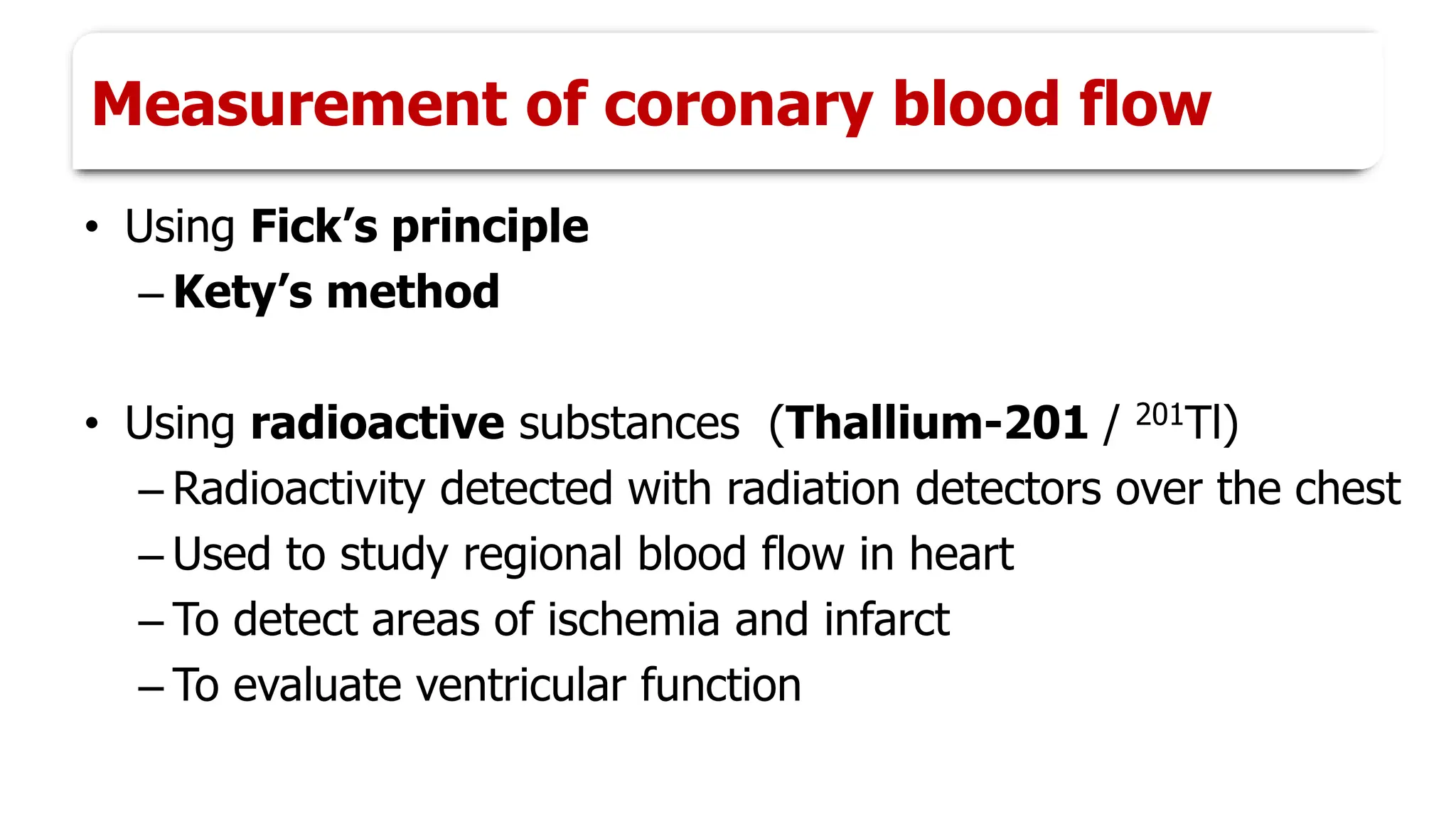
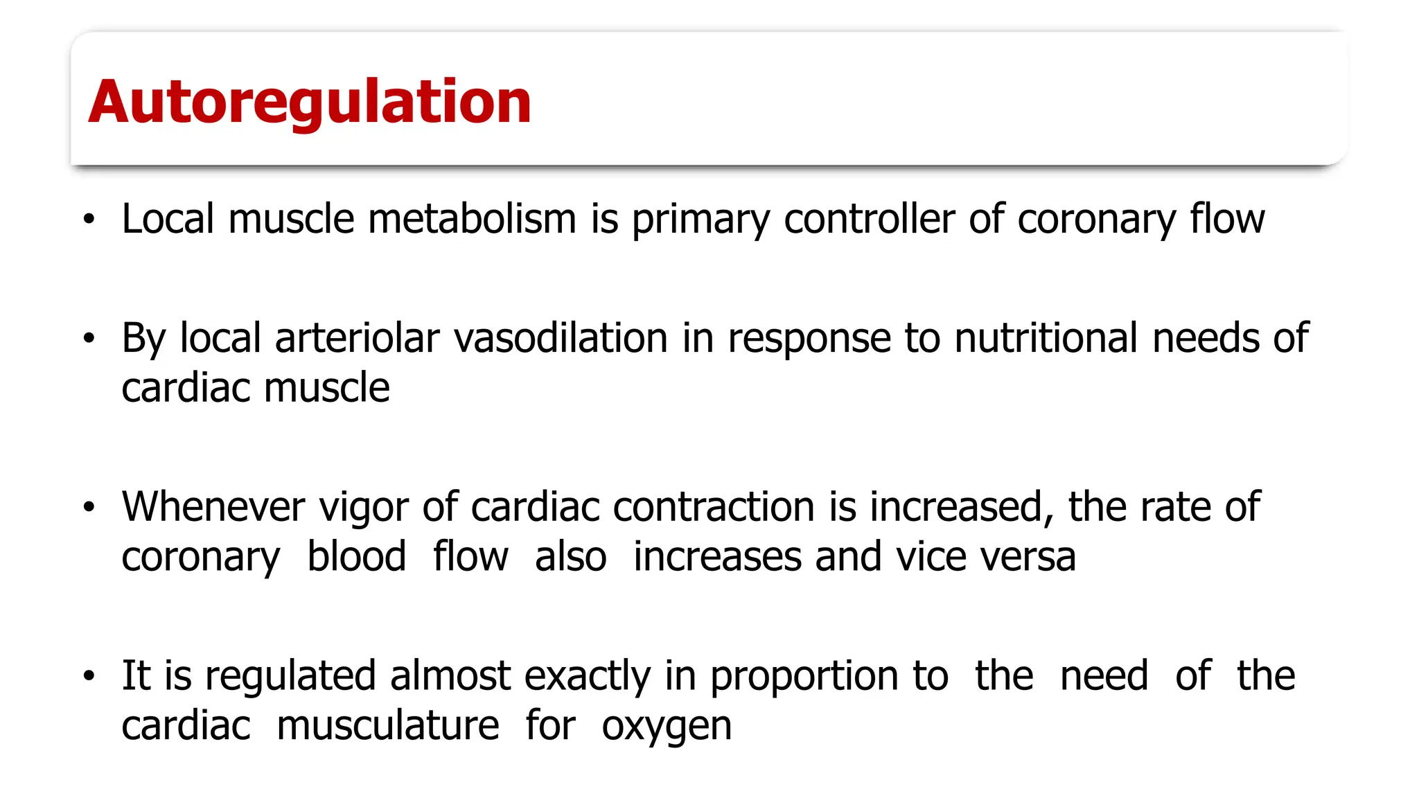
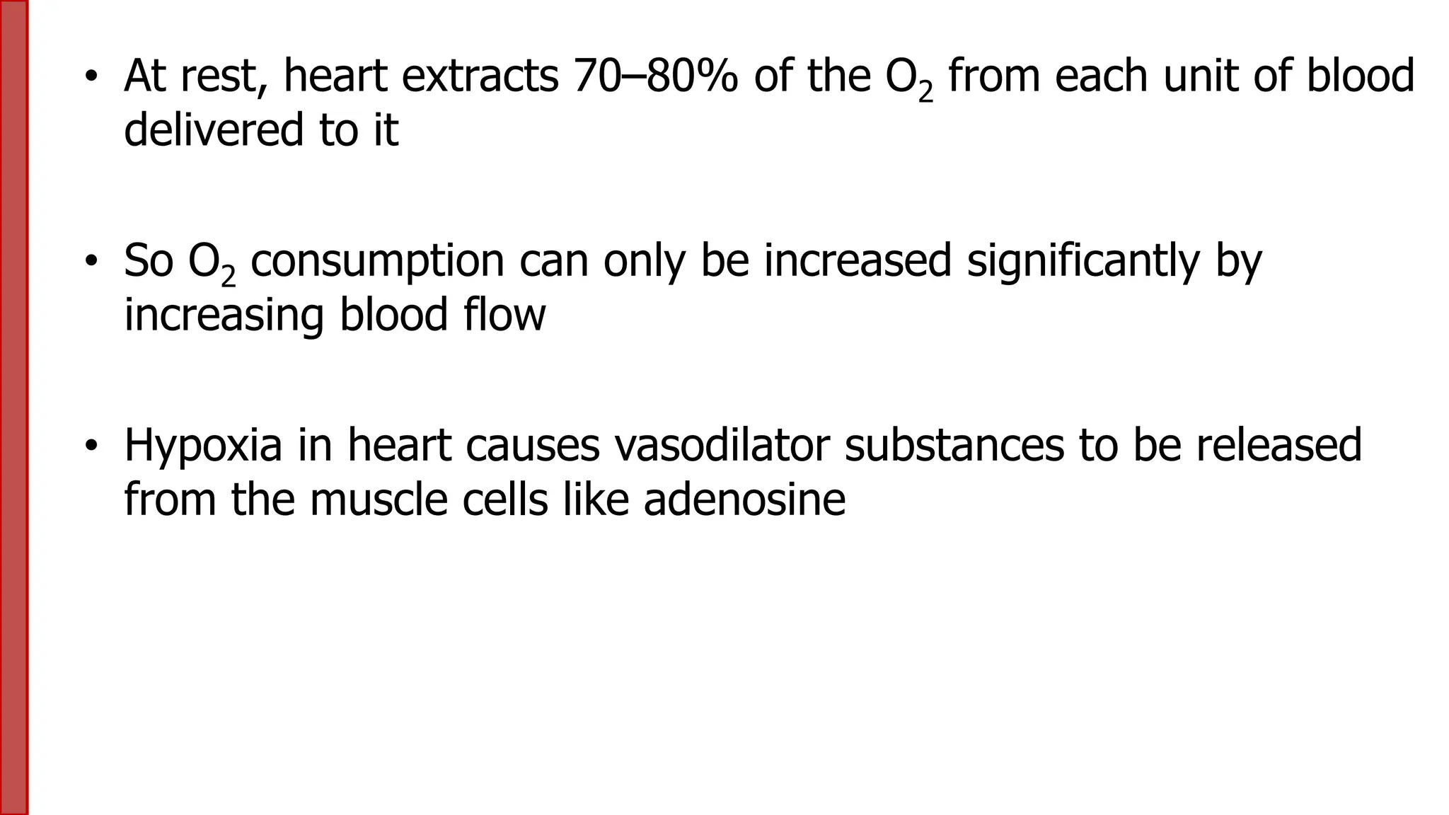
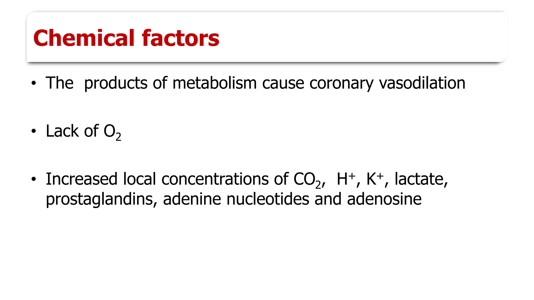
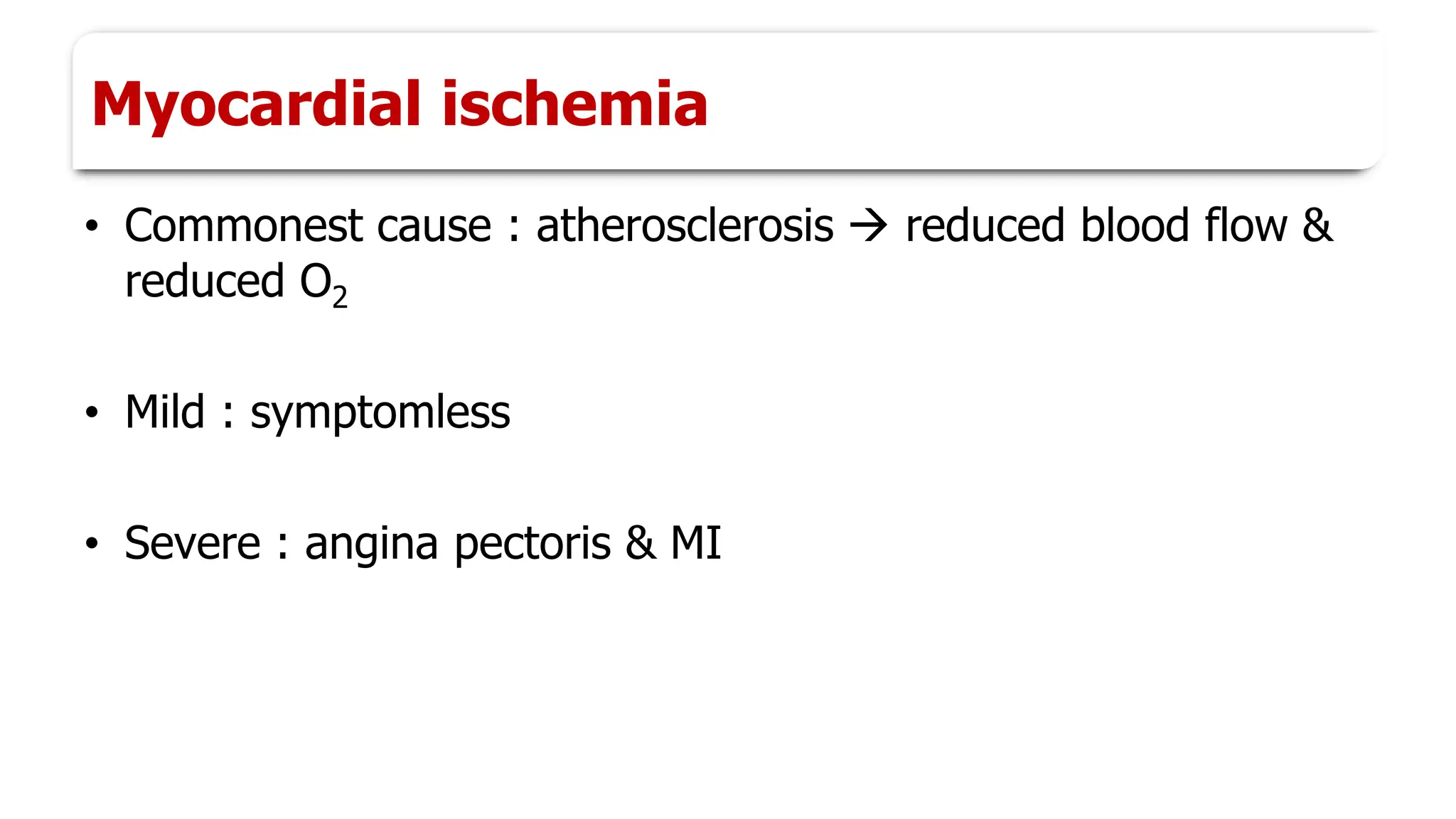
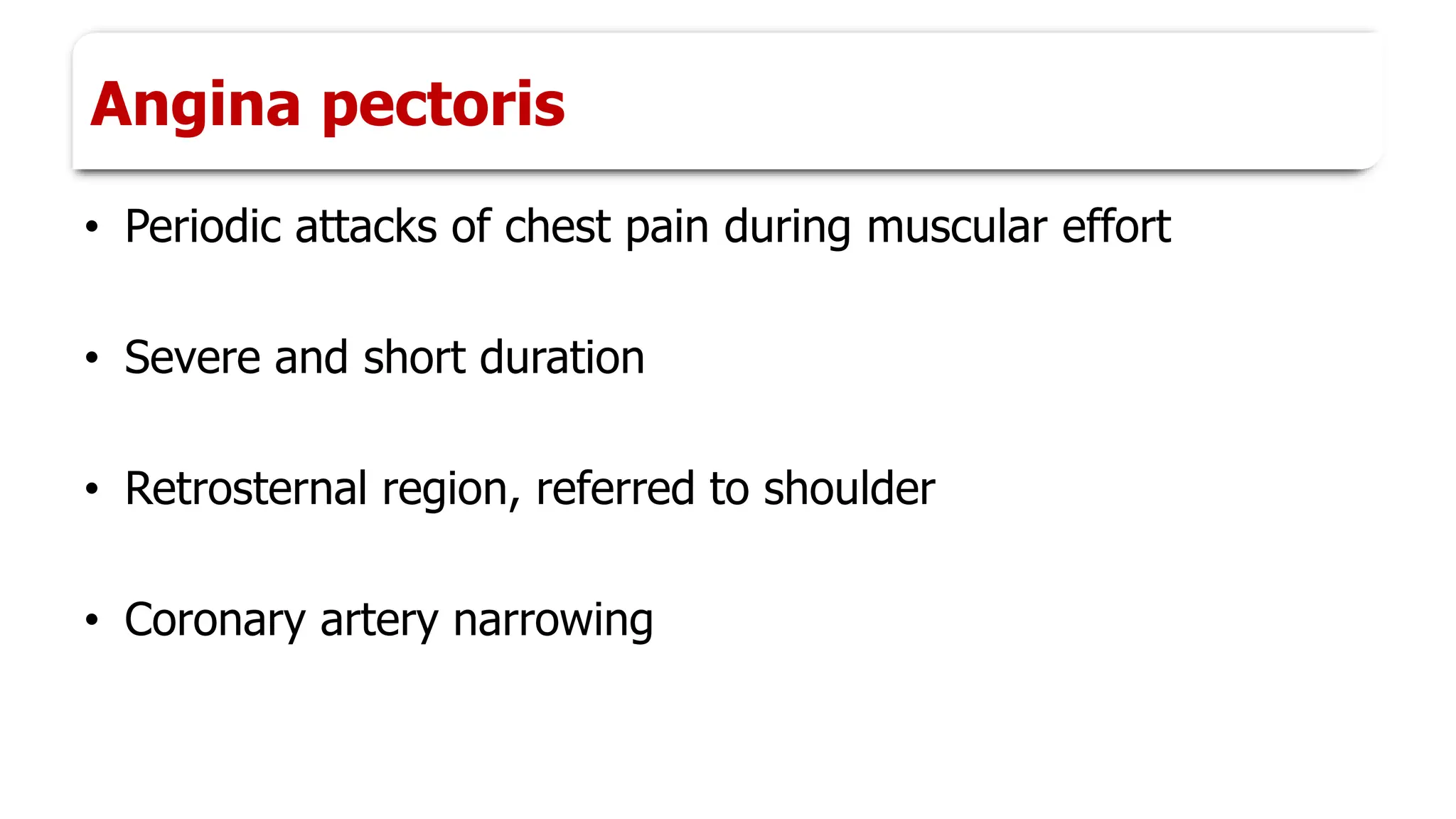
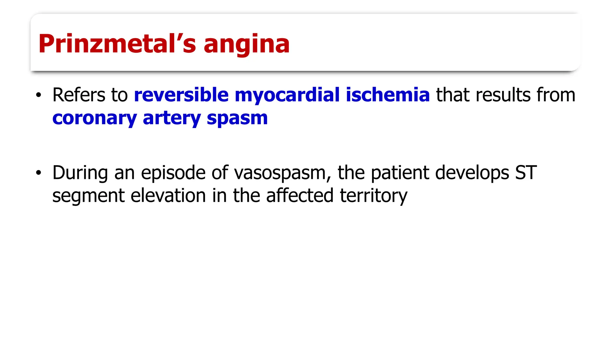
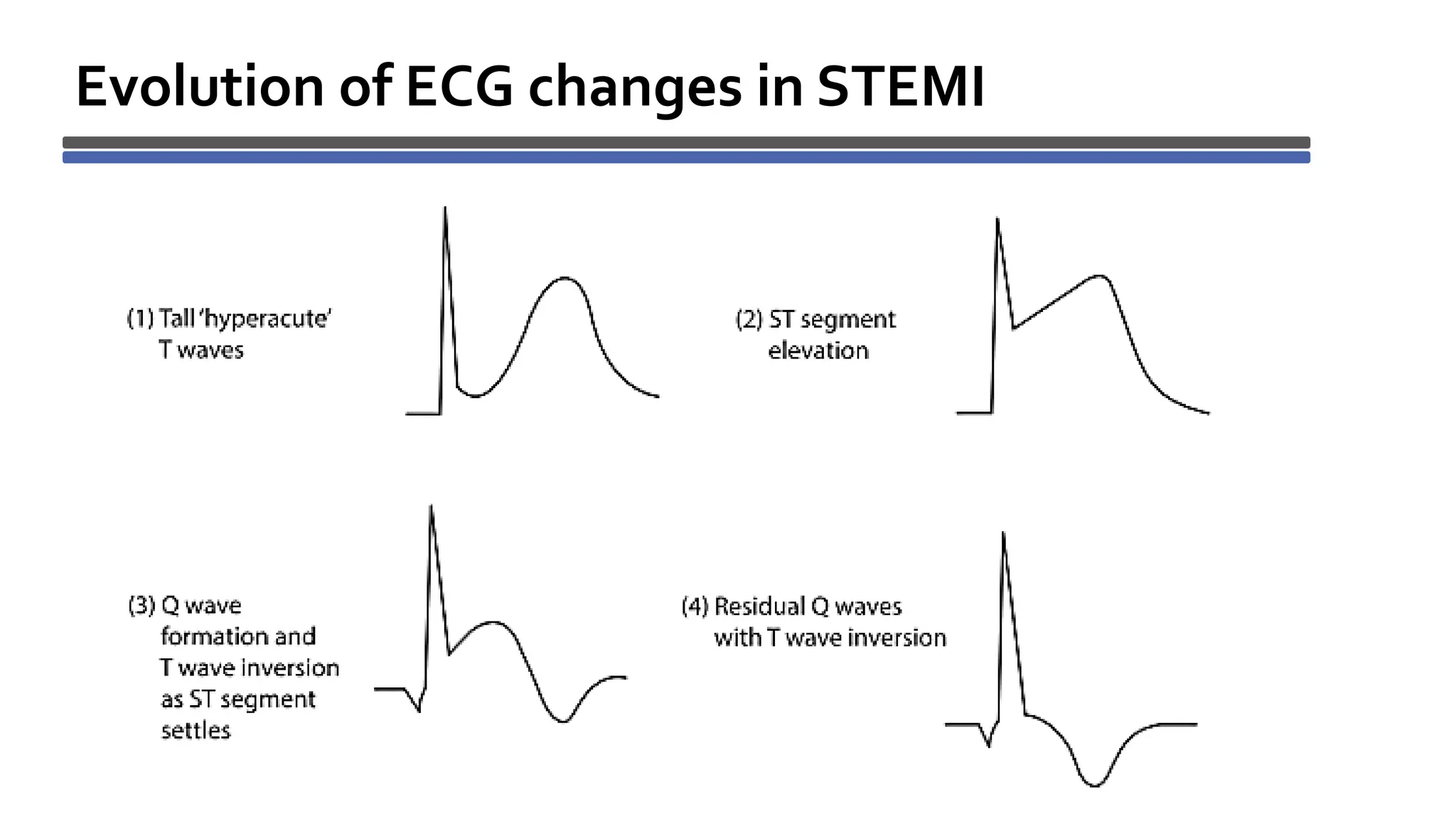
Coronary circulation
What we will discuss today…
• Functional Anatomy
• Special features of CBF
• Measurement of CBF
• Regulation of CBF
• Insufficient CBF
Blood supply to the heart
Right coronary artery supply
• Right atrium
• Greater part of RV except area near ant. inter ventricular
groove
• Small part of LV near posterior inter ventricular groove
• Posterior part of inter ventricular septum
• Whole of conducting system of heart except a part of
left branch of AV bundle
Left coronary artery supply
• Left atrium
• Greater part of LV except area near post. inter ventricular
groove
• Small part of RV near anterior inter ventricular groove
• A part of left branch of the AV bundle
Coronary veins
• Coronary sinus
• Anterior cardiac veins
• Thebesian veins
What we will discuss today…
• Functional Anatomy
• Special features of CBF
• Measurement of CBF
• Regulation of CBF
• Insufficient CBF
What we will discuss today…
• Functional Anatomy
• Special features of CBF
• Measurement of CBF
• Regulation of CBF
• Insufficient CBF
Special features of Coronary Blood
Flow
Special features
• Normal coronary flow : 250 ml/min
• 70ml/100g tissue/min
• 5% of total cardiac output
Phasic changes of coronary flow
SYSTOLE
DIASTOLE
DIASTOLE
SYSTOLE
DIASTOLE
DIASTOLE
Phasic flow
• Intramural vessels are compressed in Iso Volumet. Contract.
• Aortic pressure will be very low during IVC
• So, no flow during IVC
• Variable flow during rest of systole
• Maximal flow occurs in diastole as vessels dilate→ 80%
Left coronary artery
• LV pressure is high, sharp fall in flow in IVC phase
• During rapid ejection phase→ aortic pressure rises → flow
increases rapidly
• During reduced ejection phase →aortic pressure falls →
flow decreases
• During diastole, maximum flow , remains high and falls
gradually
Right coronary artery
• Similar phasic changes occur
• RV systolic pressure is low : 25 mm Hg
• Reduction in flow during IVC
• Flow increases first and declines later in systole
• Flow increases during diastole
Subendocardial region
• Pressure in sub-endocardial region higher than outer portions
of heart muscle during systole
• Sub-endocardial region of LV receives blood flow only
during diastole
• More prone for ischemia Physiological
basis
Other regions
• Pressure differential between aorta & RV, and also
between aorta & atria, are more during systole
• So coronary flow in those parts of the heart is not
appreciably reduced during systole
Heart rate and CBF
• Tachycardia reduces diastole
• Blood flow reduced especially to LV
• Blood flow to left ventricle is decreased in AS patients
• Pressure in left ventricle must be much higher than
that in aorta to eject the blood
• So coronary vessels are severely compressed during
systole
Aortic stenosis
• Patients with AS are particularly prone to develop symptoms
of myocardial ischemia because of
– Compression of coronary vessels
– Myocardium requires more O2 to work more to expel blood
through stenotic aortic valve
Aortic stenosis
What we will discuss today…
• Functional Anatomy
• Special features of CBF
• Measurement of CBF
• Regulation of CBF
• Insufficient CBF
What we will discuss today…
• Functional Anatomy
• Special features of CBF
• Measurement of CBF
• Regulation of CBF
• Insufficient CBF
Measurement of Coronary Blood
Flow
Measurement of coronary blood flow
• Using Fick’s principle
– Kety’s method
• Using radioactive substances (Thallium-201 / 201Tl)
– Radioactivity detected with radiation detectors over the chest
– Used to study regional blood flow in heart
– To detect areas of ischemia and infarct
– To evaluate ventricular function
What we will discuss today…
• Functional Anatomy
• Special features of CBF
• Measurement of CBF
• Regulation of CBF
• Insufficient CBF
What we will discuss today…
• Functional Anatomy
• Special features of CBF
• Measurement of CBF
• Regulation of CBF
• Insufficient CBF
Regulation of Coronary Blood Flow
Autoregulation
• Local muscle metabolism is primary controller of coronary flow
• By local arteriolar vasodilation in response to nutritional needs of
cardiac muscle
• Whenever vigor of cardiac contraction is increased, the rate of
coronary blood flow also increases and vice versa
• It is regulated almost exactly in proportion to the need of the
cardiac musculature for oxygen
• At rest, heart extracts 70–80% of the O2 from each unit of blood
delivered to it
• So O2 consumption can only be increased significantly by
increasing blood flow
• Hypoxia in heart causes vasodilator substances to be released
from the muscle cells like adenosine
Chemical factors
• The products of metabolism cause coronary vasodilation
• Lack of O2
• Increased local concentrations of CO2, H+, K+, lactate,
prostaglandins, adenine nucleotides and adenosine
Reactive hyperemia
• If coronary artery is briefly occluded → release of obstruction
→ increase in blood flow
• Release of adenosine by hypoxia
Neural factors
• Coronary arterioles contain
– α-adrenergic receptors – mediate vasoconstriction
– β-adrenergic receptors – mediate vasodilation
• Direct effect of noradrenergic stimulation is constriction
• But activity in noradrenergic nerves to heart → coronary
vasodilation
• Norepinephrine increases the heart rate and the force of
contraction
• Vasodilation is due to production of vasodilator metabolites
in the myocardium secondary to the increase in its activity
• When BP falls, overall effect of reflex increase in noradrenergic
discharge is increased coronary blood flow
• Simultaneously cutaneous, renal, and splanchnic vessels are
constricted
• In this way circulation of the heart is preserved when flow to
other organs is compromised
• Direct effect of stimulation of vagal fibers to heart is coronary
vasodilation
What we will discuss today…
• Functional Anatomy
• Special features of CBF
• Measurement of CBF
• Regulation of CBF
• Insufficient CBF
What we will discuss today…
• Functional Anatomy
• Special features of CBF
• Measurement of CBF
• Regulation of CBF
• Insufficient CBF
Insufficient Coronary Blood Flow
Myocardial ischemia
• Commonest cause : atherosclerosis → reduced blood flow &
reduced O2
• Mild : symptomless
• Severe : angina pectoris & MI
Angina pectoris
• Periodic attacks of chest pain during muscular effort
• Severe and short duration
• Retrosternal region, referred to shoulder
• Coronary artery narrowing
Prinzmetal’s angina
• Refers to reversible myocardial ischemia that results from
coronary artery spasm
• During an episode of vasospasm, the patient develops ST
segment elevation in the affected territory
Role of Collateral Circulation in Heart
• Anastomoses exist among the smaller arteries sized 20 to 250
micrometers in diameter
• Damage to heart muscle is determined mainly by the degree
of collateral circulation
– that has already developed or
– that can open within minutes after the occlusion
Role of Collateral Circulation in Heart
• These developing collateral channels help in almost complete
recovery, when area of muscle involved is not too great
• Regular aerobic exercise promotes formation of collaterals
Myocardial infarction
• Rupture of an atheromatous plaque
• Coronary thrombosis → occlusion→ necrosis →death of
tissue
• Fibrosis and scar formation
Symptoms
• Angina pectoris
• Profuse sweating
• Cold extremities
Diagnosis
• ECG
• Serum enzymes
– CK-MB (MB isomer of creatine kinase)
– Troponin T
– Troponin I
Evolution of ECG changes in STEMI
Intensive coronary care unit
• Sedation : opioid analgesics
• Complete rest to prevent “coronary steal” syndrome
• If heart becomes excessively active, vessels of normal
musculature become greatly dilated
• This allows most of blood to flow through normal muscle
tissue
• So blood flow through collateral channels into ischemic area
decreases and ischemia worsens
• Nitrates like nitroglycerine
• Vasodilator used in the treatment of angina
• Acts by releasing NO
Thrombolysis
• Streptokinase : fibrinolysis by plasmin
• Tissue plasminogen activator ( t-PA)
Surgical procedures
• Coronary artery angioplasty
• Coronary artery bypass graft
Role of low dose aspirin
• Aspirin inhibits thromboxane A2
• Inhibits platelet aggregation
• Useful in preventing MI
Role of beta blockers
• Block sympathetic beta-adrenergic receptors
• So prevents sympathetic enhancement of heart rate &
cardiac metabolism during exercise or emotional episodes
• Helps in reducing the number of anginal attacks, as well as
their severity
Complications
• Cardiogenic shock
• Cardiac arrhythmias
• Left ventricular failure → Pulmonary edema
• Rupture of heart
What we will discuss today…
• Functional Anatomy
• Special features of CBF
• Measurement of CBF
• Regulation of CBF
• Insufficient CBF
What we will discuss today…
• Functional Anatomy
• Special features of CBF
• Measurement of CBF
• Regulation of CBF
• Insufficient CBF
CVS 2022 - Coronary circulation.pdf functional anatomt,special features of cbf.regulation of cbf

CVS 2022 - Coronary circulation.pdf functional anatomt,special features of cbf.regulation of cbf

  • 1.
  • 2.
    What we willdiscuss today… • Functional Anatomy • Special features of CBF • Measurement of CBF • Regulation of CBF • Insufficient CBF
  • 3.
    Blood supply tothe heart
  • 4.
    Right coronary arterysupply • Right atrium • Greater part of RV except area near ant. inter ventricular groove • Small part of LV near posterior inter ventricular groove • Posterior part of inter ventricular septum • Whole of conducting system of heart except a part of left branch of AV bundle
  • 5.
    Left coronary arterysupply • Left atrium • Greater part of LV except area near post. inter ventricular groove • Small part of RV near anterior inter ventricular groove • A part of left branch of the AV bundle
  • 6.
    Coronary veins • Coronarysinus • Anterior cardiac veins • Thebesian veins
  • 7.
    What we willdiscuss today… • Functional Anatomy • Special features of CBF • Measurement of CBF • Regulation of CBF • Insufficient CBF
  • 8.
    What we willdiscuss today… • Functional Anatomy • Special features of CBF • Measurement of CBF • Regulation of CBF • Insufficient CBF
  • 9.
    Special features ofCoronary Blood Flow
  • 10.
    Special features • Normalcoronary flow : 250 ml/min • 70ml/100g tissue/min • 5% of total cardiac output
  • 11.
    Phasic changes ofcoronary flow SYSTOLE DIASTOLE DIASTOLE
  • 12.
  • 14.
    Phasic flow • Intramuralvessels are compressed in Iso Volumet. Contract. • Aortic pressure will be very low during IVC • So, no flow during IVC • Variable flow during rest of systole • Maximal flow occurs in diastole as vessels dilate→ 80%
  • 15.
    Left coronary artery •LV pressure is high, sharp fall in flow in IVC phase • During rapid ejection phase→ aortic pressure rises → flow increases rapidly • During reduced ejection phase →aortic pressure falls → flow decreases • During diastole, maximum flow , remains high and falls gradually
  • 16.
    Right coronary artery •Similar phasic changes occur • RV systolic pressure is low : 25 mm Hg • Reduction in flow during IVC • Flow increases first and declines later in systole • Flow increases during diastole
  • 18.
    Subendocardial region • Pressurein sub-endocardial region higher than outer portions of heart muscle during systole • Sub-endocardial region of LV receives blood flow only during diastole • More prone for ischemia Physiological basis
  • 19.
    Other regions • Pressuredifferential between aorta & RV, and also between aorta & atria, are more during systole • So coronary flow in those parts of the heart is not appreciably reduced during systole
  • 20.
    Heart rate andCBF • Tachycardia reduces diastole • Blood flow reduced especially to LV
  • 21.
    • Blood flowto left ventricle is decreased in AS patients • Pressure in left ventricle must be much higher than that in aorta to eject the blood • So coronary vessels are severely compressed during systole Aortic stenosis
  • 22.
    • Patients withAS are particularly prone to develop symptoms of myocardial ischemia because of – Compression of coronary vessels – Myocardium requires more O2 to work more to expel blood through stenotic aortic valve Aortic stenosis
  • 23.
    What we willdiscuss today… • Functional Anatomy • Special features of CBF • Measurement of CBF • Regulation of CBF • Insufficient CBF
  • 24.
    What we willdiscuss today… • Functional Anatomy • Special features of CBF • Measurement of CBF • Regulation of CBF • Insufficient CBF
  • 25.
  • 26.
    Measurement of coronaryblood flow • Using Fick’s principle – Kety’s method • Using radioactive substances (Thallium-201 / 201Tl) – Radioactivity detected with radiation detectors over the chest – Used to study regional blood flow in heart – To detect areas of ischemia and infarct – To evaluate ventricular function
  • 27.
    What we willdiscuss today… • Functional Anatomy • Special features of CBF • Measurement of CBF • Regulation of CBF • Insufficient CBF
  • 28.
    What we willdiscuss today… • Functional Anatomy • Special features of CBF • Measurement of CBF • Regulation of CBF • Insufficient CBF
  • 29.
  • 30.
    Autoregulation • Local musclemetabolism is primary controller of coronary flow • By local arteriolar vasodilation in response to nutritional needs of cardiac muscle • Whenever vigor of cardiac contraction is increased, the rate of coronary blood flow also increases and vice versa • It is regulated almost exactly in proportion to the need of the cardiac musculature for oxygen
  • 31.
    • At rest,heart extracts 70–80% of the O2 from each unit of blood delivered to it • So O2 consumption can only be increased significantly by increasing blood flow • Hypoxia in heart causes vasodilator substances to be released from the muscle cells like adenosine
  • 32.
    Chemical factors • Theproducts of metabolism cause coronary vasodilation • Lack of O2 • Increased local concentrations of CO2, H+, K+, lactate, prostaglandins, adenine nucleotides and adenosine
  • 33.
    Reactive hyperemia • Ifcoronary artery is briefly occluded → release of obstruction → increase in blood flow • Release of adenosine by hypoxia
  • 34.
    Neural factors • Coronaryarterioles contain – α-adrenergic receptors – mediate vasoconstriction – β-adrenergic receptors – mediate vasodilation • Direct effect of noradrenergic stimulation is constriction • But activity in noradrenergic nerves to heart → coronary vasodilation
  • 35.
    • Norepinephrine increasesthe heart rate and the force of contraction • Vasodilation is due to production of vasodilator metabolites in the myocardium secondary to the increase in its activity • When BP falls, overall effect of reflex increase in noradrenergic discharge is increased coronary blood flow
  • 36.
    • Simultaneously cutaneous,renal, and splanchnic vessels are constricted • In this way circulation of the heart is preserved when flow to other organs is compromised • Direct effect of stimulation of vagal fibers to heart is coronary vasodilation
  • 37.
    What we willdiscuss today… • Functional Anatomy • Special features of CBF • Measurement of CBF • Regulation of CBF • Insufficient CBF
  • 38.
    What we willdiscuss today… • Functional Anatomy • Special features of CBF • Measurement of CBF • Regulation of CBF • Insufficient CBF
  • 39.
  • 40.
    Myocardial ischemia • Commonestcause : atherosclerosis → reduced blood flow & reduced O2 • Mild : symptomless • Severe : angina pectoris & MI
  • 41.
    Angina pectoris • Periodicattacks of chest pain during muscular effort • Severe and short duration • Retrosternal region, referred to shoulder • Coronary artery narrowing
  • 42.
    Prinzmetal’s angina • Refersto reversible myocardial ischemia that results from coronary artery spasm • During an episode of vasospasm, the patient develops ST segment elevation in the affected territory
  • 43.
    Role of CollateralCirculation in Heart • Anastomoses exist among the smaller arteries sized 20 to 250 micrometers in diameter • Damage to heart muscle is determined mainly by the degree of collateral circulation – that has already developed or – that can open within minutes after the occlusion
  • 44.
    Role of CollateralCirculation in Heart • These developing collateral channels help in almost complete recovery, when area of muscle involved is not too great • Regular aerobic exercise promotes formation of collaterals
  • 45.
    Myocardial infarction • Ruptureof an atheromatous plaque • Coronary thrombosis → occlusion→ necrosis →death of tissue • Fibrosis and scar formation
  • 46.
    Symptoms • Angina pectoris •Profuse sweating • Cold extremities
  • 47.
    Diagnosis • ECG • Serumenzymes – CK-MB (MB isomer of creatine kinase) – Troponin T – Troponin I
  • 48.
    Evolution of ECGchanges in STEMI
  • 49.
    Intensive coronary careunit • Sedation : opioid analgesics • Complete rest to prevent “coronary steal” syndrome • If heart becomes excessively active, vessels of normal musculature become greatly dilated • This allows most of blood to flow through normal muscle tissue • So blood flow through collateral channels into ischemic area decreases and ischemia worsens
  • 50.
    • Nitrates likenitroglycerine • Vasodilator used in the treatment of angina • Acts by releasing NO
  • 51.
    Thrombolysis • Streptokinase :fibrinolysis by plasmin • Tissue plasminogen activator ( t-PA)
  • 52.
    Surgical procedures • Coronaryartery angioplasty • Coronary artery bypass graft
  • 55.
    Role of lowdose aspirin • Aspirin inhibits thromboxane A2 • Inhibits platelet aggregation • Useful in preventing MI
  • 56.
    Role of betablockers • Block sympathetic beta-adrenergic receptors • So prevents sympathetic enhancement of heart rate & cardiac metabolism during exercise or emotional episodes • Helps in reducing the number of anginal attacks, as well as their severity
  • 57.
    Complications • Cardiogenic shock •Cardiac arrhythmias • Left ventricular failure → Pulmonary edema • Rupture of heart
  • 58.
    What we willdiscuss today… • Functional Anatomy • Special features of CBF • Measurement of CBF • Regulation of CBF • Insufficient CBF
  • 59.
    What we willdiscuss today… • Functional Anatomy • Special features of CBF • Measurement of CBF • Regulation of CBF • Insufficient CBF