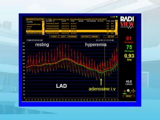
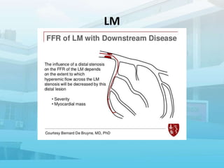
This document discusses various techniques for assessing the physiological significance of coronary artery lesions seen on angiography. It describes:
1. Borderline or intermediate lesions that appear between 40-70% blocked on angiography but their physiological significance is unclear.
2. Various pressure-based indices that can be measured invasively using a wire, such as FFR, iFR, and others, to determine if a lesion is hemodynamically significant.
3. Additional modalities like IVUS and OCT that provide anatomical data to complement the functional assessment of lesions.
Various coronary physiological measurements can be made in the cardiac catheterization laboratory using sensor-tipped guidewires; they include the measurement of poststenotic absolute coronary flow reserve, the relative coronary flow reserve, and the pressure-derived fractional flow reserve of the myocardium. Ambiguity regarding abnormal microcirculation has been reduced or eliminated with measurements of relative coronary flow reserve and fractional flow reserve. The role of microvascular flow impairment can be separately determined with coronary flow velocity reserve measurements. In addition to lesion assessment before and after intervention, emerging applications of coronary physiology include the determination of physiological responses to new pharmacological agents, such as glycoprotein IIb/IIIa blockers, in patients with acute myocardial infarction. Measurements of coronary physiology in the catheterization laboratory provide objective data that complement angiography for clinical decision-making
LECTURE ON ATRIAL FIBRILLATION TO 9TH TERM MEDICAL STUDENTS REFERENCES: DAVIDSON(2018) HARRISON 20TH ED OF MEDICINE AND 2020 EUROPEAN HEART GUIDELINES ON AF
Echocardiography for Cardiothoracic Surgeons | IACTS SCORE 2020IACTSWeb
These slides illustrates the basics of transthoracic and transoesophageal echocardiography in the cardiac operating room.
Competent surgical results in shunt lesions, valve repairs, surgery for heart failure, establishment of peripheral bypass are derivatives of good knowledge in assessing gross and finer details of the heart intro-operatively. Assessment of myocardial viability, contractility and patency of repair are few aspects that are covered under this subject. They illustrate some of the basic must-knows for cardiothoracic surgeons from the perspective of a cardiac anaesthetist.
This is courtesy of Dr. Parimala Prasanna Simha, Professor of Cardiac Anaesthesiology and Critical Care at Jayadeva Institute of Cardiovascular Sciences and Research, Bengaluru.
This presetation is part of a video which belongs to the lecture series of IACTS SCORE 2020 held at the Sri Sathya Sai Institute of Higher Medical Sciences Whitefield, Bengaluru between 7th and 8th March, 2020.
Local Advanced Lung Cancer: Artificial Intelligence, Synergetics, Complex Sys...Oleg Kshivets
Overall life span (LS) was 1671.7±1721.6 days and cumulative 5YS reached 62.4%, 10 years – 50.4%, 20 years – 44.6%. 94 LCP lived more than 5 years without cancer (LS=2958.6±1723.6 days), 22 – more than 10 years (LS=5571±1841.8 days). 67 LCP died because of LC (LS=471.9±344 days). AT significantly improved 5YS (68% vs. 53.7%) (P=0.028 by log-rank test). Cox modeling displayed that 5YS of LCP significantly depended on: N0-N12, T3-4, blood cell circuit, cell ratio factors (ratio between cancer cells-CC and blood cells subpopulations), LC cell dynamics, recalcification time, heparin tolerance, prothrombin index, protein, AT, procedure type (P=0.000-0.031). Neural networks, genetic algorithm selection and bootstrap simulation revealed relationships between 5YS and N0-12 (rank=1), thrombocytes/CC (rank=2), segmented neutrophils/CC (3), eosinophils/CC (4), erythrocytes/CC (5), healthy cells/CC (6), lymphocytes/CC (7), stick neutrophils/CC (8), leucocytes/CC (9), monocytes/CC (10). Correct prediction of 5YS was 100% by neural networks computing (error=0.000; area under ROC curve=1.0).
These simplified slides by Dr. Sidra Arshad present an overview of the non-respiratory functions of the respiratory tract.
Learning objectives:
1. Enlist the non-respiratory functions of the respiratory tract
2. Briefly explain how these functions are carried out
3. Discuss the significance of dead space
4. Differentiate between minute ventilation and alveolar ventilation
5. Describe the cough and sneeze reflexes
Study Resources:
1. Chapter 39, Guyton and Hall Textbook of Medical Physiology, 14th edition
2. Chapter 34, Ganong’s Review of Medical Physiology, 26th edition
3. Chapter 17, Human Physiology by Lauralee Sherwood, 9th edition
4. Non-respiratory functions of the lungs https://academic.oup.com/bjaed/article/13/3/98/278874
ABDOMINAL TRAUMA in pediatrics part one.drhasanrajab
Abdominal trauma in pediatrics refers to injuries or damage to the abdominal organs in children. It can occur due to various causes such as falls, motor vehicle accidents, sports-related injuries, and physical abuse. Children are more vulnerable to abdominal trauma due to their unique anatomical and physiological characteristics. Signs and symptoms include abdominal pain, tenderness, distension, vomiting, and signs of shock. Diagnosis involves physical examination, imaging studies, and laboratory tests. Management depends on the severity and may involve conservative treatment or surgical intervention. Prevention is crucial in reducing the incidence of abdominal trauma in children.
Integrating Ayurveda into Parkinson’s Management: A Holistic ApproachAyurveda ForAll
Explore the benefits of combining Ayurveda with conventional Parkinson's treatments. Learn how a holistic approach can manage symptoms, enhance well-being, and balance body energies. Discover the steps to safely integrate Ayurvedic practices into your Parkinson’s care plan, including expert guidance on diet, herbal remedies, and lifestyle modifications.
Rasamanikya is a excellent preparation in the field of Rasashastra, it is used in various Kushtha Roga, Shwasa, Vicharchika, Bhagandara, Vatarakta, and Phiranga Roga. In this article Preparation& Comparative analytical profile for both Formulationon i.e Rasamanikya prepared by Kushmanda swarasa & Churnodhaka Shodita Haratala. The study aims to provide insights into the comparative efficacy and analytical aspects of these formulations for enhanced therapeutic outcomes.
Muktapishti is a traditional Ayurvedic preparation made from Shoditha Mukta (Purified Pearl), is believed to help regulate thyroid function and reduce symptoms of hyperthyroidism due to its cooling and balancing properties. Clinical evidence on its efficacy remains limited, necessitating further research to validate its therapeutic benefits.
Title: Sense of Taste
Presenter: Dr. Faiza, Assistant Professor of Physiology
Qualifications:
MBBS (Best Graduate, AIMC Lahore)
FCPS Physiology
ICMT, CHPE, DHPE (STMU)
MPH (GC University, Faisalabad)
MBA (Virtual University of Pakistan)
Learning Objectives:
Describe the structure and function of taste buds.
Describe the relationship between the taste threshold and taste index of common substances.
Explain the chemical basis and signal transduction of taste perception for each type of primary taste sensation.
Recognize different abnormalities of taste perception and their causes.
Key Topics:
Significance of Taste Sensation:
Differentiation between pleasant and harmful food
Influence on behavior
Selection of food based on metabolic needs
Receptors of Taste:
Taste buds on the tongue
Influence of sense of smell, texture of food, and pain stimulation (e.g., by pepper)
Primary and Secondary Taste Sensations:
Primary taste sensations: Sweet, Sour, Salty, Bitter, Umami
Chemical basis and signal transduction mechanisms for each taste
Taste Threshold and Index:
Taste threshold values for Sweet (sucrose), Salty (NaCl), Sour (HCl), and Bitter (Quinine)
Taste index relationship: Inversely proportional to taste threshold
Taste Blindness:
Inability to taste certain substances, particularly thiourea compounds
Example: Phenylthiocarbamide
Structure and Function of Taste Buds:
Composition: Epithelial cells, Sustentacular/Supporting cells, Taste cells, Basal cells
Features: Taste pores, Taste hairs/microvilli, and Taste nerve fibers
Location of Taste Buds:
Found in papillae of the tongue (Fungiform, Circumvallate, Foliate)
Also present on the palate, tonsillar pillars, epiglottis, and proximal esophagus
Mechanism of Taste Stimulation:
Interaction of taste substances with receptors on microvilli
Signal transduction pathways for Umami, Sweet, Bitter, Sour, and Salty tastes
Taste Sensitivity and Adaptation:
Decrease in sensitivity with age
Rapid adaptation of taste sensation
Role of Saliva in Taste:
Dissolution of tastants to reach receptors
Washing away the stimulus
Taste Preferences and Aversions:
Mechanisms behind taste preference and aversion
Influence of receptors and neural pathways
Impact of Sensory Nerve Damage:
Degeneration of taste buds if the sensory nerve fiber is cut
Abnormalities of Taste Detection:
Conditions: Ageusia, Hypogeusia, Dysgeusia (parageusia)
Causes: Nerve damage, neurological disorders, infections, poor oral hygiene, adverse drug effects, deficiencies, aging, tobacco use, altered neurotransmitter levels
Neurotransmitters and Taste Threshold:
Effects of serotonin (5-HT) and norepinephrine (NE) on taste sensitivity
Supertasters:
25% of the population with heightened sensitivity to taste, especially bitterness
Increased number of fungiform papillae
Title: Sense of Smell
Presenter: Dr. Faiza, Assistant Professor of Physiology
Qualifications:
MBBS (Best Graduate, AIMC Lahore)
FCPS Physiology
ICMT, CHPE, DHPE (STMU)
MPH (GC University, Faisalabad)
MBA (Virtual University of Pakistan)
Learning Objectives:
Describe the primary categories of smells and the concept of odor blindness.
Explain the structure and location of the olfactory membrane and mucosa, including the types and roles of cells involved in olfaction.
Describe the pathway and mechanisms of olfactory signal transmission from the olfactory receptors to the brain.
Illustrate the biochemical cascade triggered by odorant binding to olfactory receptors, including the role of G-proteins and second messengers in generating an action potential.
Identify different types of olfactory disorders such as anosmia, hyposmia, hyperosmia, and dysosmia, including their potential causes.
Key Topics:
Olfactory Genes:
3% of the human genome accounts for olfactory genes.
400 genes for odorant receptors.
Olfactory Membrane:
Located in the superior part of the nasal cavity.
Medially: Folds downward along the superior septum.
Laterally: Folds over the superior turbinate and upper surface of the middle turbinate.
Total surface area: 5-10 square centimeters.
Olfactory Mucosa:
Olfactory Cells: Bipolar nerve cells derived from the CNS (100 million), with 4-25 olfactory cilia per cell.
Sustentacular Cells: Produce mucus and maintain ionic and molecular environment.
Basal Cells: Replace worn-out olfactory cells with an average lifespan of 1-2 months.
Bowman’s Gland: Secretes mucus.
Stimulation of Olfactory Cells:
Odorant dissolves in mucus and attaches to receptors on olfactory cilia.
Involves a cascade effect through G-proteins and second messengers, leading to depolarization and action potential generation in the olfactory nerve.
Quality of a Good Odorant:
Small (3-20 Carbon atoms), volatile, water-soluble, and lipid-soluble.
Facilitated by odorant-binding proteins in mucus.
Membrane Potential and Action Potential:
Resting membrane potential: -55mV.
Action potential frequency in the olfactory nerve increases with odorant strength.
Adaptation Towards the Sense of Smell:
Rapid adaptation within the first second, with further slow adaptation.
Psychological adaptation greater than receptor adaptation, involving feedback inhibition from the central nervous system.
Primary Sensations of Smell:
Camphoraceous, Musky, Floral, Pepperminty, Ethereal, Pungent, Putrid.
Odor Detection Threshold:
Examples: Hydrogen sulfide (0.0005 ppm), Methyl-mercaptan (0.002 ppm).
Some toxic substances are odorless at lethal concentrations.
Characteristics of Smell:
Odor blindness for single substances due to lack of appropriate receptor protein.
Behavioral and emotional influences of smell.
Transmission of Olfactory Signals:
From olfactory cells to glomeruli in the olfactory bulb, involving lateral inhibition.
Primitive, less old, and new olfactory systems with different path
2. Borderline Lesions
• Intermediate lesions
• On angiography as a luminal narrowing with
a diameter stenosis more than 40% but less
than 70%
3. Coronary Physiology
• Limitation of Angiogram
– Esp Physiology not assessed
– 40 – 70% lesions (borderline)
• NOCAD (Non Obstructive CAD)
– Poor prognosis
– MI in 40-70% lesions
• Some angiographically severe lesions are not ischemic
• Some angiographically mild lesions are hemodynamically
significant
• Some angiographically mild lesions heavily burdened by
plaque
• Assesment of Physiology important
10. Indices for Physiology Assessment
1. Hyperemic Indices
2. Nonhyperemic pressure ratio
3. Angiography-based indices
4. Epicardial resistance and conductance indices
5. Measures of microvascular resistance
6. Measurement of Coronary Blood Flow
7. Indices of endothelial function
8. Novel fluid dynamics based FFR modalities
11. Hyperemic Indices
• Needs creation of hyperemia
• FFR (Fractional Flow Reserve)
– Mean Pd/Pa ratio during maximum hyperemia
– Gold standard still
– Cut off 0.8
• cFFR (Contrast Fractional Flow Reserve)
– Mean Pd/Pa ratio during hyperemia induced by conventional
nonionic radiographic contrast
– 85% accuracy
– Cut off 0.83
• FFRMC (Micro Catheter based FFR)
– May overestimate stenosis severity owing to its larger profile
– Cut off 0.78
12. Nonhyperemic pressure ratio
• Pd/Pa
– Resting whole-cycle Pd/Pa
– Average Pd/Pa during the entire cardiac cycle
– Cut off 0.91
• iFR
– Instantaneous wave-free ratio
– Average Pd/Pa during wave-free period with no compression
or expansile waves
– From 25% into diastole to 5sec before end of diastole
– Cut off 0.89
– Requires a good quality stable ECG
– Quiet respiration
– No contrast or saline flushes
13. Nonhyperemic pressure ratio (contd)
• DPR (Diastolic pressure ratio )
– Average Pd/Pa during the entire diastole
– Cut off 0.89
• dPR (Diastolic pressure ratio)
– Pd/Pa during the flat period of the dP/dt signal
– Cut off 0.89
• RFR (Resting full-cycle ratio )
– Lowest mean Pd/Pa ratio during the entire cardiac cycle
– No ECG required
– Cut off 0.89
– 4 consecutive cardiac cycles
• DFR (Diastolic hyperemia-free ratio)
– Average Pd/Pa during downsloping Pa
– No ECG needed
– Cut off 0.89
14. Angiography-based indices
• QFR (Quantitative flow ratio)
– Computational fluid dynamics using 3-
dimensional angio
– Cut off 0.80
15. Epicardial resistance and conductance
indices
• HSR (Hyperemic stenosis resistance index)
– Ratio of hyperemic stenosis pressure gradient to
hyperemic average peak velocity
– Cut off 0.8
• BSR (Basal stenosis resistance)
– (Pa - Pd)/average peak velocity without
hyperemia
16. Measures of microvascular resistance
• HMR (Hyperemic microvascular resistance)
– Pd/ APV
– APV is the average peak velocity
– >2mmHg x s/cm
• IMR (Index of microcirculatory resistance)
– IMR = Pd x Tmn. Tmn = mean transit time
– >25 mmHg x s
17. Measurement of Coronary Blood Flow
• CFR (Coronary Flow Reserve)
– Ratio between coronary blood flow at maximal
hyperemia and at baseline condition
– < 2 indicates microvascular dysfunction
22. • TTDE
• MPI
• Novel fluid dynamics based FFR modalities
• High Resolution CMR perfusion (3 Tesla)
23.
24.
25. MPI
• MPI: stress and rest Tc99m sestaMIBI
myocardial perfusion SPECT
• Significant concordance, as well as high
sensitivity, specificity, PPV, and NPV when
compared with invasive IFR
26.
27. Novel fluid dynamics based FFR
modalities
• Computational fluid dynamics calculated from
CAG
• CASS-vFFR
– Cardiovascular Angiographic Analysis Systems for
vessel FFR
• FFR angio
– FFR derived from angio
• PET FFR
• FFRCT
• OCT FFR
35. Introduction
• CAG – anatomy of stenosis
• Physiologic assessment of stenosis severity is a
critical component
• Coronary autoregulation
– flow remains constant as stenosis severity increases
• Imaging resting perfusion cannot identify
hemodynamically significant stenoses
• Maximally vasodilated pressure-flow relation is
much more sensitive
36. CORONARY FLOW RESERVE
• Ratio of the maximal or hyperemic flow down a
coronary vessel to the resting flow
• Maximal flow / Resting flow
• CFR is a measure of the entire coronary circulation
• Interrogates the epicardial vessel as well as the
coronary microvasculature
• Doppler wire
• Difficult to measure with a Doppler wire because of
the challenge in obtaining a suitable Doppler signal
• Identifies coronary microvascular dysfunction
• Normal CFR is considered to be greater than 2.0
• < 2 indicates microvascular dysfunction
38. FRACTIONAL FLOW RESERVE
• Method for assessing the functional significance
of epicardial CAD
• FFR is defined as the maximum myocardial
blood flow in the presence of an epicardial
stenosis compared with the maximum flow in
the hypothetical absence of the stenosis
• Coronary pressure wire to measure mean distal
pressure during maximal hyperemia and
dividing that by the mean proximal coronary or
aortic pressure measured simultaneously
40. Unique Attributes of Fractional Flow
Reserve
1. Normal value of 1.0 in every patient and
every vessel
2. Well-defined ischemic cut-off value
3. Independent of hemodynamic perturbations
4. Extremely reproducible
5. Relatively easy to measure
6. Specific for the epicardial vessel
7. Independent of the microvasculature
41. Hyperemia
• Adenosine (either IV or intracoronary)
• Papaverine (10 mg)
• Nitroprusside (50 to 100 μg)
• Adenosine triphosphate (ATP, 50 to 100 μg)
• Regadenosone 400 ugm bolus
– Immediate effect
– No Bradycardia
42. Indications
• Intermediate coronary lesions (40%-70%)
– useful for guiding revascularization
– FFR ≥0.75 – defer
• FFR-guided PCI in patients with multivessel
disease
• Left Main Stenosis
• Ostial and Side Branch Lesions
• Saphenous Vein Graft Lesions
– FFR greater than 0.80 – no graft
43.
44.
45.
46. IV vs IC
IV IC
Dose 140 μg/kg/min continuous infusion or
Incremental dose until 160-180 μg/kg/min
Bolus injection of 20 - 30 μg/kg
for RCA and 60-100 μg/kg for
LCA
Effect peak ≤2 minutes after administration via central
vein
<10 seconds
Effect duration <2 minutes <20 seconds
Side effect Bradycardia , AV block (rare)
Bronchospasm (especially in patients with
asthma)
Decrease in blood pressure
Increase in heart rate
Angina-like symptoms and chest sensations
AV block, especially when
administered in RCA
Benefit/limitati
on
In 8% of patients, only suboptimal
hyperemia
In 10%-15% of patients, only
suboptimal maximal
hyperemia
Underestimation of values after caffeine or
theophylline intake.
Underestimation of values
after caffeine or theophylline
intake.
70. False-Negative FFR
• Insufficient hyperemia
• Small perfusion territory
• Myocardial infarction scar
• Small vessel
• Abundant collaterals
• Guiding catheter too large, resulting in ostial
occlusion
• Severe left ventricular hypertrophy
• Spasm
71. Newer developments
• Wireless connections
• Complete Integration of FFR with IVUS/ OCT
• Regadenoson
– IV: 0.4 mg over ~10 seconds, followed
immediately by a 5 mL saline flush
• Non-invasive FFR
– CTffr
72. Instantaneous Wave-Free Ratio(iFR)
• Based on a specific period in diastole called
the wave-free period
• During which resistance at rest is stable
• Beginning 25% into diastole ending 5
milliseconds before the end of diastole
• Does not require vasodilators
• iFR cutoff is 0.89
76. Study design
DEFINE FLAIR. https://clinicaltrials.gov/ct2/show/NCT02053038.
FFR >0.8
Defer PCI
FFR ≤0.8
Perform PCI
FFR-guided
revascularization
iFR ≤0.89
Perform PCI
iFR >0.89
Defer PCI
Coronary stenosis in which physiological
severity was in question
1:1 Randomization
iFR-guided
revascularization
30 day, 1-, 2- and 5-year follow-up
Primary endpoint to be reported at 1-year
MACE composite endpoint of:
• Death
• Non-fatal myocardial
infarction
• Unplanned
revascularization
Non-inferiority margin for risk
difference: 3.4%
77. Procedural characteristics
iFR (N=1242) FFR (N=1250) p-value
Radial access, N (%) 896 (72.1) 888 (71.0) 0.54
Vessels evaluated, N (%)
All 1575 1608 0.58
LAD 844 (53.6) 845 (52.5) 0.56
LCx 323 (20.5) 333 (20.7) 0.89
RCA 374 (23.7) 393 (24.4) 0.65
Other 33 (2.1) 31 (1.9) 0.74
Unknown 1 (0.1) 6 (0.4) 0.06
Hyperemic agents, N (%)
IC adenosine - 455 (28.3)
IV adenosine - 950 (59.1)
Other agents - 203 (12.6)
Multi-vessel disease, N (%) 505 (40.7) 519 (41.5) 0.66
Vessels evaluated or treated, N 1879 1940 0.42
Functionally significant lesions, N 451 557 0.004
Treated or evaluated vessels/patient; mean (sd) 1.51 (0.76) 1.55 (0.80) 0.42
78. Primary end point
0 1 2 3 4
<3.4%
Margin
iFR Non-Inferior to FFR
iFR not non-Inferior
to FFR
95% CI
95% CI
Risk Difference (%)
Hypothesis confirmed
Hypothesis rejected
✓
✘
79. FFR or iFR
• Complementary
• FFR requires adenosine
• iFR – no adenosine
– Less cost
– Less side effects
• iFR measurements greater than 0.93 -> defer
• Less than 0.86 – treat
• 0.86 and 0.93 - “grey” zone do FFR