Downloaded 124 times


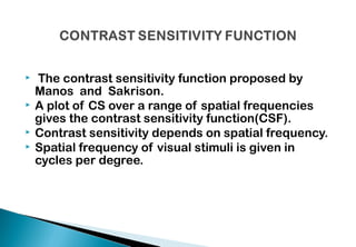


The document discusses contrast sensitivity and its importance in visual performance, detailing key concepts such as contrast threshold, spatial frequency, and the contrast sensitivity function (CSF). It explains how various factors, including age, cataracts, glaucoma, and contact lenses, impact contrast sensitivity and describes methods for measuring it. Multiple studies and tests are referenced to emphasize the clinical implications of contrast sensitivity in diagnosing and monitoring visual impairments.


![Contrast sensitivity and glare discomfort [Autosaved].pptx](https://cdn.slidesharecdn.com/ss_thumbnails/contrastsensitivityandglarediscomfortautosaved-250419142217-06d6a009-thumbnail.jpg?width=640&height=640&fit=bounds)


