Learning Objectives
At theend of this lecture you are supposed to:
1. understand what connective tissues are.
2. to able to state the functions of connective tissues.
3. be able to describe the different types of
connective tissues.
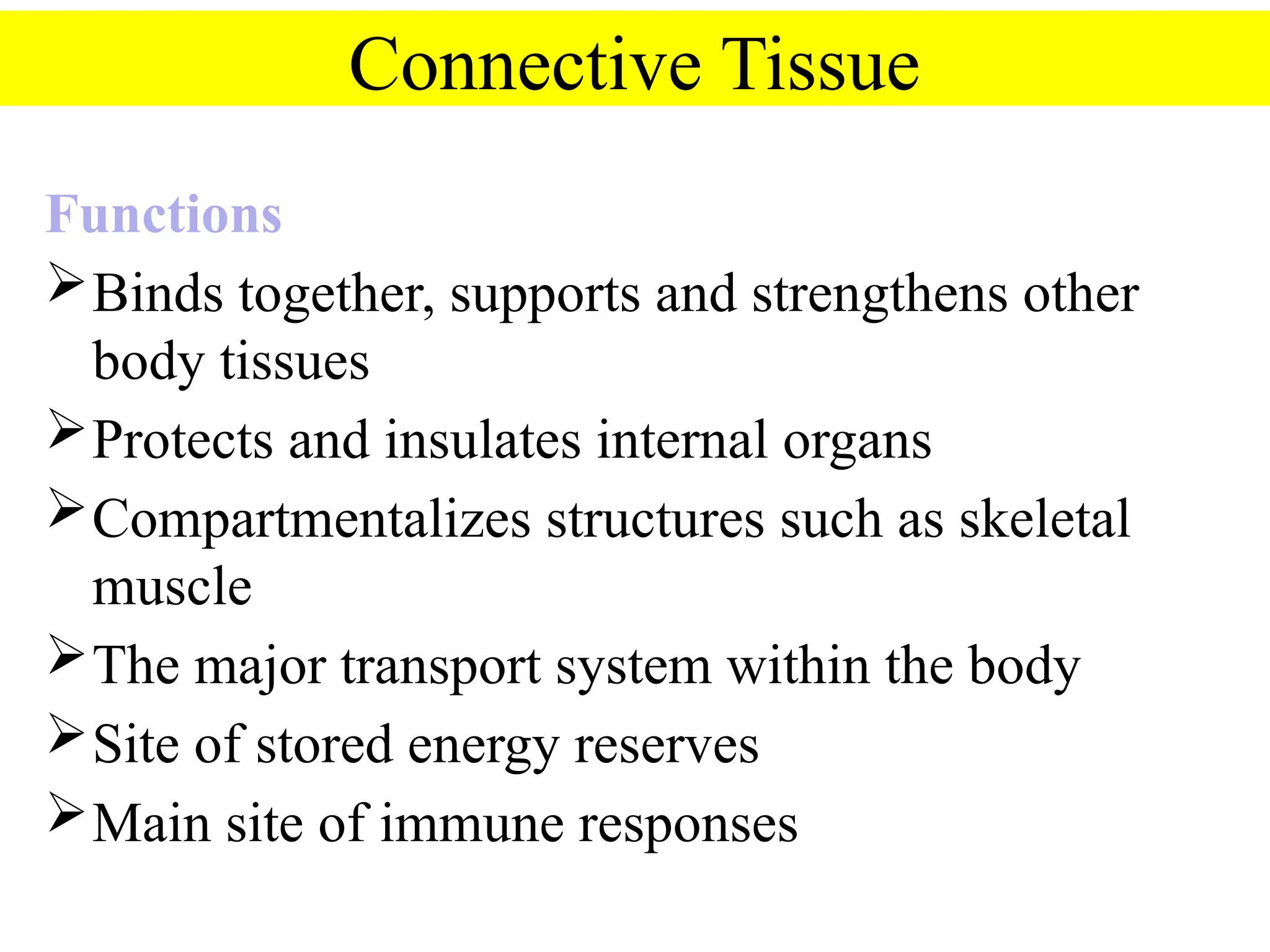
Connective Tissue
Functions
Binds together,supports and strengthens other
body tissues
Protects and insulates internal organs
Compartmentalizes structures such as skeletal
muscle
The major transport system within the body
Site of stored energy reserves
Main site of immune responses
5.
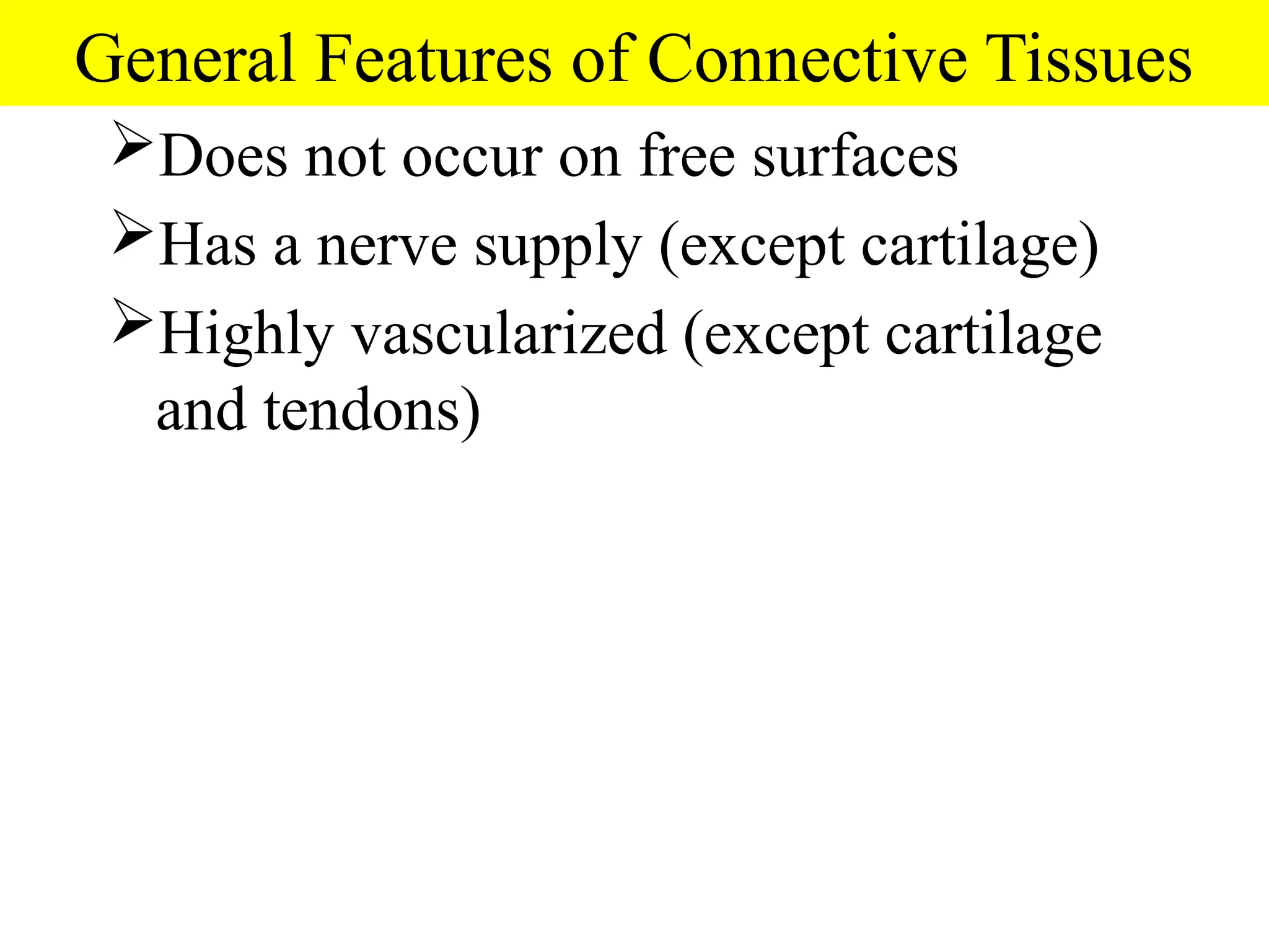
General Features ofConnective Tissues
Does not occur on free surfaces
Has a nerve supply (except cartilage)
Highly vascularized (except cartilage
and tendons)
6.
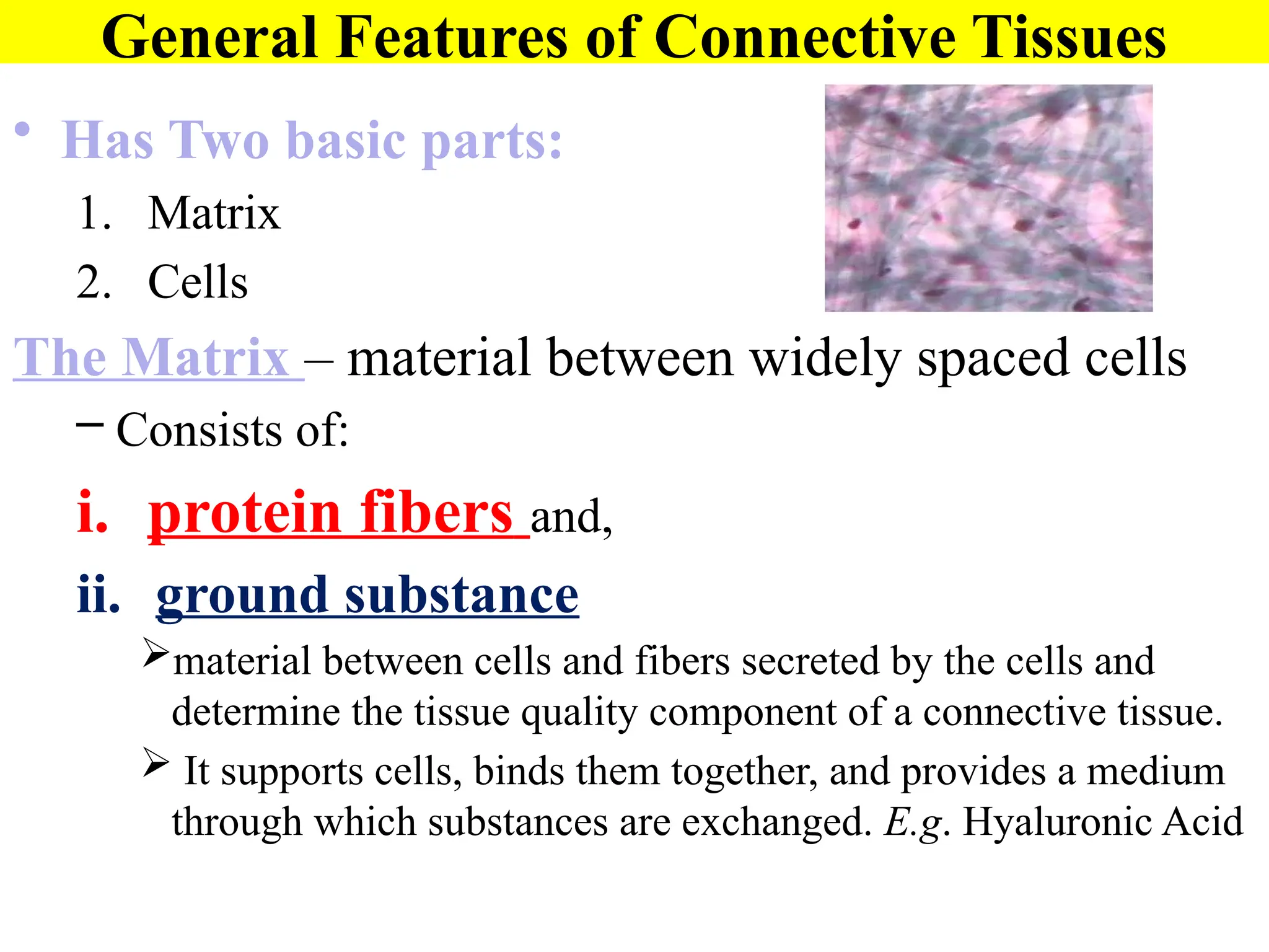
General Features ofConnective Tissues
• Has Two basic parts:
1. Matrix
2. Cells
The Matrix – material between widely spaced cells
– Consists of:
i. protein fibers and,
ii. ground substance
material between cells and fibers secreted by the cells and
determine the tissue quality component of a connective tissue.
It supports cells, binds them together, and provides a medium
through which substances are exchanged. E.g. Hyaluronic Acid
7.
Matrix: Ground Substance
HyaluronicAcid: Complex combination of
polysaccharides and proteins found in “true” or
proper connective tissue.
Chondroitin sulfate: Jellylike ground substance of
cartilage, bone, skin and blood vessels.
Other ground Substances:
Dermatin sulfate, keratin sulfate, and adhesion
proteins
8.
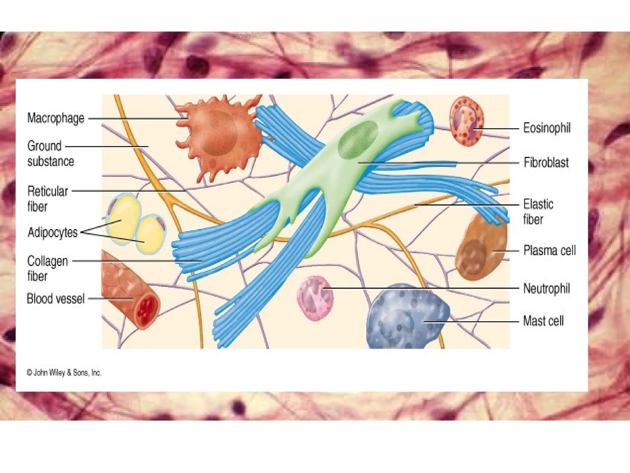
Connective Tissue Matrix–
(protein) Fibers
Strengthens and supports connective tissue
Collagen Fibers
Strong, resist pulling forces, flexible
Made of the protein collagen which is the most
abundant protein in your body
Elastic Fibers
smaller in diameter than collagen fibers, branch to
form network
Made of the protein elastin
9.
Connective Tissue Matrix–
Fibers
Reticular Fibers
Small delicate, branched fibers that have same
chemical composition of collagen.
Provide support for the walls of blood vessels
Made of collagen with a glycoprotein covering
Forms structural framework for organs such as
spleen and lymph nodes.
11.
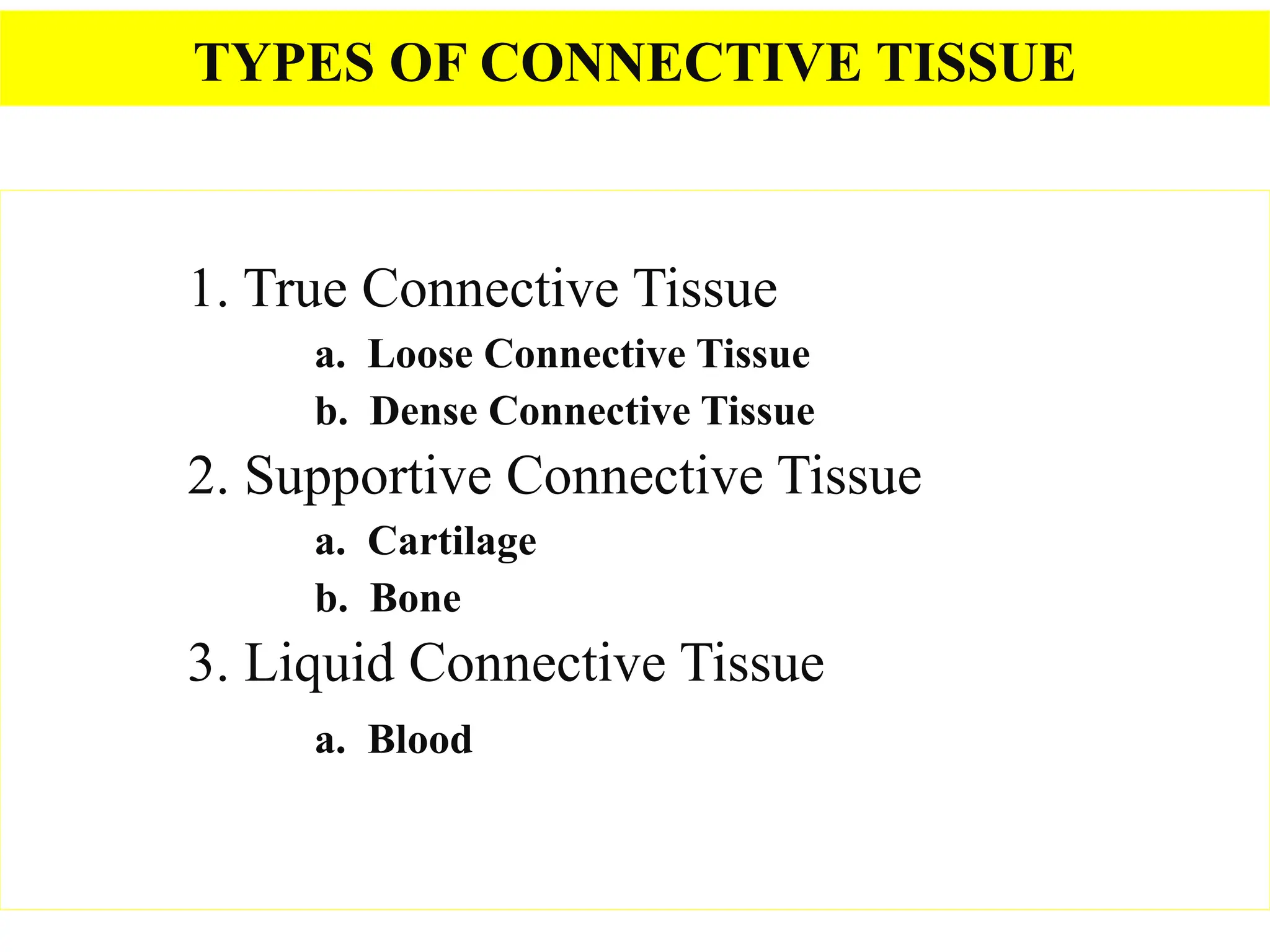
TYPES OF CONNECTIVETISSUE
1. True Connective Tissue
a. Loose Connective Tissue
b. Dense Connective Tissue
2. Supportive Connective Tissue
a. Cartilage
b. Bone
3. Liquid Connective Tissue
a. Blood
12.
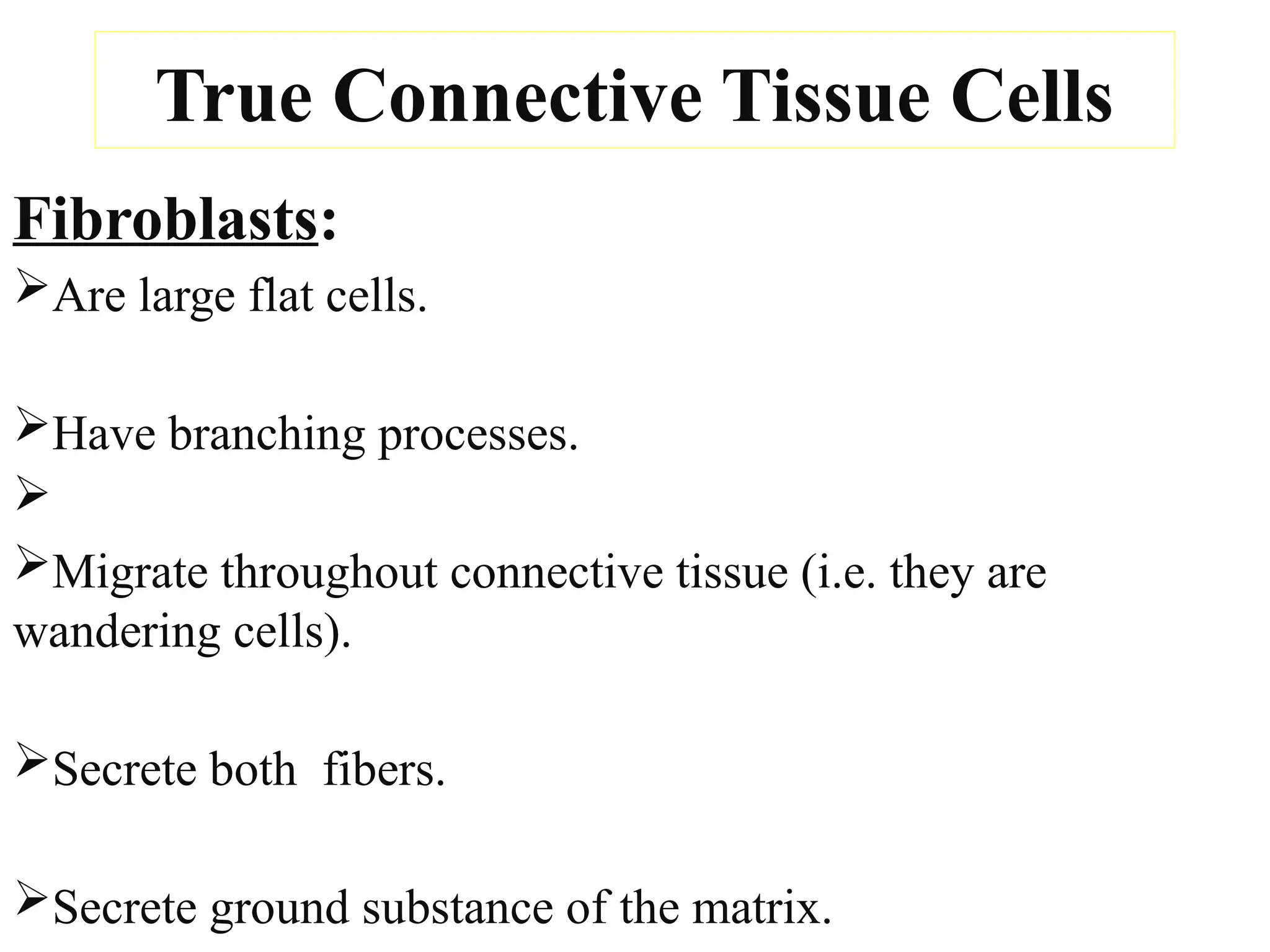
True Connective TissueCells
Fibroblasts:
Are large flat cells.
Have branching processes.
Migrate throughout connective tissue (i.e. they are
wandering cells).
Secrete both fibers.
Secrete ground substance of the matrix.
13.
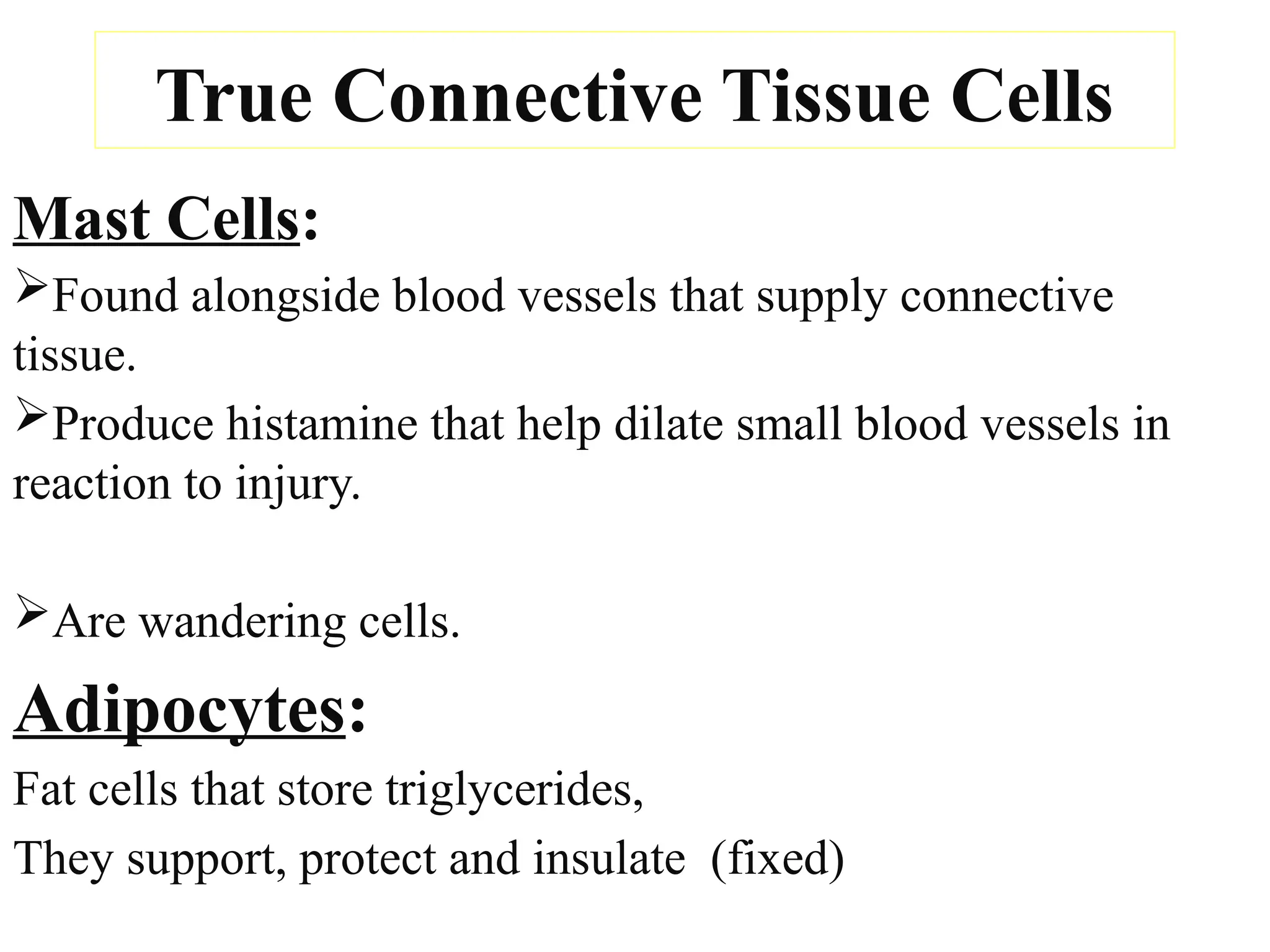
True Connective TissueCells
Mast Cells:
Found alongside blood vessels that supply connective
tissue.
Produce histamine that help dilate small blood vessels in
reaction to injury.
Are wandering cells.
Adipocytes:
Fat cells that store triglycerides,
They support, protect and insulate (fixed)
14.
True Connective TissueCells
Macrophages:
Developed from white blood cells (monocytes).
Surround and engulf material by phagocytosis.
Are wandering cells
Plasma Cells:
Are Antibody secreting cells that develop from B
Lymphocytes (wandering)
15.
Types of ConnectiveTissue(Cont’d)
(A). Loose Connective Tissue
Fibers are loosely intertwined among many cells.
Three types of loose connective tissue
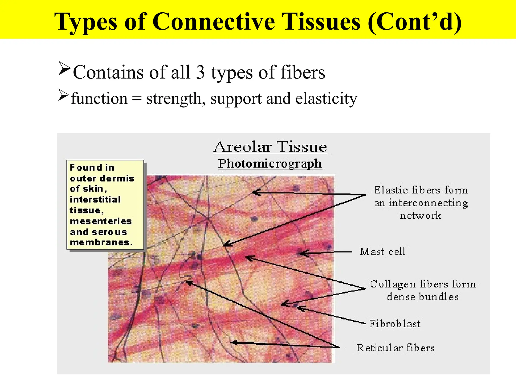
1. Areolar Connective Tissue –
one of the most widely distributed connective tissues in the
body.
Found in subcutaneous layer and mucous membranes,
and around blood vessels, nerves and organs
Contains fibroblasts, macrophages, plasma cells, mast
cells, adipocytes, and a few white blood cells.
16.
Contains of all3 types of fibers
function = strength, support and elasticity
Types of Connective Tissues (Cont’d)
17.
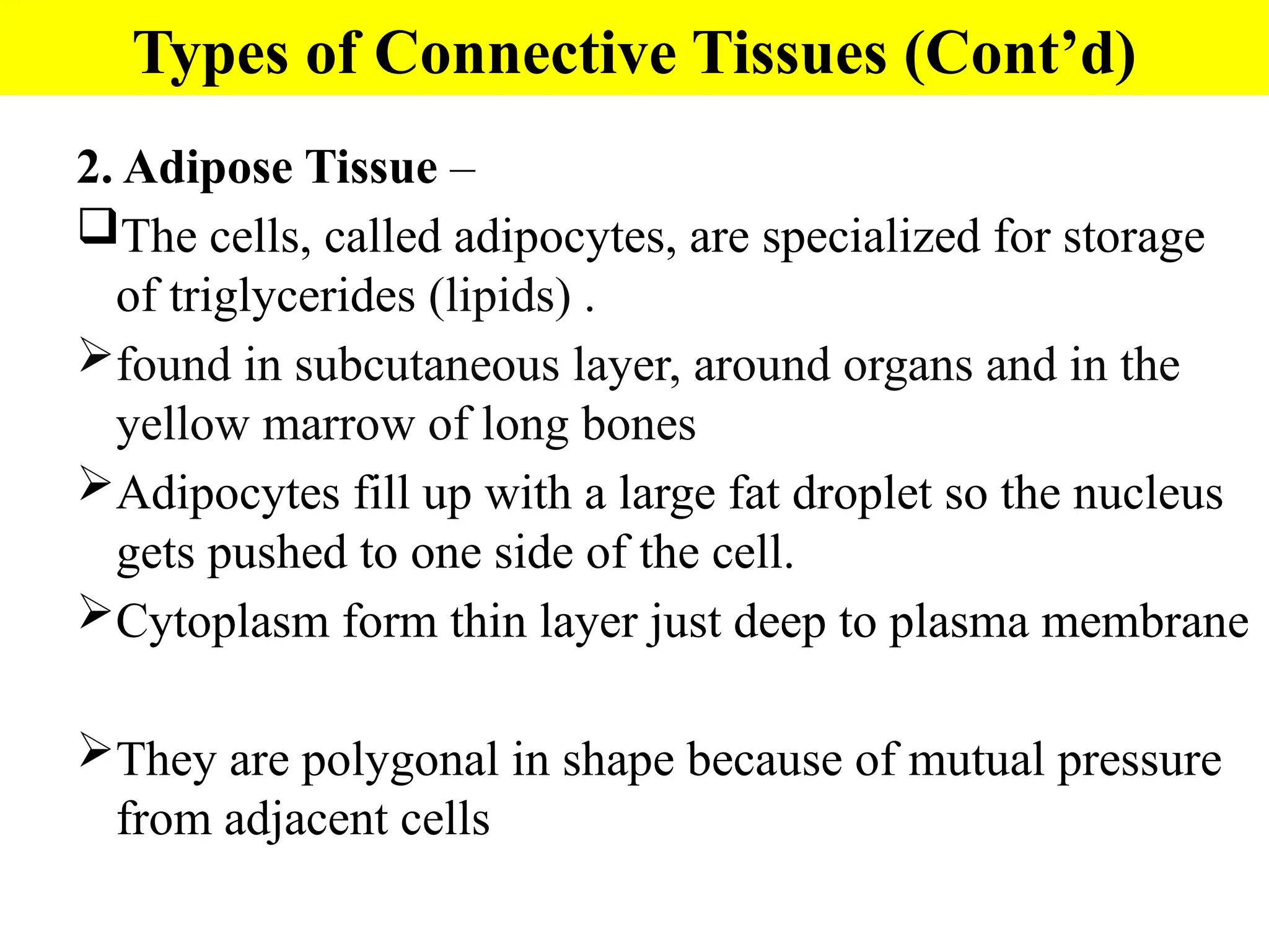
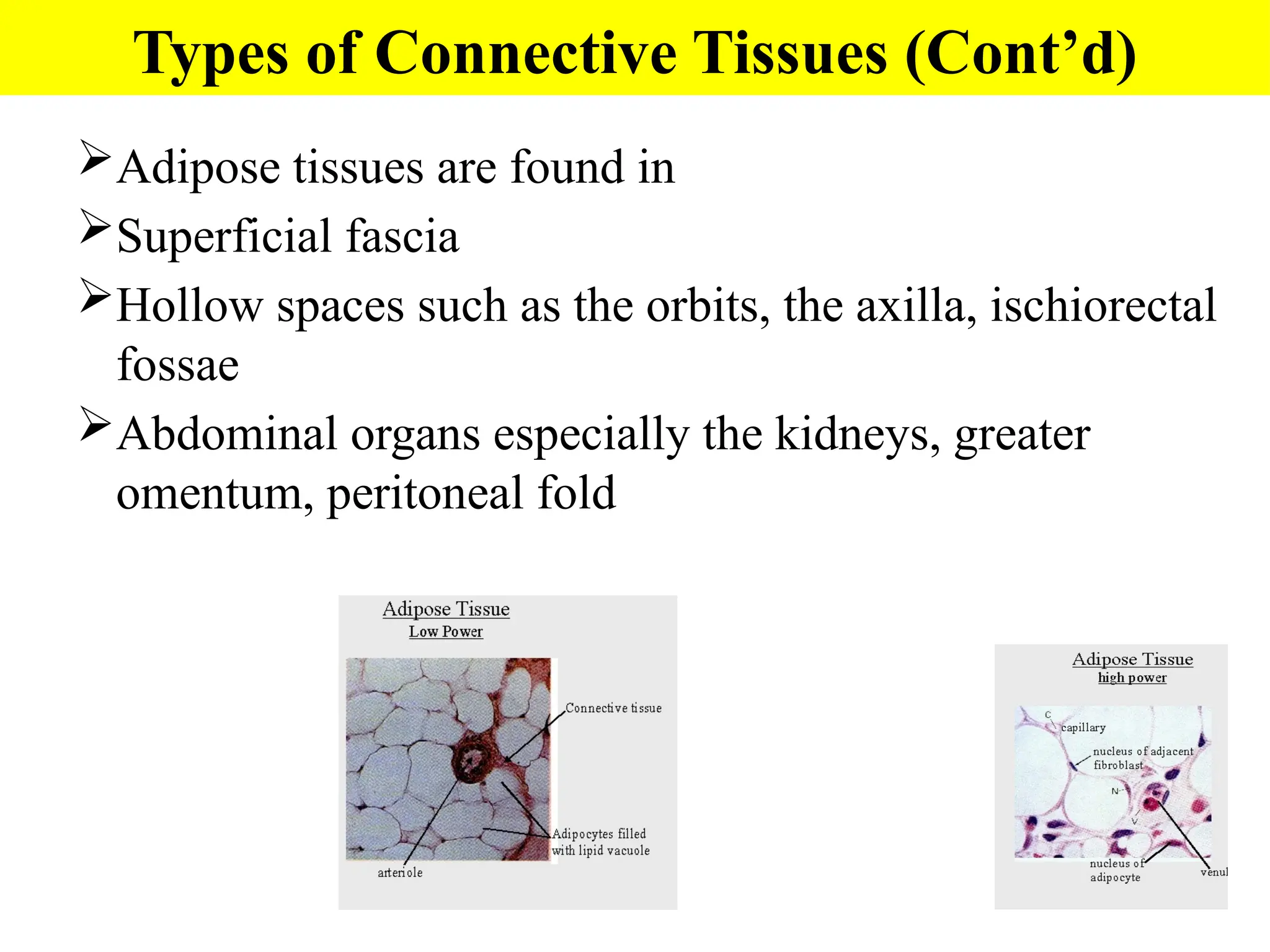
2. Adipose Tissue–
The cells, called adipocytes, are specialized for storage
of triglycerides (lipids) .
found in subcutaneous layer, around organs and in the
yellow marrow of long bones
Adipocytes fill up with a large fat droplet so the nucleus
gets pushed to one side of the cell.
Cytoplasm form thin layer just deep to plasma membrane
They are polygonal in shape because of mutual pressure
from adjacent cells
Types of Connective Tissues (Cont’d)
18.
Adipose tissues arefound in
Superficial fascia
Hollow spaces such as the orbits, the axilla, ischiorectal
fossae
Abdominal organs especially the kidneys, greater
omentum, peritoneal fold
Types of Connective Tissues (Cont’d)
3. Reticular ConnectiveTissue –
Made of interlacing reticular fibers and reticular
cells that connect to each other to form a network.
Found in liver, spleen and lymph nodes
Used to bind together smooth muscle cells and to
filter out worn out blood cells and bacteria
Function = forms the framework (stroma) of
organs and binds together smooth muscle tissue
cells
Types of Connective Tissues (Cont’d)
(B). Dense ConnectiveTissue
Contains more numerous, thicker and denser fibers
but fewer cells than loose connective tissue.
consists of bundles of collagen fibers and fibroblasts
forms tendons, ligaments and aponeuroses
Function = provide strong attachment between various
structures
3 types:
1. Dense regular connective tissue
2. Dense Irregular connective tissue
3. Elastic Connective Tissue
Types of Connective Tissues (Cont’d)
23.
1. Dense RegularConnective Tissue
Bundles of collagen fibers are arranged regularly in
parallel patterns that give it strength.
Withstands pulling from the ends, but unravels when
pulled from the side
Silvery white in appearance. Tough and pliable
Found in tendons and ligaments
Types of Connective Tissues (Cont’d)
24.
2. Dense IrregularConnective Tissue
Collagen fibers are packed closely together in an
irregular, random pattern
Found in parts of the body where pulling forces are
exerted in various directions
Usually found in sheets
Examples: Dermis of the skin, heart valves,
perichondrium and periosteum
Types of Connective Tissues (Cont’d)
25.
3. Elastic ConnectiveTissue
• Contains branching elastic fibers and fibroblasts
• Yellowish in color
• Strong, can regain shape after stretching
• Found in lungs and arteries
Types of Connective Tissues (Cont’d)
26.
Supportive Connective Tissue:
CARTILAGE:
Jelly-likematrix (chondroitin sulfate) containing
collagen and elastic fibers and chondrocytes
surrounded by a membrane called the
perichondrium.
Unlike other CT, cartilage has NO blood vessels or
nerves except in the perichondrium.
The strength of cartilage is due to collagen fibers
and the resilience is due to the presence of
chondroitin sulfate.
Chondrocytes occur within spaces in the matrix
called lacunae.
Types of Connective Tissues (Cont’d)
27.
Types of Cartilages
1.Hyaline cartilage
2. Fibrocartilage
3. Elastic cartilage
Types of Connective Tissues (Cont’d)
28.
1. Hyaline Cartilage(most abundant type)
(hyalos= glass= transparent)
fine collagen fibers embedded in a gel-type matrix.
Chondrocytes are large and in group toward middle of
hyaline cartilage
Matrix stained blue with H&E
Found in embryonic skeleton, coastal cartilages at the
ends of long bones, in the nose and in respiratory
structures.
Function= flexible, provides support, allows movement
at joints
Types of Connective Tissues (Cont’d)
29.
Supportive Connective Tissue:
2.Fibrocartilage
contains bundles of collagen in the matrix that
are usually more visible under microscopy.
Found in the pubic symphysis, intervertebral
discs, and menisci of the knee.
Function = support and fusion, and absorbs
shocks.
30.
Supportive Connective Tissue:
3.Elastic Cartilage
threadlike network of elastic fibers within the
matrix.
found in external ear, auditory tubes, epiglottis.
function = gives support, maintains shape,
allows flexibility
31.
Bone Tissue (osseoustissue)
• 2 types – compact and spongy
Compact
Osteon – basic unit of compact bone
Lamellae – concentric circles of
matrix
Lacunae – spaces in the matrix that
house cells
Osteocytes – mature bone cells
Spongy
Trabeculae – columns of bone with
spaces filled with red bone marrow
Types of Connective Tissue(Cont’d)
32.
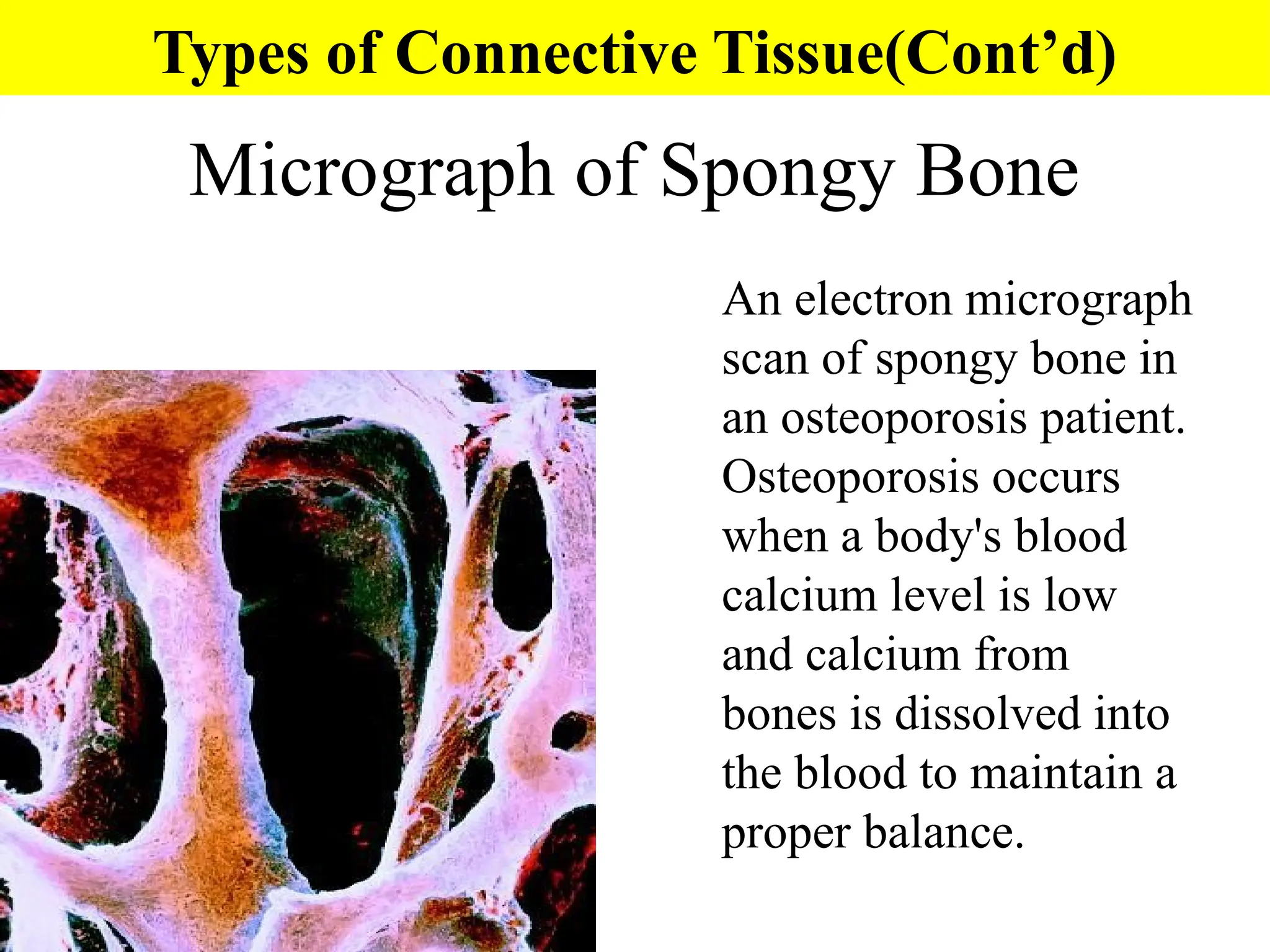
Micrograph of SpongyBone
An electron micrograph
scan of spongy bone in
an osteoporosis patient.
Osteoporosis occurs
when a body's blood
calcium level is low
and calcium from
bones is dissolved into
the blood to maintain a
proper balance.
Types of Connective Tissue(Cont’d)
33.
Blood Tissue
Connective Tissuewith a liquid matrix
Red Blood Cells (erythrocytes) – transport
oxygen
White Blood Cells – function in immunity
Neutrophils, Eosinophils, Basophils, T and B
leukocytes, natural killer cells and Monocytes
Platelets – participate in blood clotting
Types of Connective Tissue(Cont’d)
35.
Marfan Syndrome
An inheriteddisorder caused by a defective gene
for the glycoprotein fibrillin resulting in
abnormal development of elastic fibers.
This causes tissues that contain many elastic fibers
to be malformed or weak (including the covering of
bone, ligament that suspends the lens of the eye,
and the walls of large arteries
People with Marfan syndrome are often tall, have
long arms, legs, fingers and toes, blurred vision,
and weakened aortic walls that may burst.