
Download as PDF, PPTX


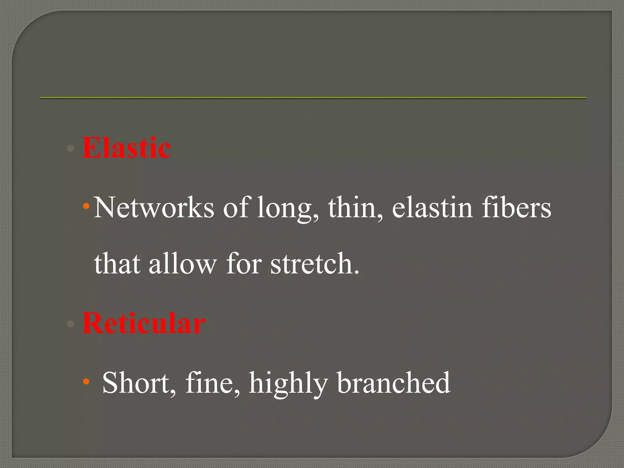


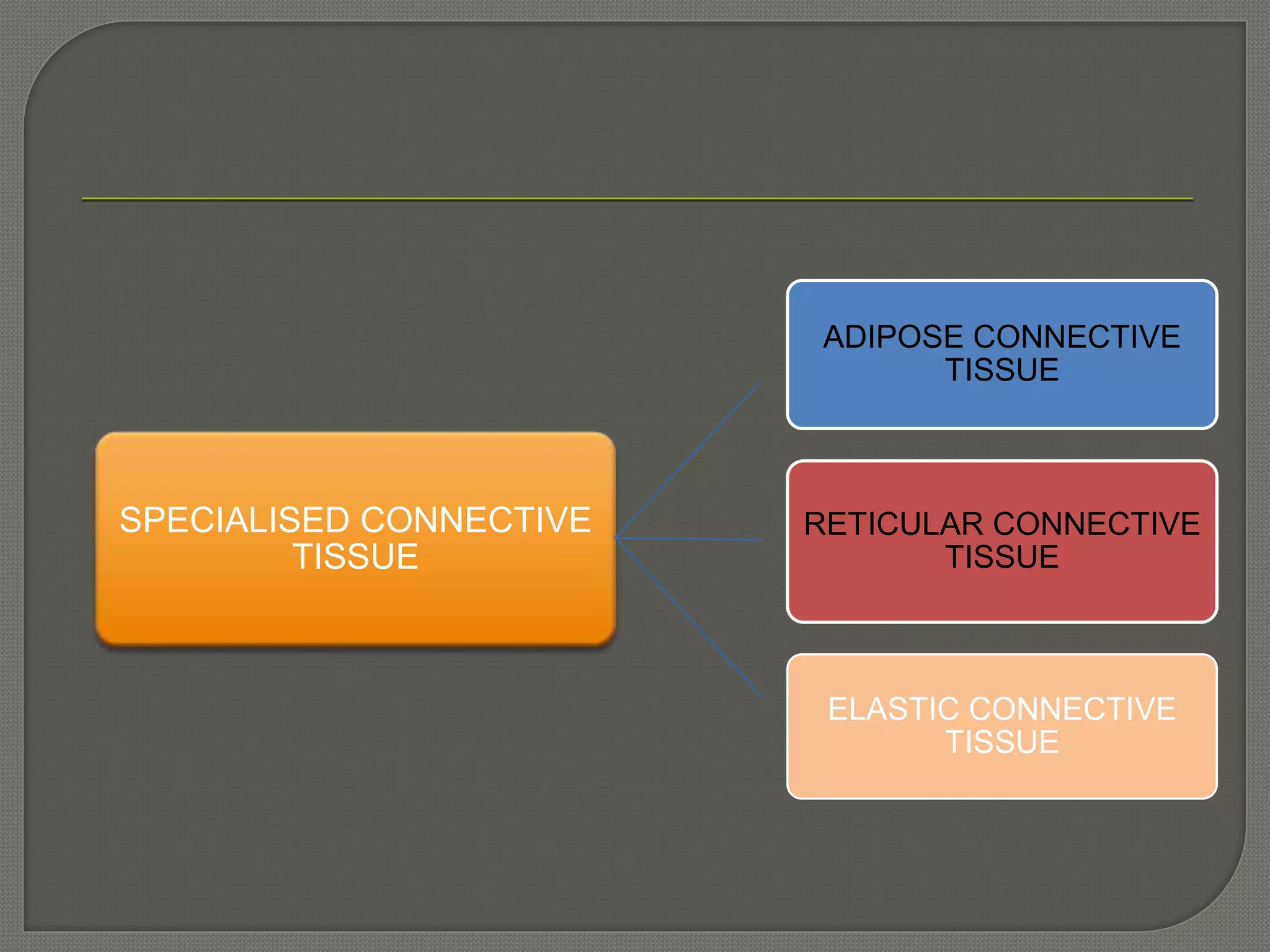

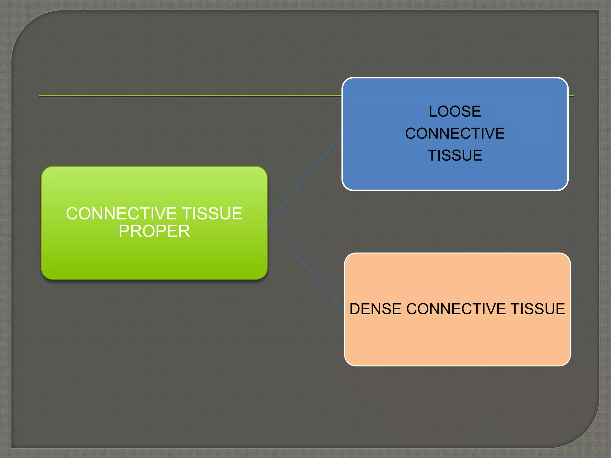


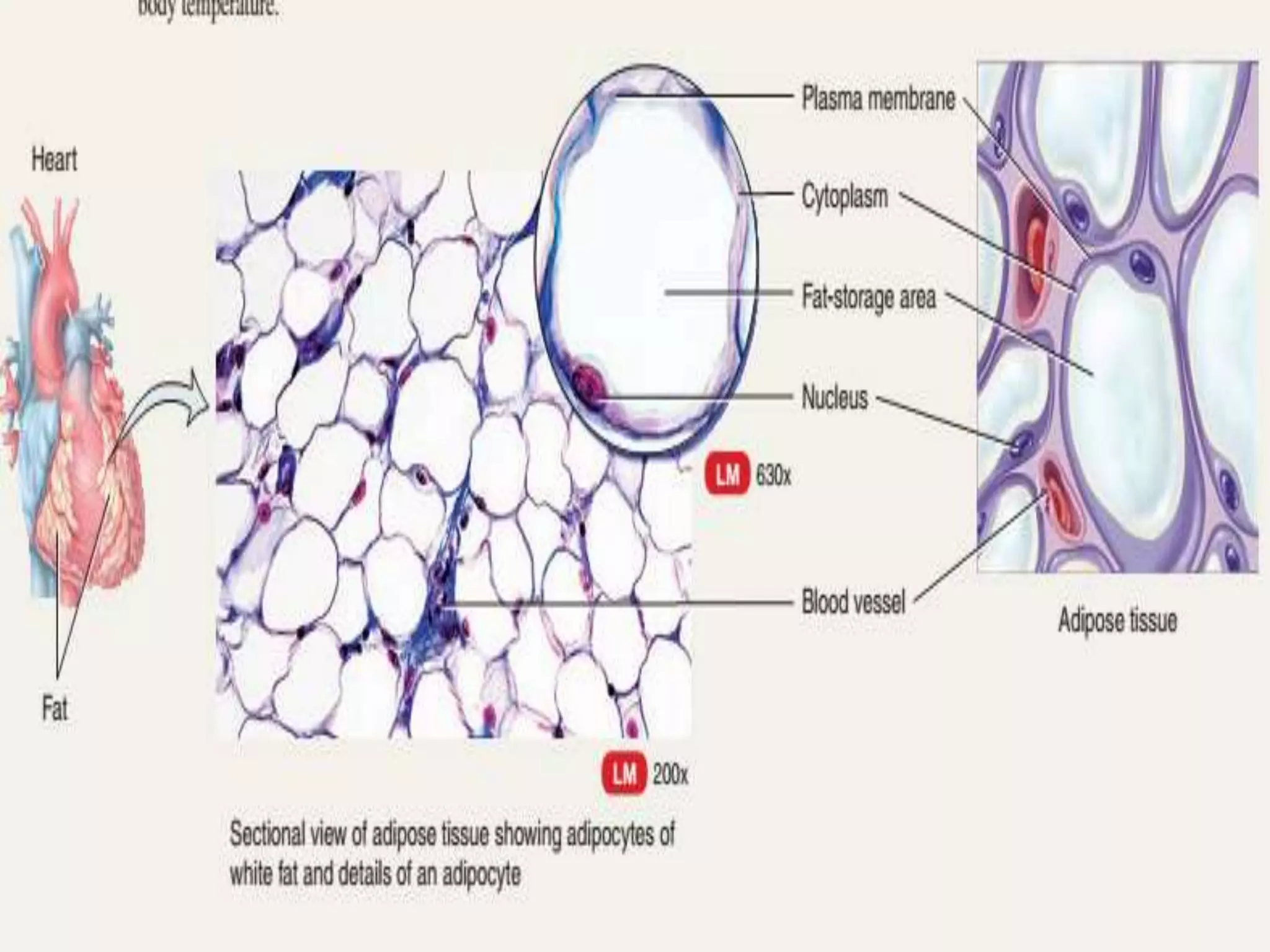


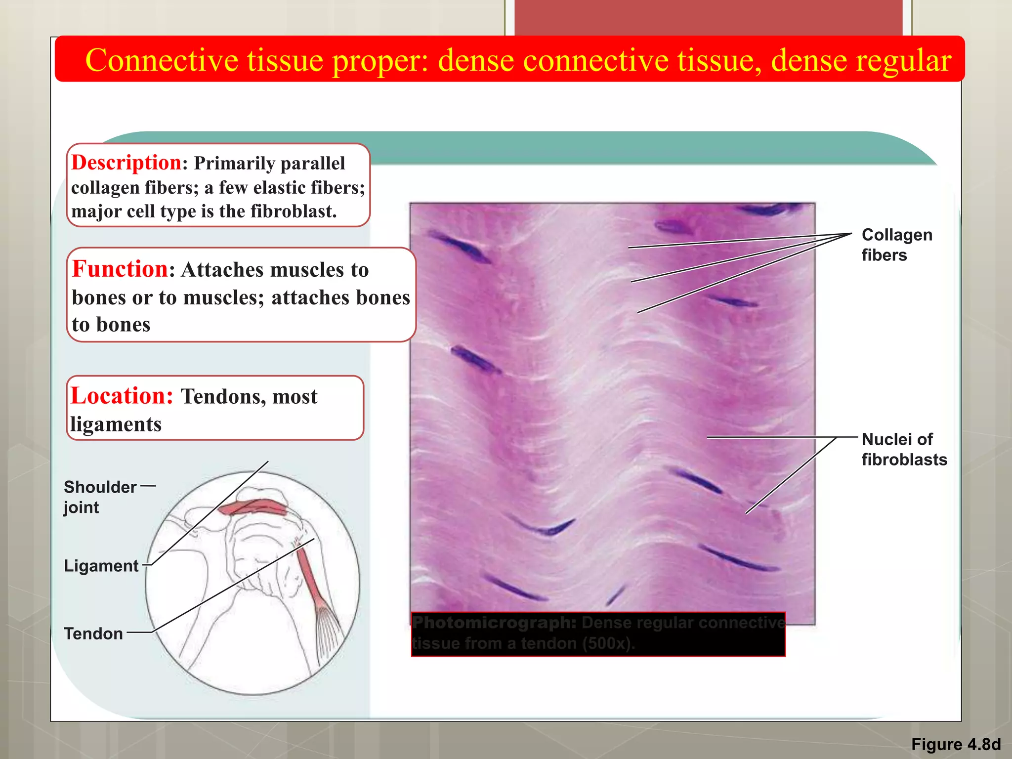

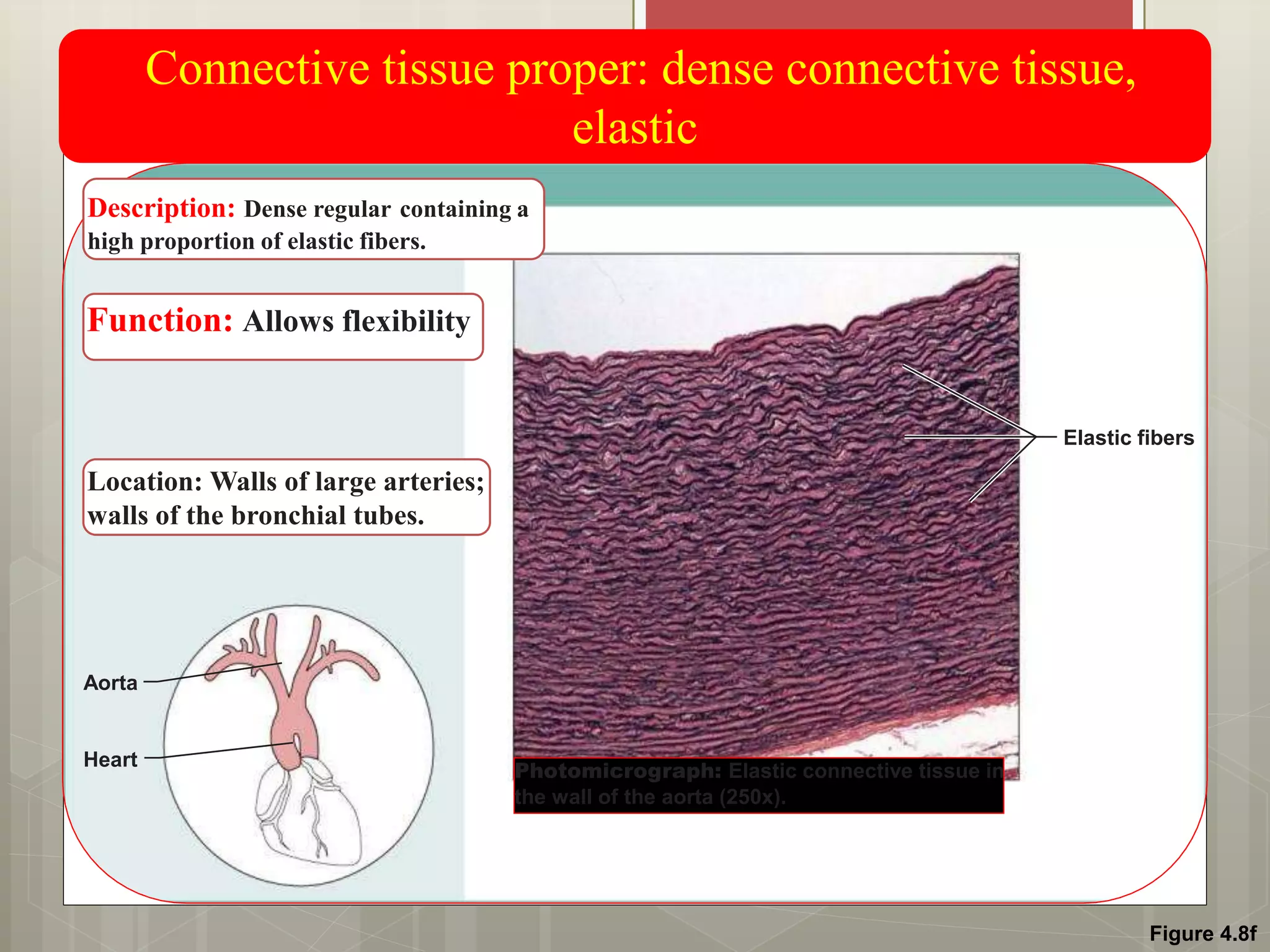


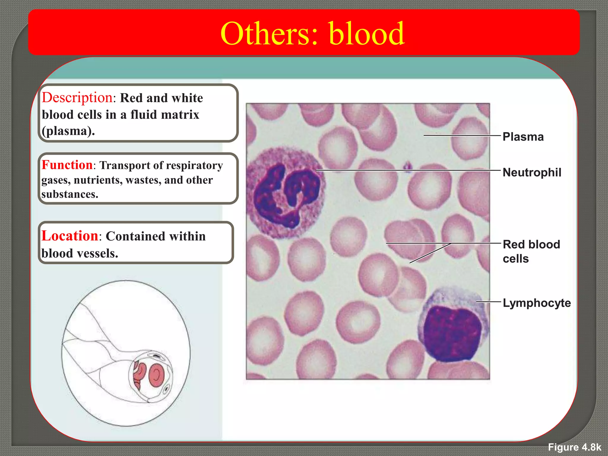


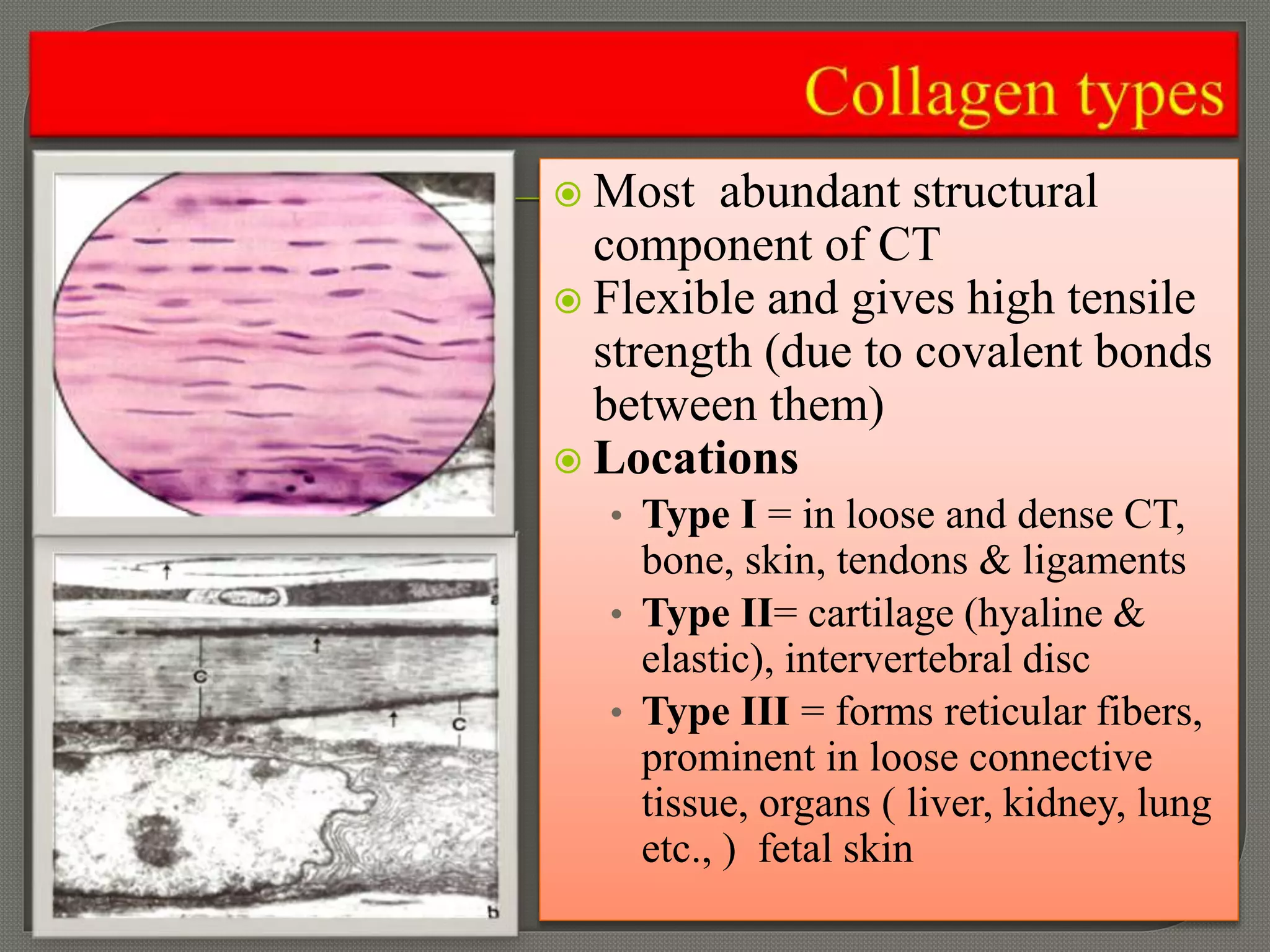


Connective tissue is one of the four primary tissue types, characterized by its abundance and diverse functions, which include supporting and connecting tissues, as well as transporting substances. It consists of an extracellular matrix composed of fibers (collagen, elastic, reticular), ground substance, and various cell types. Connective tissue can be categorized into different classes, including loose, dense, specialized, and embryonic connective tissues, each with unique structures and roles in the body.


