Outline of presentation
•Definition of connective tissue
• Functions of connective tissue
• Composition of CT
• Classification of CT
• Types of CT
3.
Definition
• A groupof tissues that provide metabolic and
structural support to other tissues
• In simplest form, it provides a biological
packaging btn other tissues
• Fxns include providing a media for exchange
of nutrients, metabolites and waste products
• Others include tensile strength as in bones,
muscles, tendons and cartilages
4.
Types of CT
•In its simplest form, it is called loose
areolar tissue and acts as a biological
packing material between cells and
different tissues
• More dense forms are for tensile strength
as in tendons and ligaments
• More rigid forms occur in bones and
cartilage
5.
Functions of CT
•White adipose tissue: synthesis and
metabolism of fat
• Brown adipose tissue: temp. regulation
• Media for exchange of materials between
cells and tissues
• Tensile strength
• Defence
• Tissue repair
6.
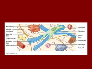
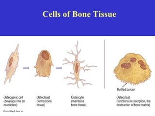
Composition of CT
•Cells: adipocytes, fibroblasts, defence
cells
• Extracellular material: ground substance,
fibres and glycoproteins
7.
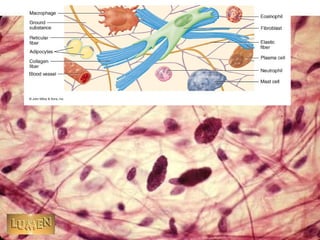
Cells of CT
•Adipocytes: Storage and metabolism of fat
• Fibroblasts: synthesis of fibres, repair and
maintainance of extracellular material
• Defence cells: mast cells, tissue
macrophages and some white blood cells
8.
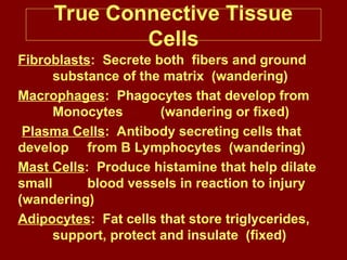
True Connective Tissue
Cells
Fibroblasts:Secrete both fibers and ground
substance of the matrix (wandering)
Macrophages: Phagocytes that develop from
Monocytes (wandering or fixed)
Plasma Cells: Antibody secreting cells that
develop from B Lymphocytes (wandering)
Mast Cells: Produce histamine that help dilate
small blood vessels in reaction to injury
(wandering)
Adipocytes: Fat cells that store triglycerides,
support, protect and insulate (fixed)
11.
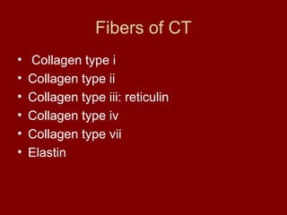
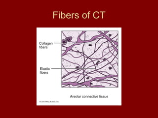
Fibers of CT
•Collagen type i
• Collagen type ii
• Collagen type iii: reticulin
• Collagen type iv
• Collagen type vii
• Elastin
12.
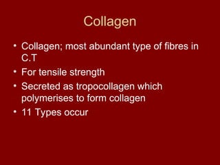
Collagen
• Collagen; mostabundant type of fibres in
C.T
• For tensile strength
• Secreted as tropocollagen which
polymerises to form collagen
• 11 Types occur
13.
• Type 1:occurs in fibrous C.T of skin
dermis, tendons , ligaments and bone
fxn: for greater tensile strength
• Type 2: found in hyaline cartilage. Has fine
fibrils interspersed in ground substance
14.
• Type 3:found in CT type called reticulin.
Has high affinity for silver salts.forms a fine
branching mesh work in highly cellular
organs like liver, spleen, bone marrow and
thymus
• Type 4: doesn’t form fibrils but a mesh work
of fibres as in basement membrane
• Type 7: forms anchoring fibers that link to
basement membranes
15.
Elastin
• Rubber likematerial arranged as fibres;
Occirs in skin, lung tissue, and blood vessels
• Fxn: confers properties of stretching and
elastic recoil
• Synthesized as tropoelastin by fibroblasts
and plumerises to form elastin
• Deposition of elastin requires presence of a
structural glycoprotein fibrillin
16.
Structural glycoprotiens
• Composedof protein bound to branched
polysaccharides molecules
Two types
• Fibril forming molecules: fibrillin and
fibronectin. These are a constituent of elastin
• Non filamentous proteins: laminin, entactin,
tenasin. Act as links between cells and
extracellular material.
17.
• Fibrillin: foundin mesangium of kidney and
elastin
• Fibronectin: controls deposition of collagen
and binding of cells to extracellular material,
also forms cytoskeleton
• Laminin: found in basement membranes.
For cell adhesion and forms links btn cell
membrane and other constituents of
extracellular matrix
18.
• Enactin: bindslaminin to type 4 collagen in
basement membranes
• Tenactin: binds integrins and is important
in the embryo in control of nerve cell
growth
19.
Ground substance
• Gellike and is responsible for turbidity
• Consists of long unbranched polysaccharide
chains of seven types
• Polysaccharides composed of repeating units
of disaccharide molecules
• Disaccharides made of a uronic acid and an
amino sugar hence called glucosaminoglycans
• Commonest GAG is hyarulonic acid, the only
P.S with no sulfate gr
20.
• Other GAGinclude; Chondrotin 4
phosphate, C-6-P, Dermatan sulfate,
Heparan sulfate, heparin sulfate, Keratan
sulfate
• These are all collectively called
proteoglycans
• Hence glycosaminoglycans are composed
of HA and proteoglycans.
TYPES OF CONNECTIVETISSUE
1. True Connective Tissue
a. Loose Connective Tissue
b. Dense Connective Tissue
2. Supportive Connective Tissue
a. Cartilage
b. Bone
5. Liquid Connective Tissue
a. Blood
26.
True or ProperConnective
Tissue
1. Loose Connective Tissue:
a. Areolar tissue
Widely distributed under
epithelia
b. Adipose tissue
Hypodermis, within abdomen,
breasts
c. Reticular connective tissue
Lymphoid organs such as lymph
nodes
27.
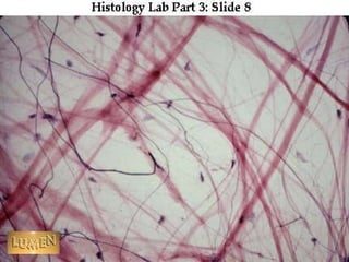
LOOSE Connective Tissue:
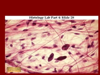
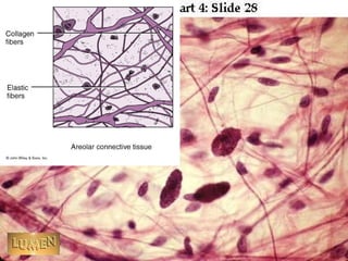
1.Areolar CT
– consists of all 3 types of fibers,
several types of cells, and semi-fluid
ground substance
– found in subcutaneous layer and
mucous membranes, and around
blood vessels, nerves and organs
– function = strength, support and
elasticity
29.
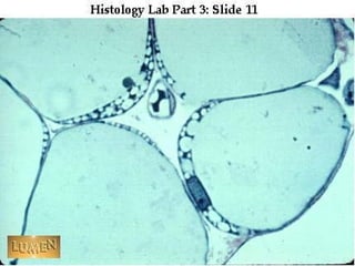
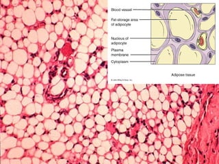
LOOSE Connective Tissue:
2.Adipose tissue
– consists of adipocytes; "signet ring"
appearing fat cells. They store energy in
the form of triglycerides (lipids).
– found in subcutaneous layer, around
organs and in the yellow marrow of long
bones
– function = supports, protects and
insulates, and serves as an energy
reserve
30.
• White adiposetissue: found in well
nourished adults forming 25% of total
weight in females and 20% in adult males.
A source of energy, insulator and shock
absorber
• Brown adipose tissue: occurs in
hibernating animals and babies. Main
function is temperature regulation.
33.
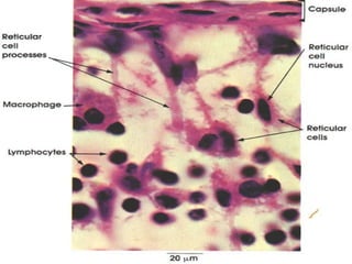
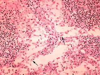
LOOSE Connective Tissue:
3.Reticular CT
– Consists of fine interlacing reticular
fibers and reticular cells
– Found in liver, spleen and lymph
nodes
– Function = forms the framework
(stroma) of organs and binds together
smooth muscle tissue cells
36.
True or ProperConnective
Tissue
2. Dense Connective Tissue:
a. Dense regular connective
tissue
Tendons and ligaments
b. Dense irregular
connective tissue
Dermis of skin, submucosa of
digestive tract
37.
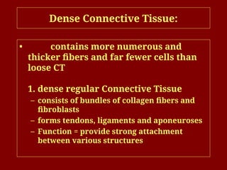
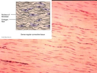
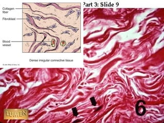
Dense Connective Tissue:
•contains more numerous and
thicker fibers and far fewer cells than
loose CT
1. dense regular Connective Tissue
– consists of bundles of collagen fibers and
fibroblasts
– forms tendons, ligaments and aponeuroses
– Function = provide strong attachment
between various structures
40.
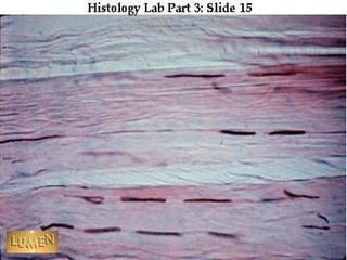
Dense Connective Tissue:
2.Dense Irregular CT
– consists of randomly-arranged collagen
fibers and a few fibroblasts
– Found in fasciae, dermis of skin, joint
capsules, and heart valves
– Function = provide strength
42.
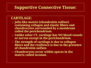
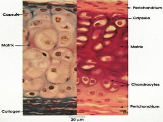
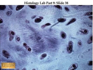
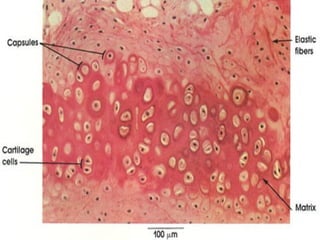
Supportive Connective Tissue:
CARTILAGE:
–Jelly-like matrix (chondroitin sulfate)
containing collagen and elastic fibers and
chondrocytes surrounded by a membrane
called the perichondrium.
– Unlike other CT, cartilage has NO blood vessels
or nerves except in the perichondrium.
– The strength of cartilage is due to collagen
fibers and the resilience is due to the presence
of chondroitin sulfate.
– Chondrocytes occur within spaces in the
matrix called lacunae.
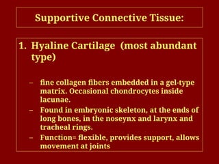
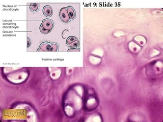
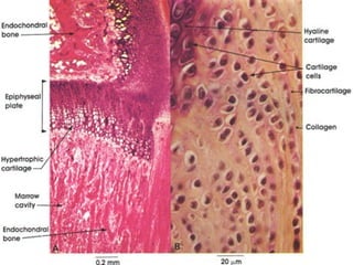
Supportive Connective Tissue:
1.Hyaline Cartilage (most abundant
type)
– fine collagen fibers embedded in a gel-type
matrix. Occasional chondrocytes inside
lacunae.
– Found in embryonic skeleton, at the ends of
long bones, in the noseynx and larynx and
tracheal rings.
– Function= flexible, provides support, allows
movement at joints
47.
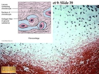
Supportive Connective Tissue:
2.Fibrocartilage
• Has features intermediate between dense
CT and cartilage
• contains bundles of collagen in the matrix
that are usually more visible under
microscopy.
• Found in the pubic symphysis,
intervertebral discs, joint capsules, tendons
and menisci of the knee.
• Function = support and fusion, and absorbs
shocks.
51.
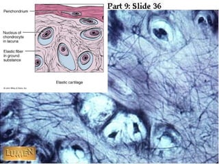
Supportive Connective Tissue:
3.Elastic Cartilage
– threadlike network of elastic fibers within
the matrix.
– found in external ear, auditory tubes,
epiglottis.
– function = gives support, maintains shape,
allows flexibility
54.
Bone Tissue
• Aspecialised type of connective tissue in
which the extracellular matrix is
mineralised
• Type of supportive connective tissue
together with cartilage
• Same compostion like connective tissue,
ie cells and extracellular matrix.
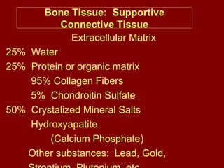
Bone Tissue: Supportive
ConnectiveTissue
Extracellular Matrix
25% Water
25% Protein or organic matrix
95% Collagen Fibers
5% Chondroitin Sulfate
50% Crystalized Mineral Salts
Hydroxyapatite
(Calcium Phosphate)
Other substances: Lead, Gold,
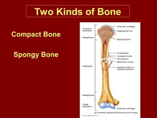
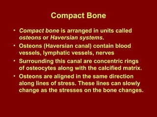
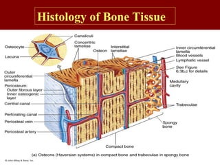
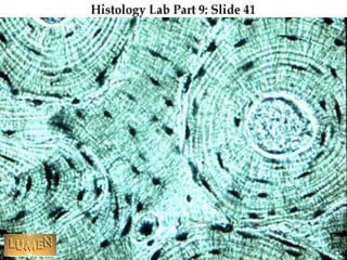
Compact Bone
• Compactbone is arranged in units called
osteons or Haversian systems.
• Osteons (Haversian canal) contain blood
vessels, lymphatic vessels, nerves
• Surrounding this canal are concentric rings
of osteocytes along with the calcified matrix.
• Osteons are aligned in the same direction
along lines of stress. These lines can slowly
change as the stresses on the bone changes.
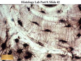
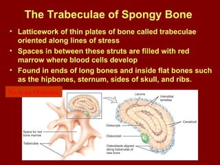
The Trabeculae ofSpongy Bone
• Latticework of thin plates of bone called trabeculae
oriented along lines of stress
• Spaces in between these struts are filled with red
marrow where blood cells develop
• Found in ends of long bones and inside flat bones such
as the hipbones, sternum, sides of skull, and ribs.
No true Osteons.
64.
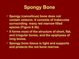
Spongy Bone
• Spongy(cancellous) bone does not
contain osteons. It consists of trabeculae
surrounding many red marrow filled
spaces (Figure 6.3b).
• It forms most of the structure of short, flat,
and irregular bones, and the epiphyses of
long bones.
• Spongy bone tissue is light and supports
and protects the red bone marrow.
65.
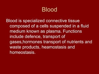
Blood
Blood is specializedconnective tissue
composed of a cells suspended in a fluid
medium known as plasma. Functions
include defence, transport of
gases,hormones transport of nutrients and
waste products, heamostasis and
homeostasis.
66.
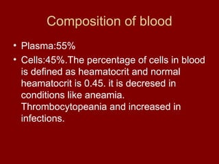
Composition of blood
•Plasma:55%
• Cells:45%.The percentage of cells in blood
is defined as heamatocrit and normal
heamatocrit is 0.45. it is decresed in
conditions like aneamia.
Thrombocytopeania and increased in
infections.
67.
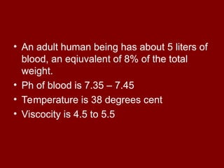
• An adulthuman being has about 5 liters of
blood, an eqiuvalent of 8% of the total
weight.
• Ph of blood is 7.35 – 7.45
• Temperature is 38 degrees cent
• Viscocity is 4.5 to 5.5
68.
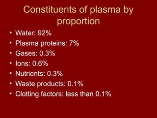
Constituents of plasmaby
proportion
• Water: 92%
• Plasma proteins: 7%
• Gases: 0.3%
• Ions: 0.6%
• Nutrients: 0.3%
• Waste products: 0.1%
• Clotting factors: less than 0.1%
69.
Plasma proteins
• Constitute7 – 9% of plasma.
• Three types: albumin, Fibrinogen and
globulins
• Albumin:( 60 – 80%) smallest in size.
Produced by the liver and serve to
maintain the osmotic pressure of blood.
• Fibrinogen (4%):produced by the liver and
play a role in blood clotting.
70.
• Globulins 38%:three types namely alpha,
beta and gamma globulins
• Alpha and beta globulins are produced by
the liver and serve as carriers for drugs,
hormones, lipids and lipid soluble vitamis
in the body.
• Gamma globulins are produced by plasma
cells and are known as antibodies.
71.
Blood cells
• Form45% of blood
• Three types:red blood cells, white blood
cells and platelets
• Red blood cells are the most abundant,
4.3 to 5.2 million in adult females and 5.1
to 5.8 million in adult males
• White blood cells are about 5000 to 9000
72.
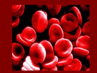
Erythrocytes
• Most abundantcells in blood.
• Responsible for the red colour of blood.
• Flattened biconcave discs, 7.2
micrometers in diameter and 2.2
micrometers thick.
• Shape allows them to pass through even
the smallest cappillaries.
• Mature cells lack a nucleus and
mitochondria and are destroyed in liver,
spleen and bone marrow.
73.
• Immature redblood cells are known as
reticulocytes and have a nucleus.
• Constitute less than 1% of circulating
erythrocytes.
• Can be increased in accelerated
heamolysis.
75.
Leucocytes
Also known aswhite blood cells
Two main types: granulocytes and
agranulocytes
Play a role in defence
Total is 5000 – 7000 cell per cubic mm of
blood.
76.
Granulocytes
Also known aspolymorhonuclear leucocytes
because they have a mutlilobbed nucleus.
Contain granules that take up three different
types of stains.
Three types namely
a) neutrophils
b) basophils
c) eosinophils.
77.
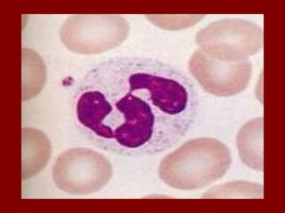
Neutrophils
Constitute 40 –75% of all circulating blood
cells.
About 12 – 14 micrometres in diameter
Have 3 – 5 lobbed nucleus
The nucleus of females has a
characteristic bar body
Are highly phagocytic and play a role in
bacterial infections.
78.
• Have threetypes of granules
• Primary granules: contain
myleperoxidases and lysosomal enzymes
• Secondary granules: contain inflammatory
mediators and complement activators
• Teritary granules: contain gelatinases
80.
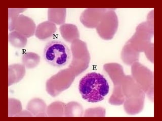
Eosinophils
Constitute 1 –6% of all circulating
leucocytes
Have a bilobbed nucleus that stains pink
12 – 17 micrometrs in diameter
Play a role in parasitic infections and
allergies
Their granules contain histaminase,
peroxidases and lysosomal enzymes
82.
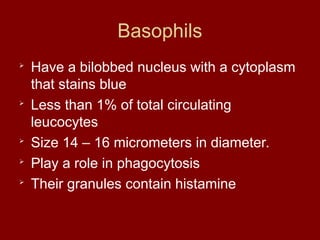
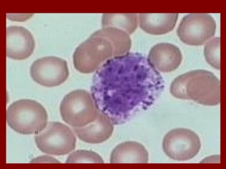
Basophils
Have a bilobbednucleus with a cytoplasm
that stains blue
Less than 1% of total circulating
leucocytes
Size 14 – 16 micrometers in diameter.
Play a role in phagocytosis
Their granules contain histamine
84.
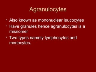
Agranulocytes
Also known asmononuclear leucocytes
Have granules hence agranulocytes is a
misnomer
Two types namely lymphocytes and
monocytes.
85.
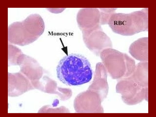
Monocytes
Constitute 2 –10% of all circulating
leucocytes
Largest of all leucocytes: 16 – 20
micrometers in diameter
Have a single lobbed nucleus that is bean
shaped
Are highly phagocytic.
87.
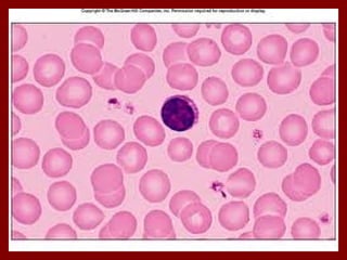
Lymphocytes
Smallest of allleucocytes
Second most abundant leucocytes(20 –
50%)
Two types, b- lymphocytes that mature in
the bone marrow and t – lymphocytes that
mature in the thymus.
B cells proliferate to become plasma cells
that produce antibodies
T cells are three types; cytotoxic, natural
killer and t helper cells
89.
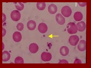
Platelets
Smallest of allblood cells, 1.5- 3.5
micrometers
Biconvex, oval or spherical
150,000 – 400,000 cells per mm cubed
Play a role in blood clotting by
a) aggregation
b) secreting coagulation factors
Providing a surface for adherence of
clotting factors