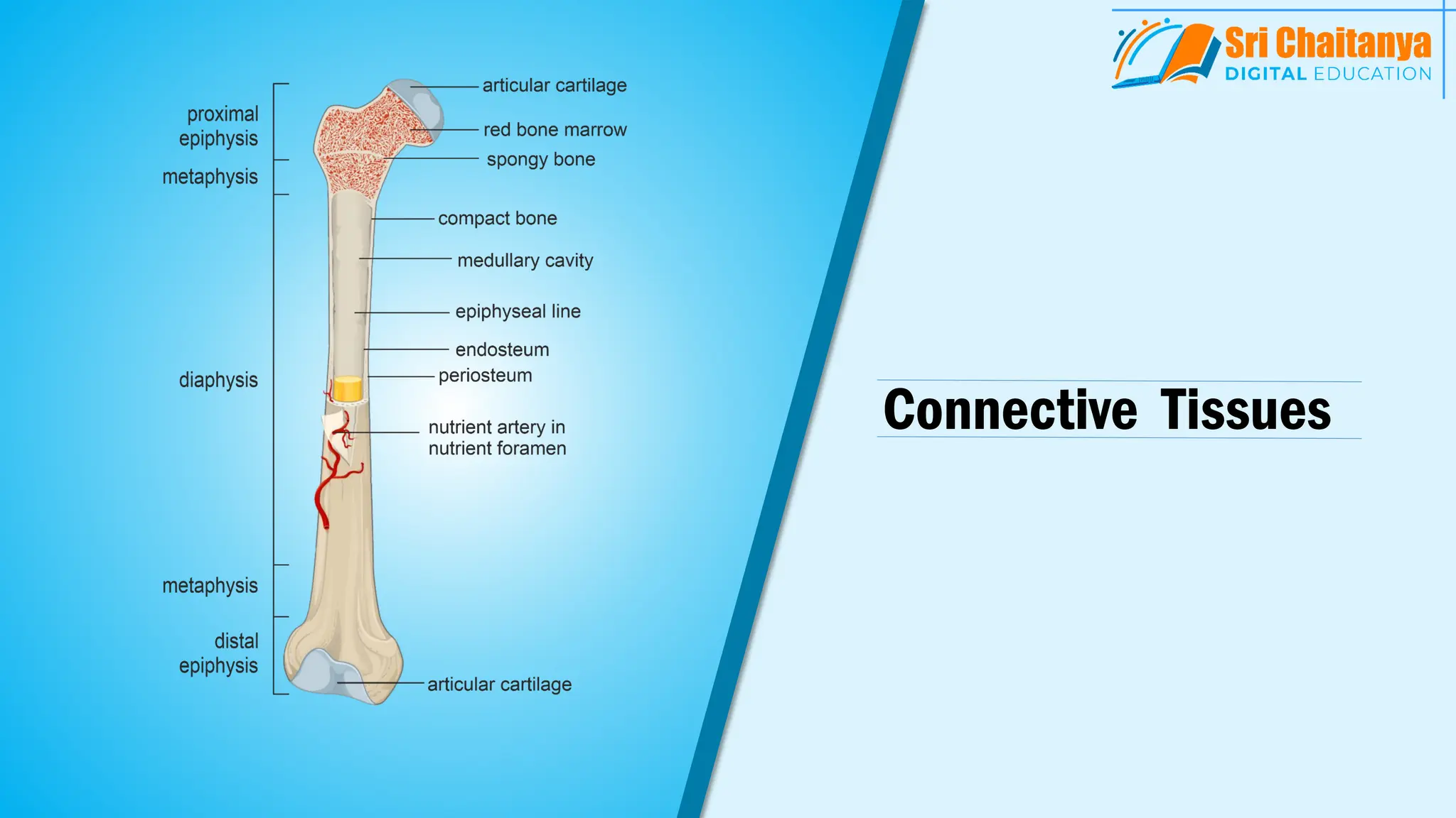
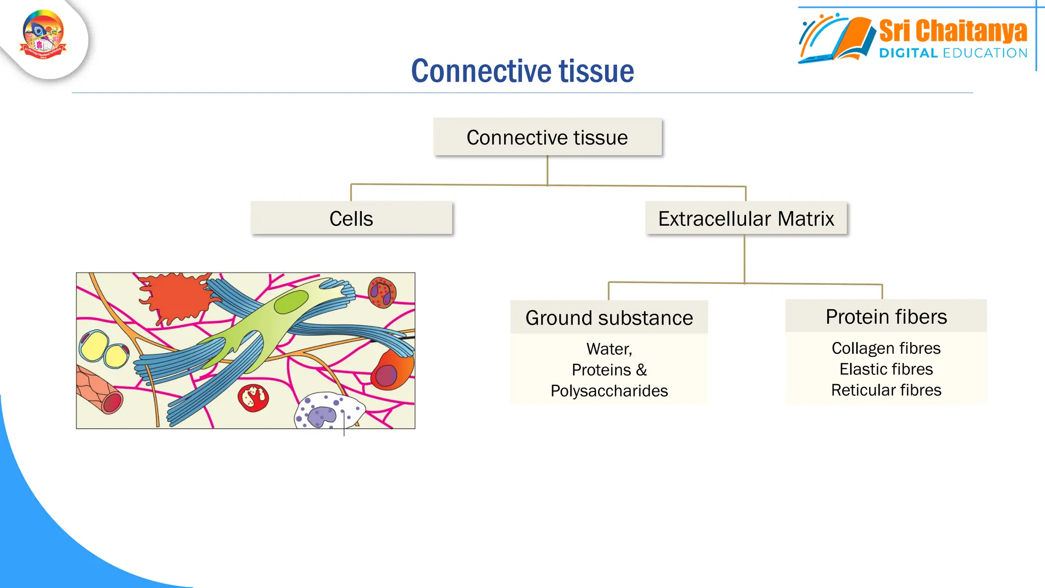
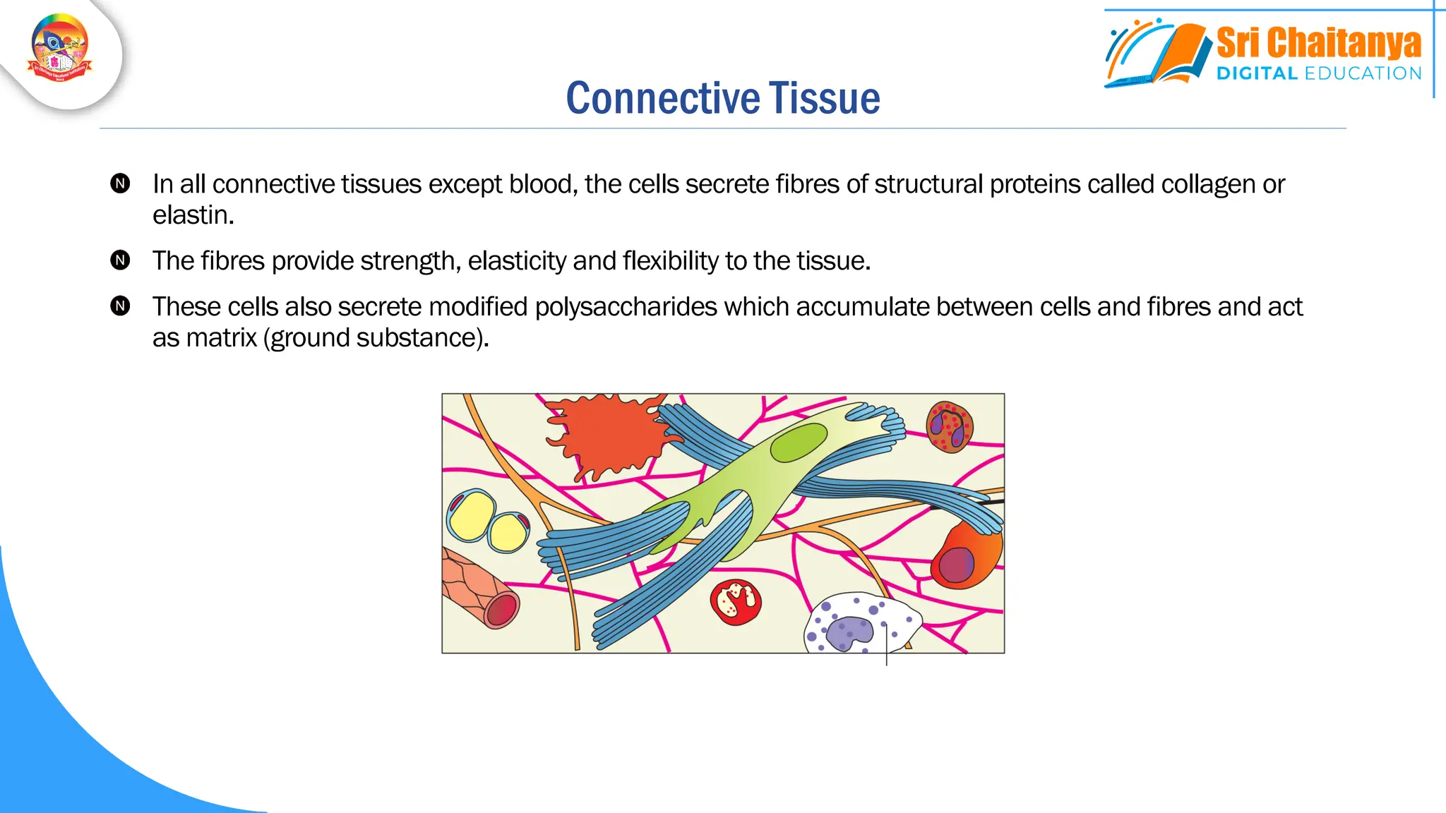
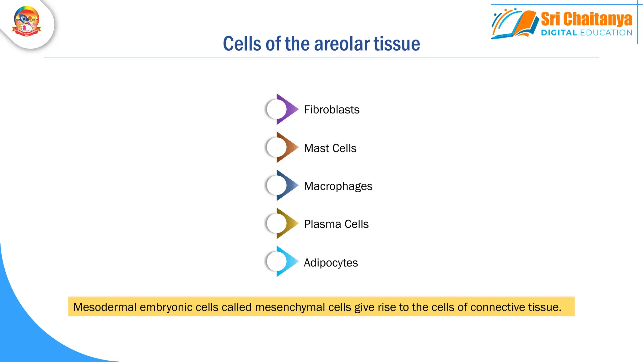
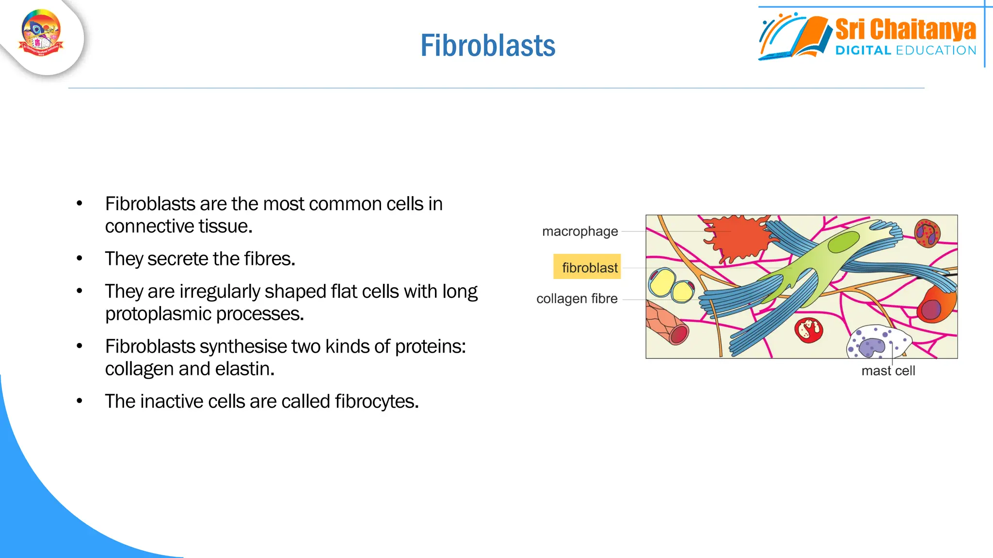
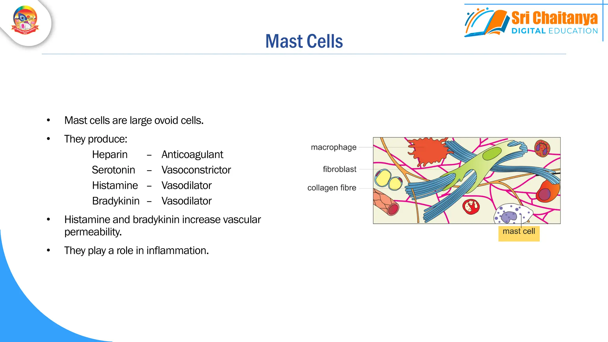
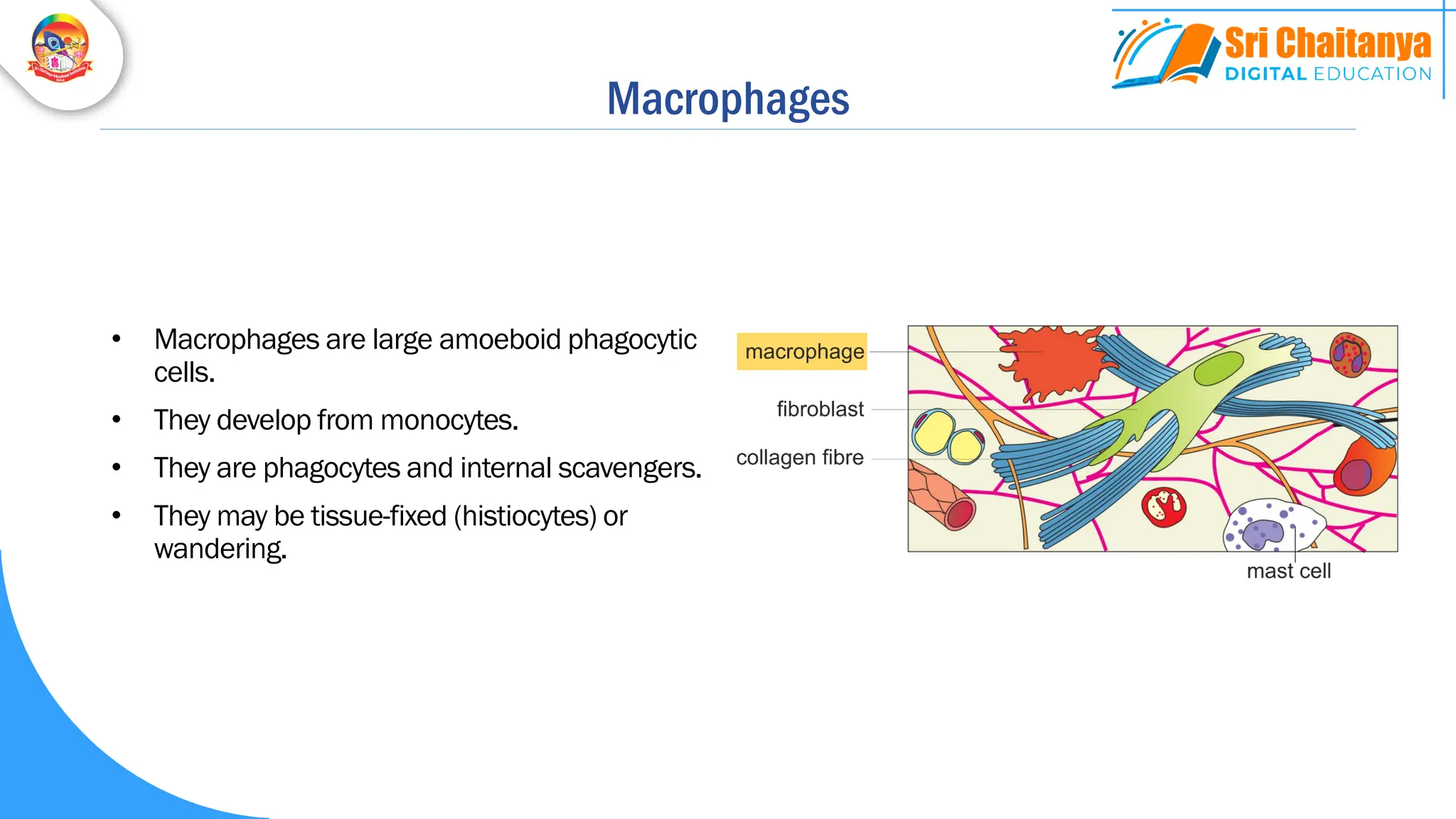
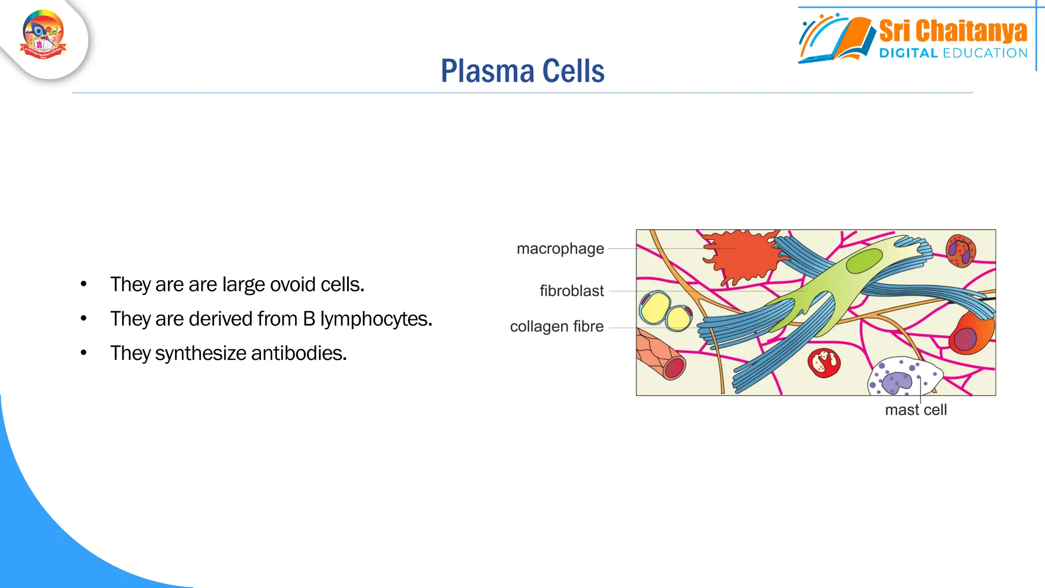
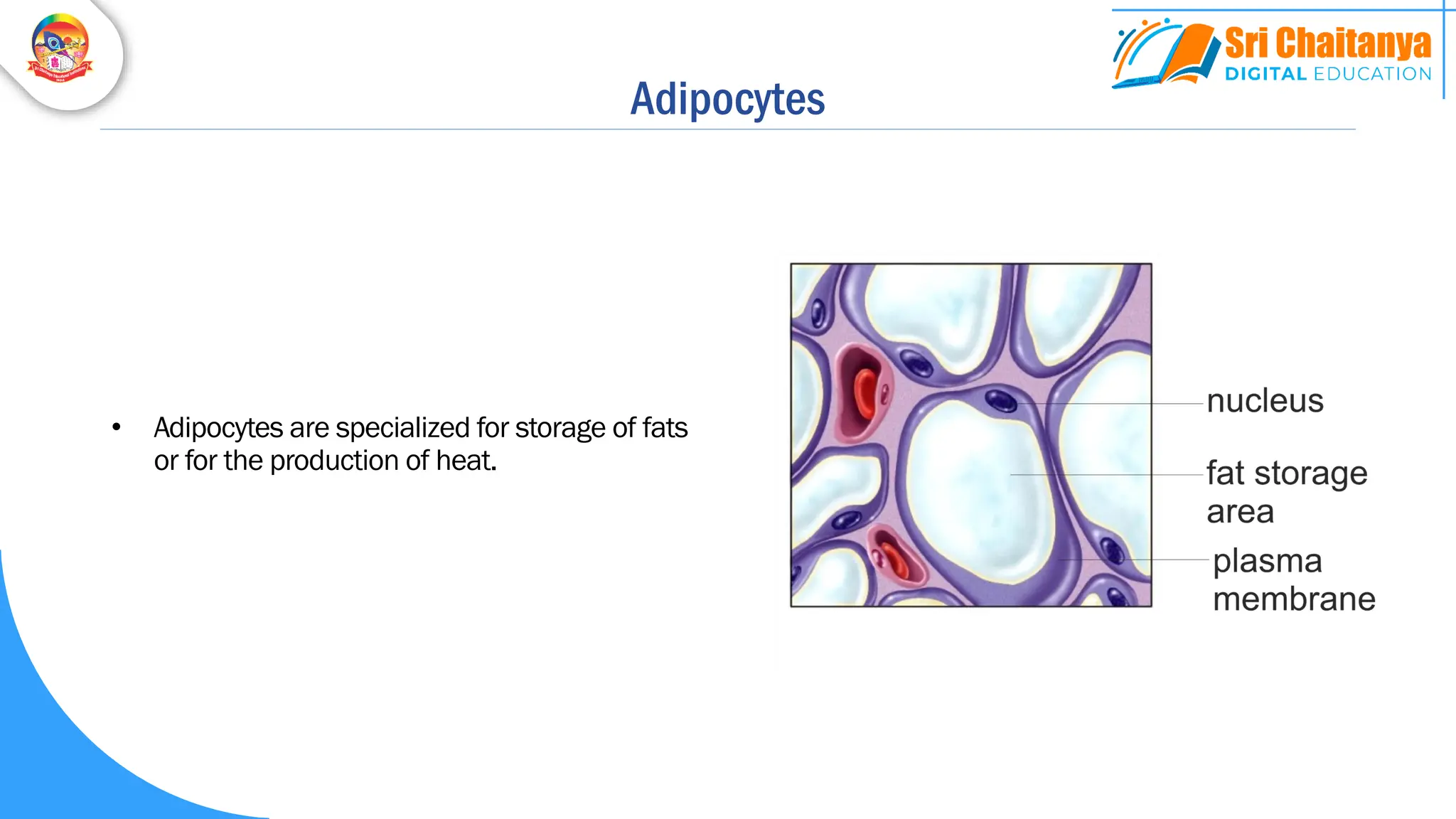
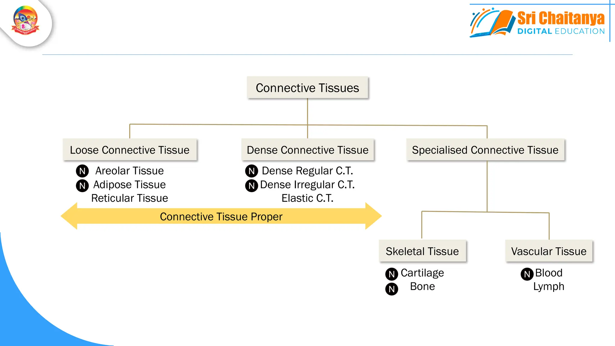
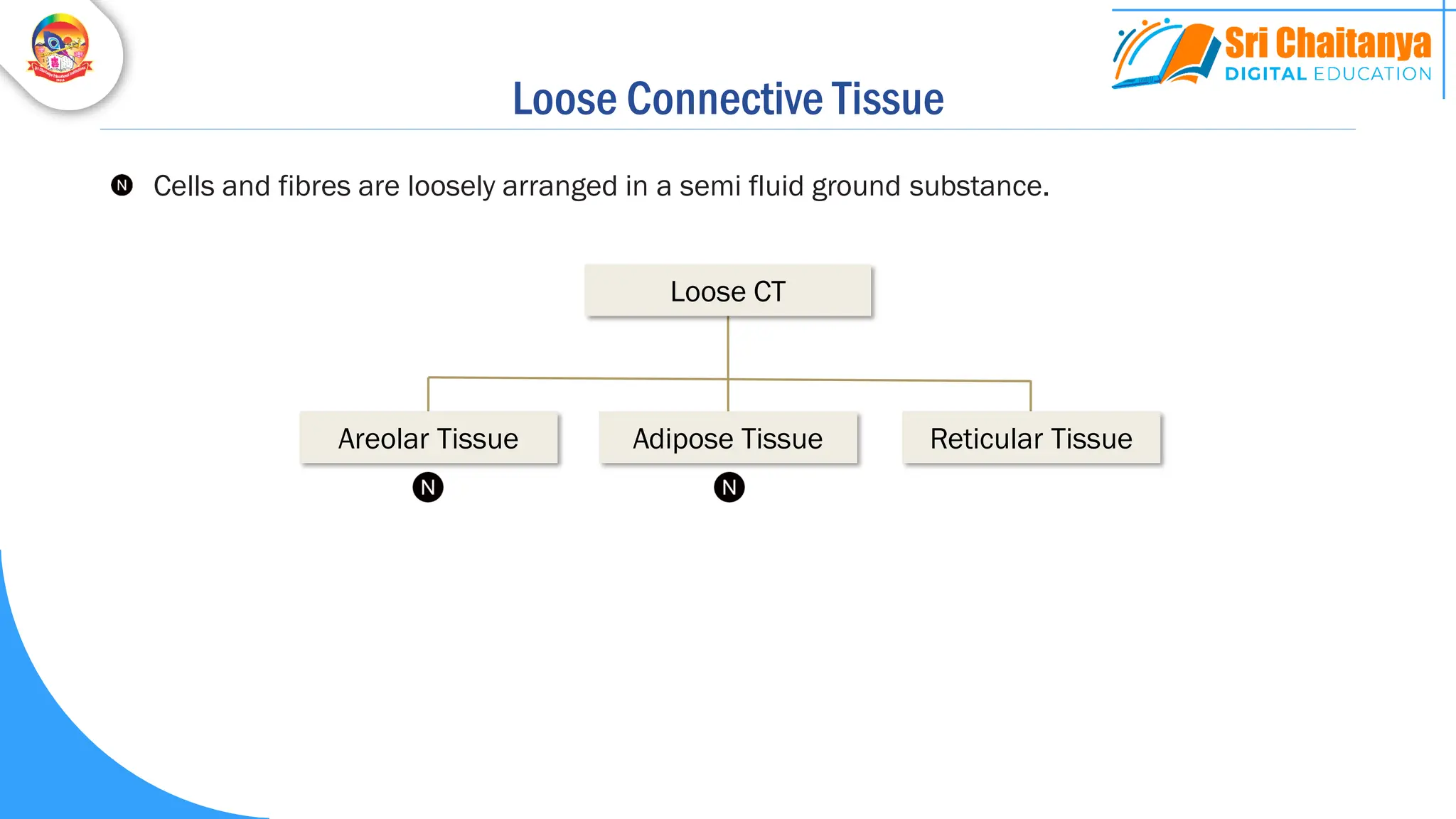
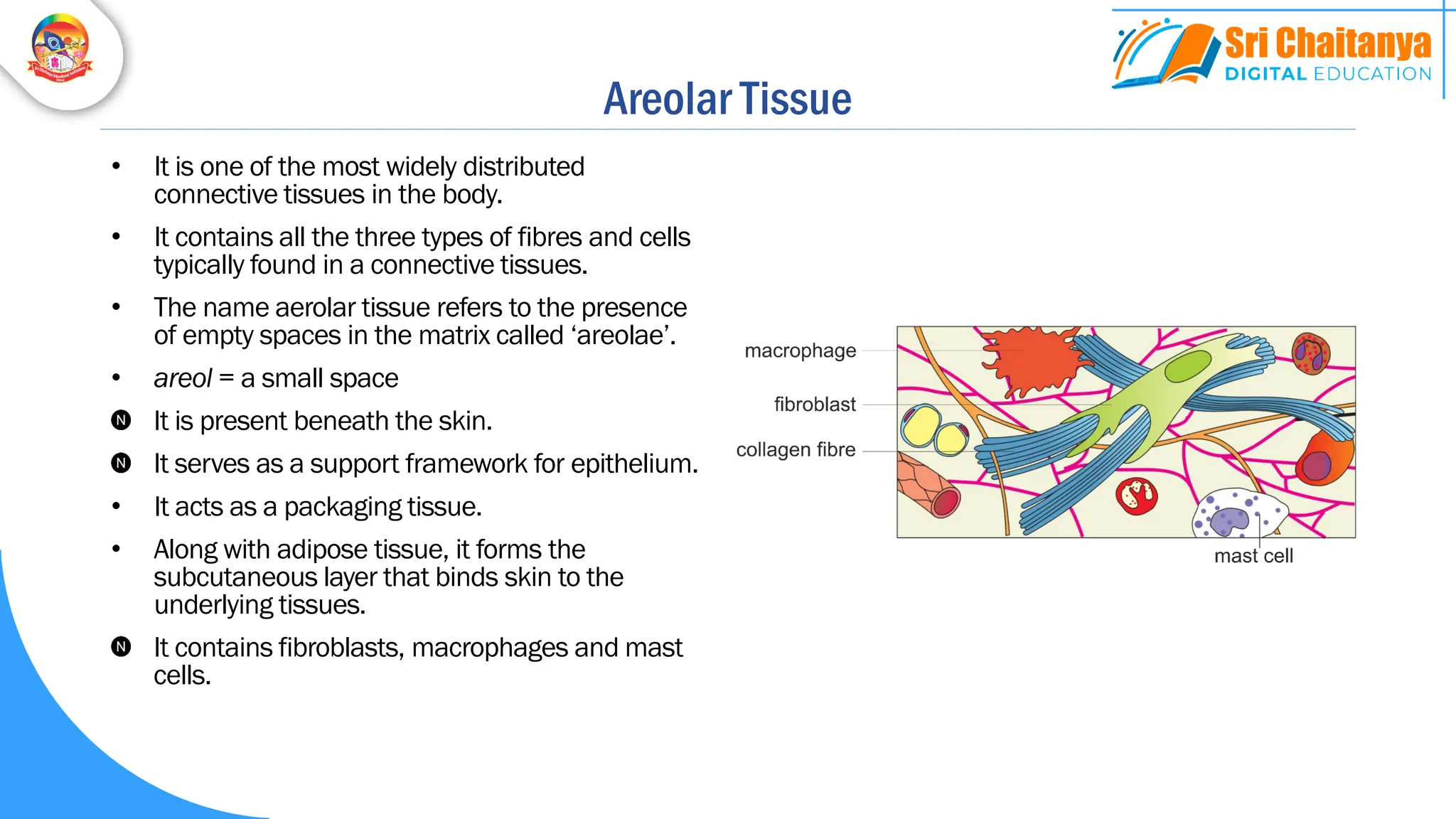
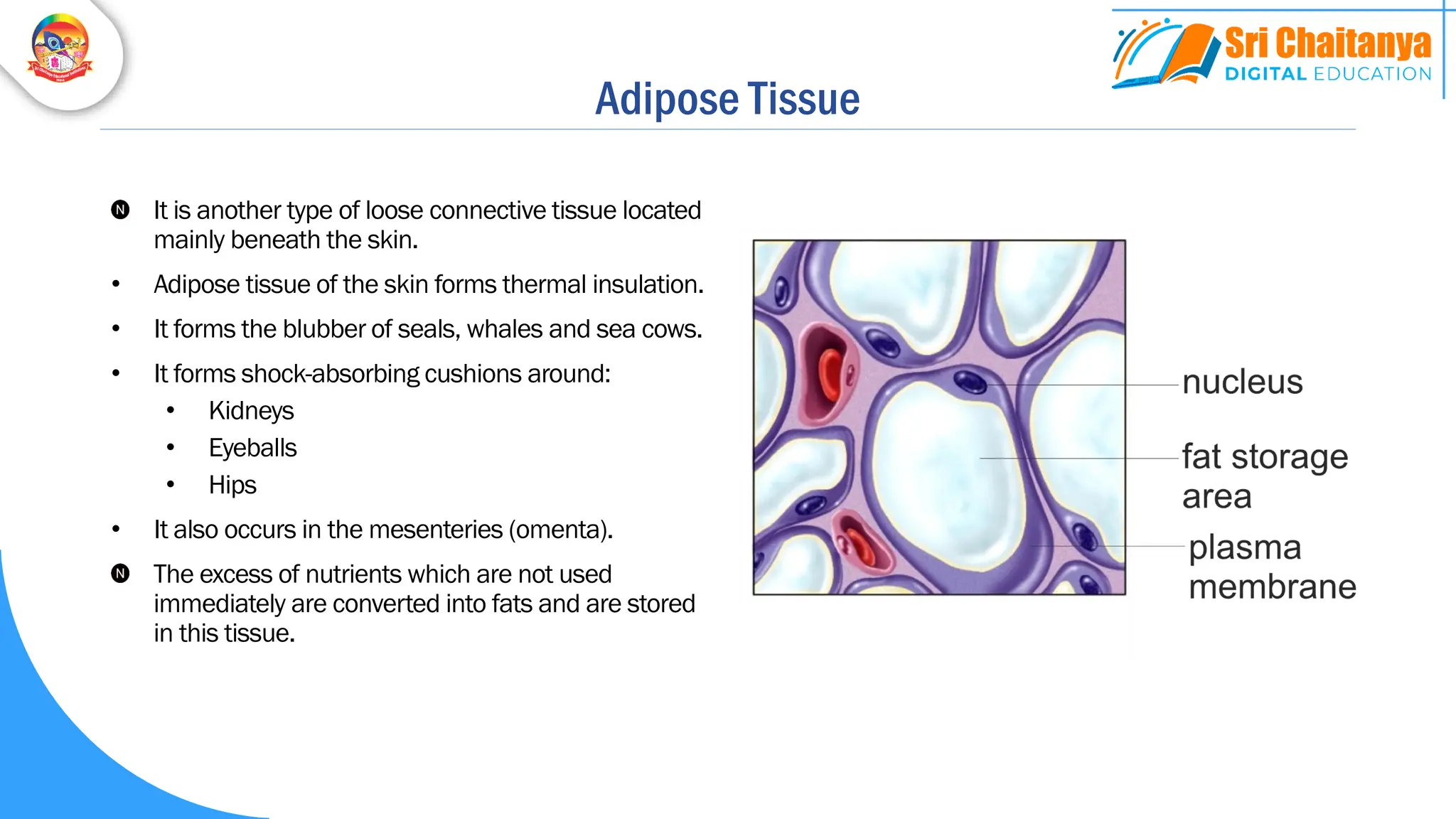
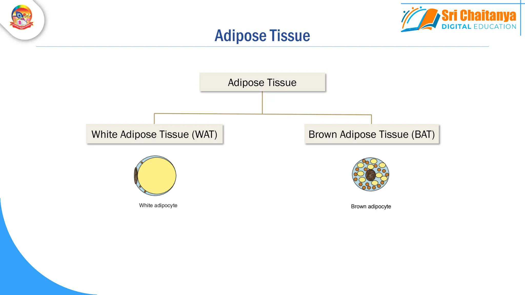
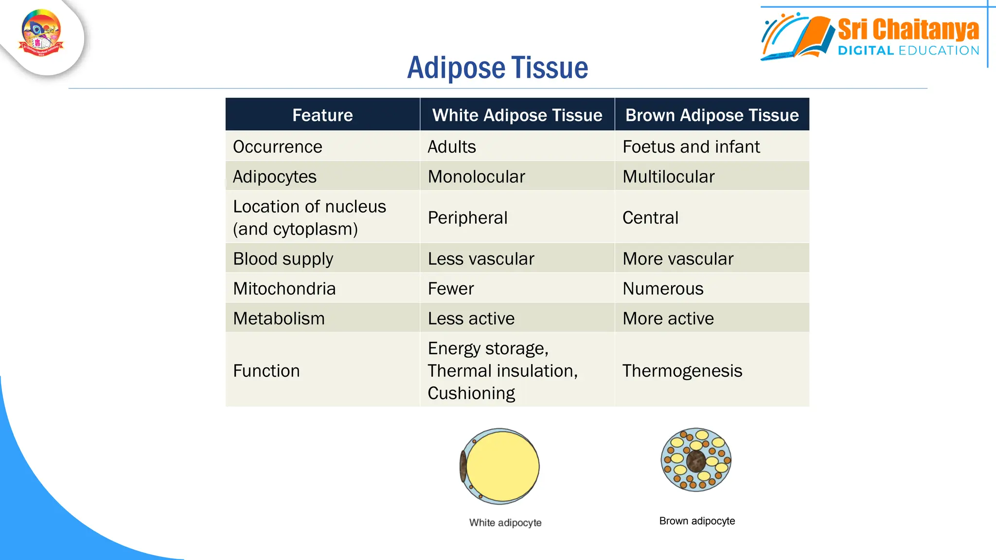
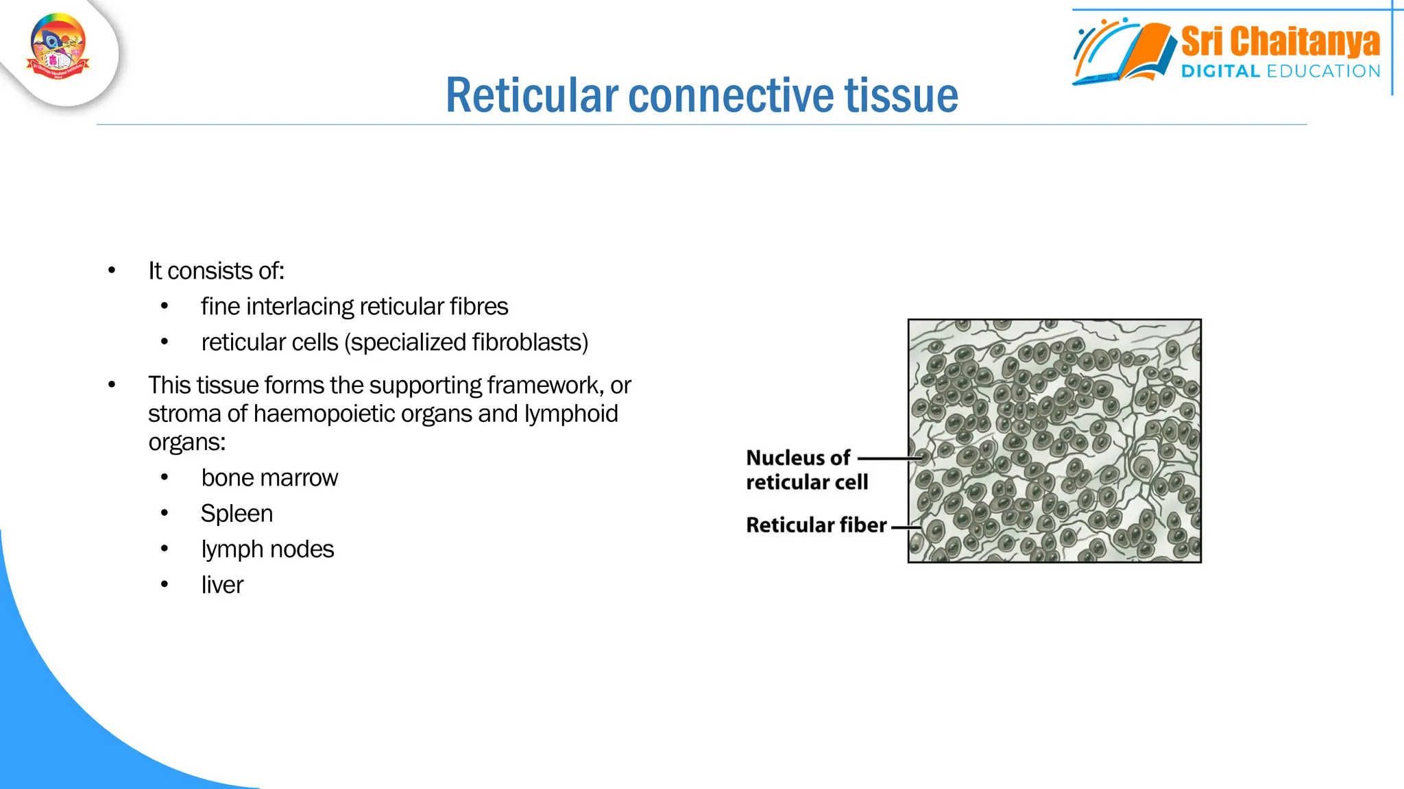
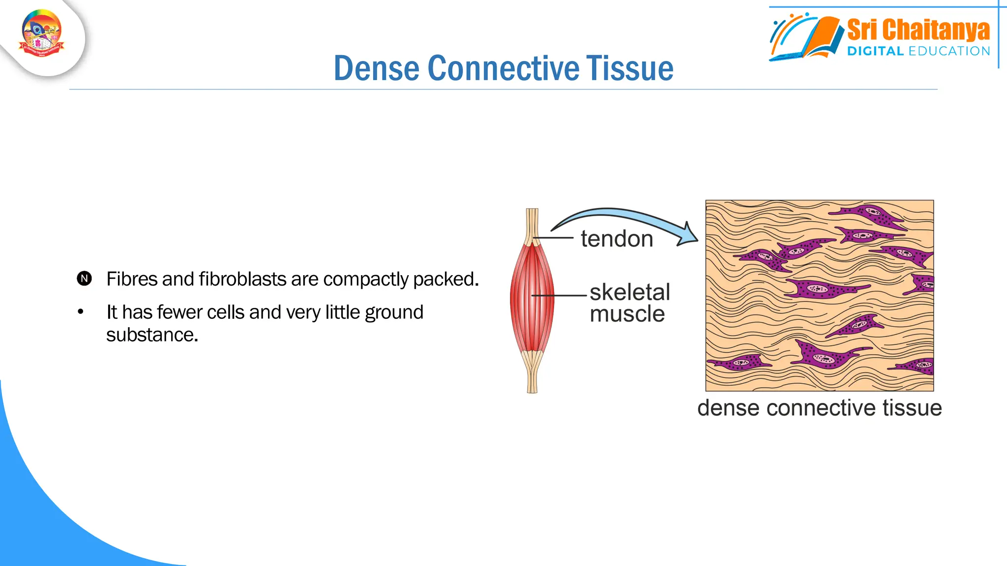
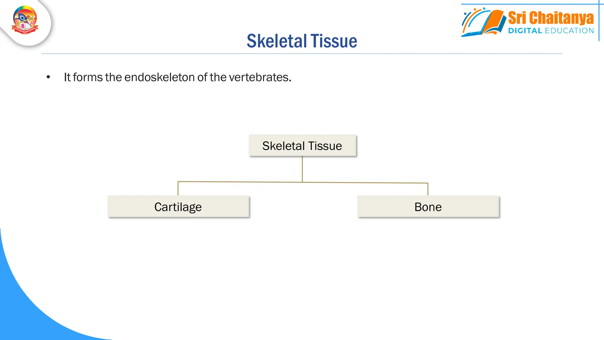
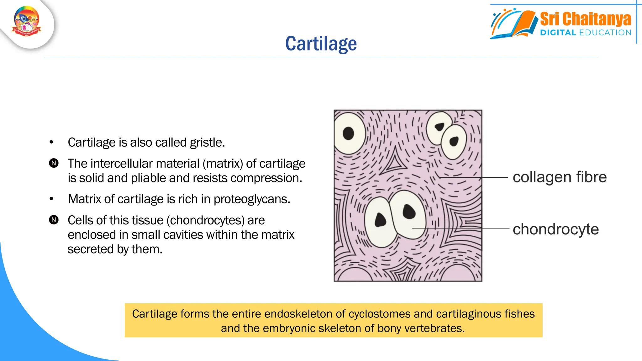
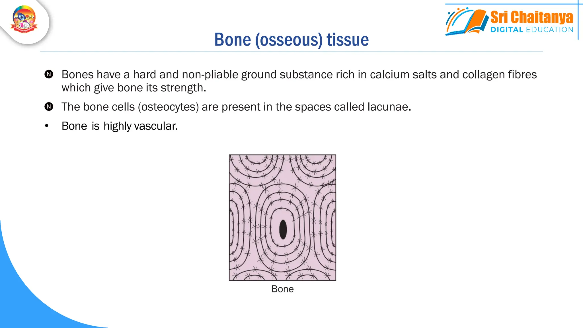
Connective Tissues
Connective Tissues
Connective tissues are most abundant and widely distributed in the body of complex animals.
They are named connective tissues because of their special function of linking and supporting
other tissues/ organs of the body.
• Connective tissues are mesodermal.
• Connective tissues are vascular, except cartilage.
Connective Tissues
They range from soft connective tissues to specialised types, which include
cartilage, bone, adipose tissue and blood
Summary:
Cartilage, bone, blood, and adipose tissue are specialised connective tissues.
Cells Extracellular Matrix
Connective tissue
Water,
Proteins &
Polysaccharides
Ground substance
Collagen fibres
Elastic fibres
Reticular fibres
Protein fibers
Connective tissue
Connective Tissue
In all connective tissues except blood, the cells secrete fibres of structural proteins called collagen or
elastin.
The fibres provide strength, elasticity and flexibility to the tissue.
These cells also secrete modified polysaccharides which accumulate between cells and fibres and act
as matrix (ground substance).
Cells of the areolar tissue
Fibroblasts
Mast Cells
Macrophages
Plasma Cells
Adipocytes
Mesodermal embryonic cells called mesenchymal cells give rise to the cells of connective tissue.
Fibroblasts
• Fibroblasts are the most common cells in
connective tissue.
• They secrete the fibres.
• They are irregularly shaped flat cells with long
protoplasmic processes.
• Fibroblasts synthesise two kinds of proteins:
collagen and elastin.
• The inactive cells are called fibrocytes.
Mast Cells
• Mast cells are large ovoid cells.
• They produce:
Heparin – Anticoagulant
Serotonin – Vasoconstrictor
Histamine – Vasodilator
Bradykinin – Vasodilator
• Histamine and bradykinin increase vascular
permeability.
• They play a role in inflammation.
Macrophages
• Macrophages are large amoeboid phagocytic
cells.
• They develop from monocytes.
• They are phagocytes and internal scavengers.
• They may be tissue-fixed (histiocytes) or
wandering.
Plasma Cells
• They are are large ovoid cells.
• They are derived from B lymphocytes.
• They synthesize antibodies.
Adipocytes
• Adipocytes are specialized for storage of fats
or for the production of heat.
Fibres of Connective Tissue
• Formed of proteins.
• They are of three types:
Collagen Fibres
Elastic Fibres
Reticular fibres
Collagen (white) fibres
• They are composed of collagen.
• They often occur in wavy bundles.
• They are strong and stretch-resistant.
Elastic (yellow) fibres
• Elastic (yellow) fibres are composed mainly of elastin.
• They are branched.
• They are strong but can be stretched.
• They are resilient (return to original shape after being stretched).
elastic fibre
Reticular Fibres
• Reticular fibres are extremely thin collagen
fibres (type III).
• They form an extensive network in certain
organs.
• They form the supporting frame work (stroma)
of organs like bone marrow.
Collagen Fibres Elastic Fibres Reticular Fibres
White fibres Yellow fibres -
Collagen Elastin Collagen (type III)
Unbranched Branched Branched
Bundles Network Network
Stretch-resistant Resilient Form stroma
Fibres of Connective Tissue
Loose Connective Tissue Specialised Connective Tissue
Connective Tissues
Dense Connective Tissue
Skeletal Tissue Vascular Tissue
Areolar Tissue
Adipose Tissue
Reticular Tissue
Dense Regular C.T.
Dense Irregular C.T.
Elastic C.T.
Cartilage
Bone
Blood
Lymph
Connective Tissue Proper
Loose Connective Tissue
Cells and fibres are loosely arranged in a semi fluid ground substance.
Areolar Tissue Reticular Tissue
Loose CT
Adipose Tissue
Areolar Tissue
• It is one of the most widely distributed
connective tissues in the body.
• It contains all the three types of fibres and cells
typically found in a connective tissues.
• The name aerolar tissue refers to the presence
of empty spaces in the matrix called ‘areolae’.
• areol = a small space
It is present beneath the skin.
It serves as a support framework for epithelium.
• It acts as a packaging tissue.
• Along with adipose tissue, it forms the
subcutaneous layer that binds skin to the
underlying tissues.
It contains fibroblasts, macrophages and mast
cells.
Adipose Tissue
It is another type of loose connective tissue located
mainly beneath the skin.
• Adipose tissue of the skin forms thermal insulation.
• It forms the blubber of seals, whales and sea cows.
• It forms shock-absorbing cushions around:
• Kidneys
• Eyeballs
• Hips
• It also occurs in the mesenteries (omenta).
The excess of nutrients which are not used
immediately are converted into fats and are stored
in this tissue.
White Adipose Tissue (WAT) Brown Adipose Tissue (BAT)
Adipose Tissue
Brown adipocyte
Adipose Tissue
Feature White Adipose Tissue Brown Adipose Tissue
Occurrence Adults Foetus and infant
Adipocytes Monolocular Multilocular
Location of nucleus
(and cytoplasm)
Peripheral Central
Blood supply Less vascular More vascular
Mitochondria Fewer Numerous
Metabolism Less active More active
Function
Energy storage,
Thermal insulation,
Cushioning
Thermogenesis
Brown adipocyte
Adipose Tissue
Reticular connective tissue
• It consists of:
• fine interlacing reticular fibres
• reticular cells (specialized fibroblasts)
• This tissue forms the supporting framework, or
stroma of haemopoietic organs and lymphoid
organs:
• bone marrow
• Spleen
• lymph nodes
• liver
Dense Connective Tissue
Fibres and fibroblasts are compactly packed.
• It has fewer cells and very little ground
substance.
Dense Regular C.T. Elastic C.T.
Dense Connective Tissue
Dense Irregular C.T.
Fibres and fibroblasts are compactly packed.
• It has fewer cells and very little ground substance.
Dense Connective Tissue
Dense Regular Connective Tissue
Collagen fibres are present in rows between many
parallel bundles of fibres.
• This tissue withstands pulling along the axis of the
fibres.
Examples:
• Tendons (attach skeletal muscles to bones)
• Ligaments (attach one bone to another).
A sprain is caused by excessive stretching of a ligament.
Dense Irregular Connective Tissue
Dense irregular connective tissue has fibroblasts
and many fibres (mostly collagen) that are oriented
differently (irregularly).
This tissue is present in the skin (dermis).
• It is found in parts of the body where pulling forces
are exerted in various directions. Other examples:
• Periosteum
• Perichondrium
• Pericardium
• Heart valves
• Joint capsules
Elastic connective tissue
• In this tissue, elastic fibres predominate
giving yellowish colour to the tissue.
• This tissue can recoil to its original shape
after being stretched and released.
• Location
• Wall of elastic arteries
• Vocal cords
• Trachea
• Bronchi
• Elastic ligaments (between vertebrae).
Mucous connective tissue
• It is an embryonic connective tissue.
• It is found mainly in the umbilical cord where
it is referred to as Wharton’s Jelly.
Specialised Connective Tissues
Cartilage, bones and blood are various types of specialised connective tissues.
Skeletal Tissue
• It forms the endoskeleton of the vertebrates.
Cartilage Bone
Skeletal Tissue
Cartilage
• Cartilage is also called gristle.
The intercellular material (matrix) of cartilage
is solid and pliable and resists compression.
• Matrix of cartilage is rich in proteoglycans.
Cells of this tissue (chondrocytes) are
enclosed in small cavities within the matrix
secreted by them.
Cartilage forms the entire endoskeleton of cyclostomes and cartilaginous fishes
and the embryonic skeleton of bony vertebrates.
Cartilage
Most of the cartilages in vertebrate embryos are
replaced by bones in adults.
Cartilage is present in the:
Tip of nose
Outer ear joints
Between adjacent bones of the vertebral
column
Limbs and hands in adults
Cartilage
• Cartilage is surrounded by perichondrium.
• Cartilage is avascular because it secretes
anti-angiogenesis factor.
• Cartilage is nourished by the diffusion of
nutrients from capillaries in perichondrium.
• During formation of cartilage, chondroblasts
secrete the matrix, and the cells themselves
get enclosed in cavities called lacunae.
• Each lacuna may contain upto eight
chondrocytes originating from mitotic
divisions of a single chondrocyte.
Hyaline cartilage
• It is the most common type of cartilage.
• It is bluish-white and translucent (glass-like).
• The matrix is homogeneous with delicate collagen fibrils.
Examples of Hyaline Cartilage
• Larynx
• Cartilaginous rings of trachea, bronchi and initial
bronchioles.
• Costal cartilage (in the ventral ends of ribs)
• Epiphyseal plate,
• Articular cartilages
Elastic cartilage
• Matrix has an abundance of yellow elastic fibres in addition to collagen fibres.
• Perichondrium is present.
• It provides strength and elasticity.
Location:
• Ear pinna
• Eustachian tubes
• Epiglottis.
Fibrocartilage (fibrous cartilage)
• Matrix contains a dense network of coarse collagen fibres.
• Perichondrium is absent.
• It is the strongest of all the three types of cartilage.
• Location: Intervertebral discs and pubic symphysis
pubic symphysis
Cartilage
Hyaline Cartilage Elastic Cartilage Fibrous Cartilage
Appearance Bluish-white, translucent
and glass-like
Yellowish Whitish
Collagen fibres Delicate Present Present (bundles)
Elastic fibres Absent Abundant Absent
Perichondrium Present except in
articular cartilage
Present Absent
Nature Weakest Strong and elastic Strongest
Calcified Cartilage
• Calcified cartilage is initially like hyaline
cartilage but later on it gets hardened like bone
due to deposition of calcium salts.
Examples:
• In the metaphyses of long bones
• Supra scapula of frog’s pectoral girdle
• Pubis of pelvic girdle of frog
Bone (osseous) tissue
Bones have a hard and non-pliable ground substance rich in calcium salts and collagen fibres
which give bone its strength.
The bone cells (osteocytes) are present in the spaces called lacunae.
• Bone is highly vascular.
Bone (osseous) tissue
Functions of Bone Tissue
It is the main tissue that provides structural frame to the body.
Bones support and protect softer tissues and organs.
Limb bones, such as the long bones of the legs, serve weight-
bearing functions.
They also interact with skeletal muscles attached to them to bring
about movements.
The bone marrow in some bones is the site of production of blood
cells.
• Storage of minerals (e.g.,. calcium, phosphorous, magnesium)
Bone (osseous) tissue
Inorganic 65% Organic 35%
Bone (dry weight)
• The major mineral is
calcium phosphate
• It is present primary in the
form of hydroxyapatite
crystals [Ca10(PO4)6(OH)2]
• The major organic
substance is collagen
Bone Cells
Osteoblasts
• Immature bone cells
• Secrete the bone matrix
Osteocytes
• Mature bone cells
• Enclosed in lacunae
Osteoclasts
• Phagocytic cells
• Involved in bone resorption
Types of bones based on the method of formation
Formed by ossification
within the cartilage.
Cartilage Bones
Formed by the
ossification in soft
tissues
Visceral Bones
Bones
Formed by the
ossification of the
embryonic
mesenchyme.
Membrane Bones
Formed by the
ossification of
tendons.
Sesamoid Bones
Bones of limbs, girdles
and vertebrae
Most of the cranial
bones
Patella
Pisiform
Os penis
Os cordis
Replacing (endochondral) bones
Dermal (membrane) bones
Types of bones based on shape and size
Types of bones based on the structure
Spongy (cancellous) bone Compact bone
Epiphyses and metaphyses of
long bones, hip bones, ribs,
sternum
Diaphyses of long bones
Matrix is spongy with
trabeculae
Dense lamellar matrix
Interspaces filled with red
bone marrow (haemopoietic)
Marrow cavity has yellow
bone marrow
Structure of a compact bone
• Diaphysis is the shaft.
• Epiphyses are the expanded ends.
• Metaphysis is between the diaphysis and
epiphysis.
• It consists of an epiphyseal plate formed by
hyaline cartilage.
• It helps in the elongation of the bone.
• In adults it is represented by a bony epiphyseal
line.
Structure of a compact bone
• Diaphysis is covered by periosteum.
• Diaphysis of a long bone has a marrow
cavity.
• Marrow cavity is lined by the endosteum.
• Haversian systems or osteons (units of
bone) are present between periosteum
and endosteum.
Structure of a compact bone
• Haversian system consists of a longitudinal
Haversian canal that contains blood vessels
and nerves.
• Haversian canal is surrounded by
concentric lamellae.
• Small fluid filled spaces called lacunae lie
in between the lamellae.
• Each lacuna encloses one osteocyte.
• Minute canaliculi radiate from lacunae.
• The Haversian canals communicate with one
another by Volkmann’scanals.

(2) Structural org - Connective Tissues.pdf

  • 1.
  • 2.
    Connective Tissues Connective tissuesare most abundant and widely distributed in the body of complex animals. They are named connective tissues because of their special function of linking and supporting other tissues/ organs of the body. • Connective tissues are mesodermal. • Connective tissues are vascular, except cartilage.
  • 3.
    Connective Tissues They rangefrom soft connective tissues to specialised types, which include cartilage, bone, adipose tissue and blood Summary: Cartilage, bone, blood, and adipose tissue are specialised connective tissues.
  • 4.
    Cells Extracellular Matrix Connectivetissue Water, Proteins & Polysaccharides Ground substance Collagen fibres Elastic fibres Reticular fibres Protein fibers Connective tissue
  • 5.
    Connective Tissue In allconnective tissues except blood, the cells secrete fibres of structural proteins called collagen or elastin. The fibres provide strength, elasticity and flexibility to the tissue. These cells also secrete modified polysaccharides which accumulate between cells and fibres and act as matrix (ground substance).
  • 6.
    Cells of theareolar tissue Fibroblasts Mast Cells Macrophages Plasma Cells Adipocytes Mesodermal embryonic cells called mesenchymal cells give rise to the cells of connective tissue.
  • 7.
    Fibroblasts • Fibroblasts arethe most common cells in connective tissue. • They secrete the fibres. • They are irregularly shaped flat cells with long protoplasmic processes. • Fibroblasts synthesise two kinds of proteins: collagen and elastin. • The inactive cells are called fibrocytes.
  • 8.
    Mast Cells • Mastcells are large ovoid cells. • They produce: Heparin – Anticoagulant Serotonin – Vasoconstrictor Histamine – Vasodilator Bradykinin – Vasodilator • Histamine and bradykinin increase vascular permeability. • They play a role in inflammation.
  • 9.
    Macrophages • Macrophages arelarge amoeboid phagocytic cells. • They develop from monocytes. • They are phagocytes and internal scavengers. • They may be tissue-fixed (histiocytes) or wandering.
  • 10.
    Plasma Cells • Theyare are large ovoid cells. • They are derived from B lymphocytes. • They synthesize antibodies.
  • 11.
    Adipocytes • Adipocytes arespecialized for storage of fats or for the production of heat.
  • 12.
    Fibres of ConnectiveTissue • Formed of proteins. • They are of three types: Collagen Fibres Elastic Fibres Reticular fibres
  • 13.
    Collagen (white) fibres •They are composed of collagen. • They often occur in wavy bundles. • They are strong and stretch-resistant.
  • 14.
    Elastic (yellow) fibres •Elastic (yellow) fibres are composed mainly of elastin. • They are branched. • They are strong but can be stretched. • They are resilient (return to original shape after being stretched). elastic fibre
  • 15.
    Reticular Fibres • Reticularfibres are extremely thin collagen fibres (type III). • They form an extensive network in certain organs. • They form the supporting frame work (stroma) of organs like bone marrow.
  • 16.
    Collagen Fibres ElasticFibres Reticular Fibres White fibres Yellow fibres - Collagen Elastin Collagen (type III) Unbranched Branched Branched Bundles Network Network Stretch-resistant Resilient Form stroma Fibres of Connective Tissue
  • 17.
    Loose Connective TissueSpecialised Connective Tissue Connective Tissues Dense Connective Tissue Skeletal Tissue Vascular Tissue Areolar Tissue Adipose Tissue Reticular Tissue Dense Regular C.T. Dense Irregular C.T. Elastic C.T. Cartilage Bone Blood Lymph Connective Tissue Proper
  • 18.
    Loose Connective Tissue Cellsand fibres are loosely arranged in a semi fluid ground substance. Areolar Tissue Reticular Tissue Loose CT Adipose Tissue
  • 19.
    Areolar Tissue • Itis one of the most widely distributed connective tissues in the body. • It contains all the three types of fibres and cells typically found in a connective tissues. • The name aerolar tissue refers to the presence of empty spaces in the matrix called ‘areolae’. • areol = a small space It is present beneath the skin. It serves as a support framework for epithelium. • It acts as a packaging tissue. • Along with adipose tissue, it forms the subcutaneous layer that binds skin to the underlying tissues. It contains fibroblasts, macrophages and mast cells.
  • 20.
    Adipose Tissue It isanother type of loose connective tissue located mainly beneath the skin. • Adipose tissue of the skin forms thermal insulation. • It forms the blubber of seals, whales and sea cows. • It forms shock-absorbing cushions around: • Kidneys • Eyeballs • Hips • It also occurs in the mesenteries (omenta). The excess of nutrients which are not used immediately are converted into fats and are stored in this tissue.
  • 21.
    White Adipose Tissue(WAT) Brown Adipose Tissue (BAT) Adipose Tissue Brown adipocyte Adipose Tissue
  • 22.
    Feature White AdiposeTissue Brown Adipose Tissue Occurrence Adults Foetus and infant Adipocytes Monolocular Multilocular Location of nucleus (and cytoplasm) Peripheral Central Blood supply Less vascular More vascular Mitochondria Fewer Numerous Metabolism Less active More active Function Energy storage, Thermal insulation, Cushioning Thermogenesis Brown adipocyte Adipose Tissue
  • 23.
    Reticular connective tissue •It consists of: • fine interlacing reticular fibres • reticular cells (specialized fibroblasts) • This tissue forms the supporting framework, or stroma of haemopoietic organs and lymphoid organs: • bone marrow • Spleen • lymph nodes • liver
  • 24.
    Dense Connective Tissue Fibresand fibroblasts are compactly packed. • It has fewer cells and very little ground substance.
  • 25.
    Dense Regular C.T.Elastic C.T. Dense Connective Tissue Dense Irregular C.T. Fibres and fibroblasts are compactly packed. • It has fewer cells and very little ground substance. Dense Connective Tissue
  • 26.
    Dense Regular ConnectiveTissue Collagen fibres are present in rows between many parallel bundles of fibres. • This tissue withstands pulling along the axis of the fibres. Examples: • Tendons (attach skeletal muscles to bones) • Ligaments (attach one bone to another). A sprain is caused by excessive stretching of a ligament.
  • 27.
    Dense Irregular ConnectiveTissue Dense irregular connective tissue has fibroblasts and many fibres (mostly collagen) that are oriented differently (irregularly). This tissue is present in the skin (dermis). • It is found in parts of the body where pulling forces are exerted in various directions. Other examples: • Periosteum • Perichondrium • Pericardium • Heart valves • Joint capsules
  • 28.
    Elastic connective tissue •In this tissue, elastic fibres predominate giving yellowish colour to the tissue. • This tissue can recoil to its original shape after being stretched and released. • Location • Wall of elastic arteries • Vocal cords • Trachea • Bronchi • Elastic ligaments (between vertebrae).
  • 29.
    Mucous connective tissue •It is an embryonic connective tissue. • It is found mainly in the umbilical cord where it is referred to as Wharton’s Jelly.
  • 30.
    Specialised Connective Tissues Cartilage,bones and blood are various types of specialised connective tissues.
  • 31.
    Skeletal Tissue • Itforms the endoskeleton of the vertebrates. Cartilage Bone Skeletal Tissue
  • 32.
    Cartilage • Cartilage isalso called gristle. The intercellular material (matrix) of cartilage is solid and pliable and resists compression. • Matrix of cartilage is rich in proteoglycans. Cells of this tissue (chondrocytes) are enclosed in small cavities within the matrix secreted by them. Cartilage forms the entire endoskeleton of cyclostomes and cartilaginous fishes and the embryonic skeleton of bony vertebrates.
  • 33.
    Cartilage Most of thecartilages in vertebrate embryos are replaced by bones in adults. Cartilage is present in the: Tip of nose Outer ear joints Between adjacent bones of the vertebral column Limbs and hands in adults
  • 34.
    Cartilage • Cartilage issurrounded by perichondrium. • Cartilage is avascular because it secretes anti-angiogenesis factor. • Cartilage is nourished by the diffusion of nutrients from capillaries in perichondrium. • During formation of cartilage, chondroblasts secrete the matrix, and the cells themselves get enclosed in cavities called lacunae. • Each lacuna may contain upto eight chondrocytes originating from mitotic divisions of a single chondrocyte.
  • 35.
    Hyaline cartilage • Itis the most common type of cartilage. • It is bluish-white and translucent (glass-like). • The matrix is homogeneous with delicate collagen fibrils.
  • 36.
    Examples of HyalineCartilage • Larynx • Cartilaginous rings of trachea, bronchi and initial bronchioles. • Costal cartilage (in the ventral ends of ribs) • Epiphyseal plate, • Articular cartilages
  • 37.
    Elastic cartilage • Matrixhas an abundance of yellow elastic fibres in addition to collagen fibres. • Perichondrium is present. • It provides strength and elasticity. Location: • Ear pinna • Eustachian tubes • Epiglottis.
  • 38.
    Fibrocartilage (fibrous cartilage) •Matrix contains a dense network of coarse collagen fibres. • Perichondrium is absent. • It is the strongest of all the three types of cartilage. • Location: Intervertebral discs and pubic symphysis pubic symphysis
  • 39.
    Cartilage Hyaline Cartilage ElasticCartilage Fibrous Cartilage Appearance Bluish-white, translucent and glass-like Yellowish Whitish Collagen fibres Delicate Present Present (bundles) Elastic fibres Absent Abundant Absent Perichondrium Present except in articular cartilage Present Absent Nature Weakest Strong and elastic Strongest
  • 40.
    Calcified Cartilage • Calcifiedcartilage is initially like hyaline cartilage but later on it gets hardened like bone due to deposition of calcium salts. Examples: • In the metaphyses of long bones • Supra scapula of frog’s pectoral girdle • Pubis of pelvic girdle of frog
  • 41.
    Bone (osseous) tissue Boneshave a hard and non-pliable ground substance rich in calcium salts and collagen fibres which give bone its strength. The bone cells (osteocytes) are present in the spaces called lacunae. • Bone is highly vascular.
  • 42.
    Bone (osseous) tissue Functionsof Bone Tissue It is the main tissue that provides structural frame to the body. Bones support and protect softer tissues and organs. Limb bones, such as the long bones of the legs, serve weight- bearing functions. They also interact with skeletal muscles attached to them to bring about movements. The bone marrow in some bones is the site of production of blood cells. • Storage of minerals (e.g.,. calcium, phosphorous, magnesium)
  • 43.
    Bone (osseous) tissue Inorganic65% Organic 35% Bone (dry weight) • The major mineral is calcium phosphate • It is present primary in the form of hydroxyapatite crystals [Ca10(PO4)6(OH)2] • The major organic substance is collagen
  • 44.
    Bone Cells Osteoblasts • Immaturebone cells • Secrete the bone matrix Osteocytes • Mature bone cells • Enclosed in lacunae Osteoclasts • Phagocytic cells • Involved in bone resorption
  • 45.
    Types of bonesbased on the method of formation Formed by ossification within the cartilage. Cartilage Bones Formed by the ossification in soft tissues Visceral Bones Bones Formed by the ossification of the embryonic mesenchyme. Membrane Bones Formed by the ossification of tendons. Sesamoid Bones Bones of limbs, girdles and vertebrae Most of the cranial bones Patella Pisiform Os penis Os cordis
  • 46.
  • 47.
  • 48.
    Types of bonesbased on shape and size
  • 49.
    Types of bonesbased on the structure Spongy (cancellous) bone Compact bone Epiphyses and metaphyses of long bones, hip bones, ribs, sternum Diaphyses of long bones Matrix is spongy with trabeculae Dense lamellar matrix Interspaces filled with red bone marrow (haemopoietic) Marrow cavity has yellow bone marrow
  • 50.
    Structure of acompact bone • Diaphysis is the shaft. • Epiphyses are the expanded ends. • Metaphysis is between the diaphysis and epiphysis. • It consists of an epiphyseal plate formed by hyaline cartilage. • It helps in the elongation of the bone. • In adults it is represented by a bony epiphyseal line.
  • 51.
    Structure of acompact bone • Diaphysis is covered by periosteum. • Diaphysis of a long bone has a marrow cavity. • Marrow cavity is lined by the endosteum. • Haversian systems or osteons (units of bone) are present between periosteum and endosteum.
  • 52.
    Structure of acompact bone • Haversian system consists of a longitudinal Haversian canal that contains blood vessels and nerves. • Haversian canal is surrounded by concentric lamellae. • Small fluid filled spaces called lacunae lie in between the lamellae. • Each lacuna encloses one osteocyte. • Minute canaliculi radiate from lacunae. • The Haversian canals communicate with one another by Volkmann’scanals.