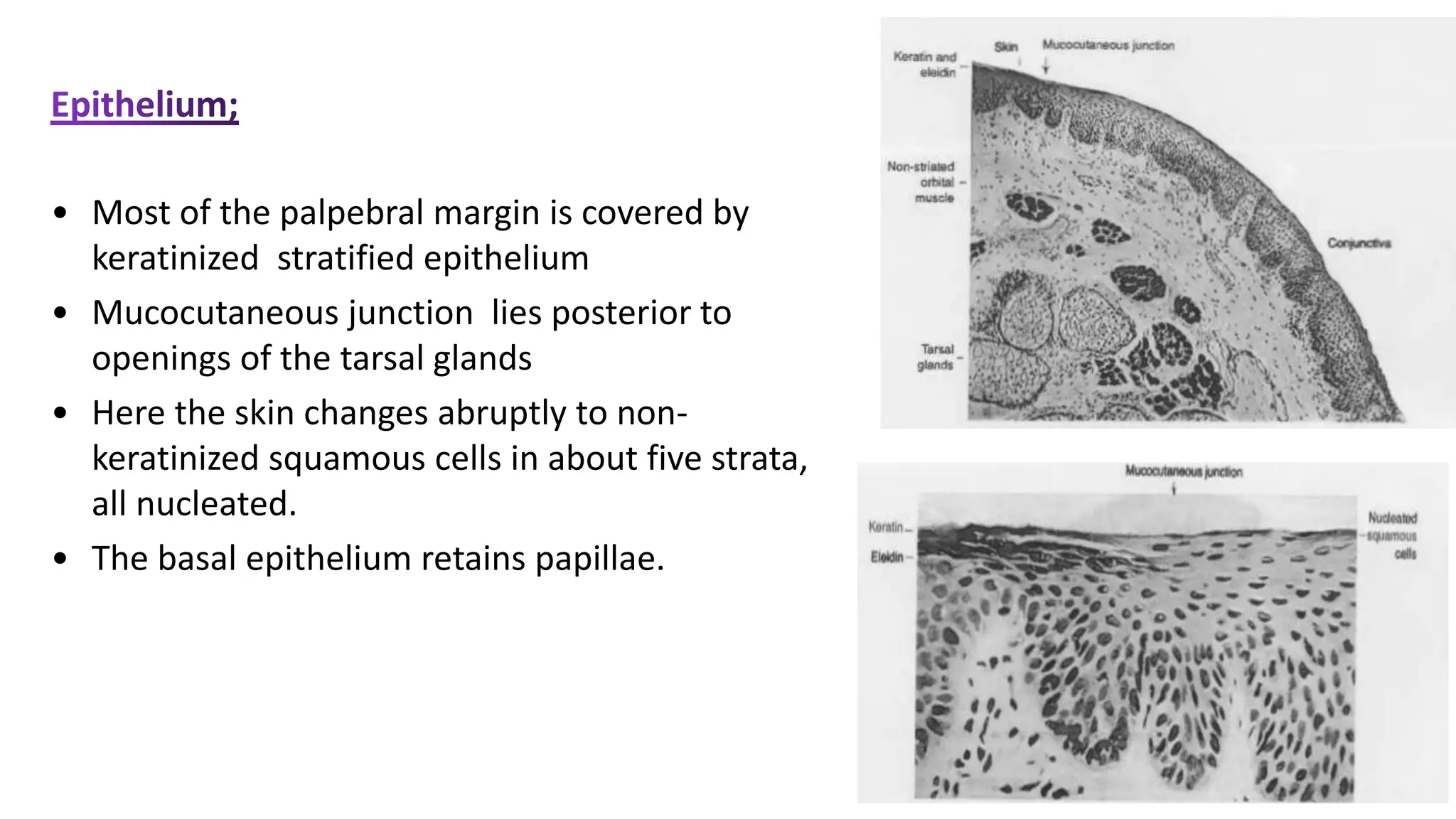
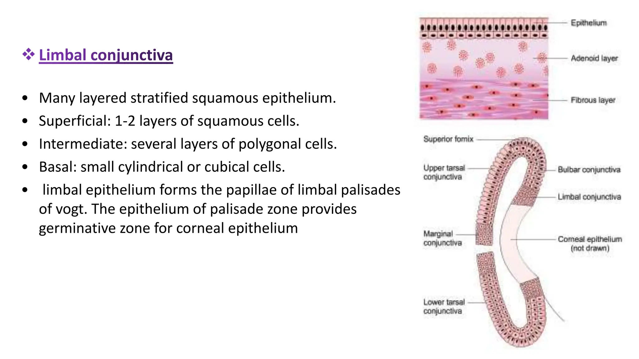
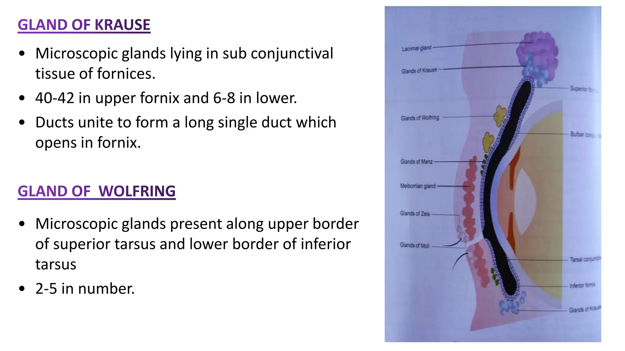
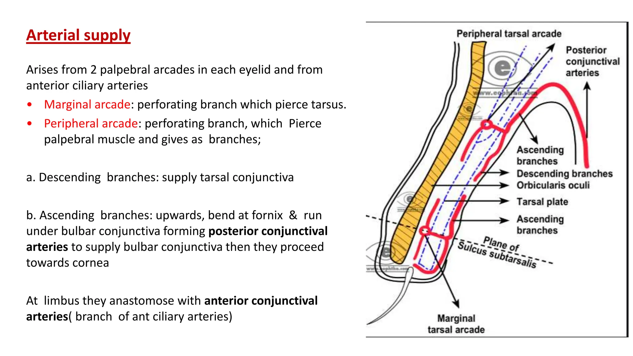
3. INTRODUCTION
• The overwhelming part of the anterior eye ball around the cornea is covered by
the conjunctiva. This is the main support tissue of the cornea in terms of wetting,
nutrition, defense and many other functions.
• Latin origin of word : ( it joins the eye ball to the eye lid)
• Transparent and Vascularized mucous membrane.
• It covers the anterior surface of the globe ( ).
• Posterior surface of the upper and lower eyelids (
4. EMBRYOLOGY
Develops from the ectoderm lining the lids and covering
the globe.
Conjunctival glands develop as a growth of the basal cells of upper
conjunctival fornix
Fewer glands develop from the lower fornix.
5. Conjunctiva covers the lids posteriorly, is reflected
anteriorly to the sclera, becoming continuous with
the corneal epithelium.
• Conjunctival sac thus formed is open at the
palpebral fissure.
• It normally contains about 7 µl of tear fluid but
can accommodate up to 30 µl. Instilled eye
drops in excess of this volume are either
drained by the lacrimal sac or overflow the lids
ANATOMY
6.
7. Subdivided into the
Transition zone between skin and the conjunctiva
proper
• Extends from back of the lid for about 2 mm to a
shallow sub tarsal fold--sulcus subtarsalis , near
which the perforating vessels traverse the tarsus
to conjunctiva
• Common site of lodgement of conjunctival foreign
body
8. • Thin, adherent and very vascular, reddish
colour
• Unlike the upper tarsal conjunctiva the lower
is adherent for only half the tarsal width
• Common site for follicular and papillary
reaction .
upper lid: between the tarsal upper border and
fornix
9. • Is a continuous annular cul-de-sac broken on
medial side by caruncle and plica seminularis
• Divided into superior, inferior, lateral and
medial fornices
a) Superior fornix:
reaches the orbital margin , 8-10 mm from the
limbus
• Glands of Krause and Muller's muscle present
in subconjunctival tissue in superior fornix
• Knife passed through sup fornix,enters fibrous
tissue between levator and SR
• FB in sup fornix seen by double version of UL
10. b) Inferior fornix
• Reaches a few mm of the inferior orbital
margin, 8 mm from the limbus
• Extension of fascial sheath of IO and IR is
attached to conjunctical fold in lower fornix
which helps maintaining the inferior fornix
• Glands of Krause are present in the
subconjunctival tissue of inferior fornix
11. c) Lateral fornix:
6 mm from the surface and 14 mm from the
limbus, extends just posterior to the equator.
d) Medial fornix :
Shallow, comprising medial ends of the superior
and inferior fornix . It’s a sac where Caruncle and
Plica semilunaris lie dipped in pool of tears = lacus
lacrimlais
12. BULBAR CONJUNCTIVA
• Thin + translucent : so underlying sclera appear
white
• Is in contact with tendon of recti, covered by
fascia bulbi (tenon’s capsule )
• Tied to adjacent structures by areolar tissue,
and is thus mobile enough to allow ocular
movement
• At about 3 mm from the cornea, conjunctiva
fascia bulbi and sclera are adheren: the limbal
conjunctiva. (Because the conjunctiva here is
less mobile, a firmer hold of the globe can be
obtained at surgery.)
13. Clinical Applications:
• Through vascularity of thin palpebral conjunctiva ---
anemia diagnosis
• Capacity of sac is 30 µl ---excess is spilled
• Most common site of foreign body lodgment is
subtarsal sulcus in upper palpebral conjunctiva
14. It consists of 3 layers as follows:-
1. Epithelium
2. Adenoid layer
3. Fibrous layer
15. • Most of the palpebral margin is covered by
keratinized stratified epithelium
• Mucocutaneous junction lies posterior to
openings of the tarsal glands
• Here the skin changes abruptly to non-
keratinized squamous cells in about five strata,
all nucleated.
• The basal epithelium retains papillae.
16. • It comprises a superficial adenoid layer and a deep fibrous layer.
• Adenoid layer contains lymphoid tissue that results in follicle formation with
appropriate stimuli. It is undeveloped in infants who cannot develop a
follicular response.
• The tarsal conjunctiva is firmly anchored to the tarsus, resulting in papillae
when there is conjunctival infiltration, whereas the bulbar conjunctiva is only
loosely attached to the globe and papillae are not seen except at the limbus.
• The fibrous layer is generally thicker than the lymphoid layer, except over the
tarsal plate,where it blends.
• It contains the conjunctival vessels and nerves and glands of Krause
17. It contains;
• 5 layered non keratinized stratified squamous
epithelium
• Superficial layer: squamous cells
• Intermediate layer: 3 layers of polyhedral cells
• Deepest layer: cylindrical cells
18. Epithelium of the superior tarsal conjunctiva is
bilaminar (2 layers)
• Deeper layer of cubical cells
• Superficial layer of cylindrical cells
Epithelium of inferior tarsal conjunctiva has three or
four
• Basal cells(deep layer) are cubical, followed by
layers of polygonal cuneiform, and conical cells.
• Surface cells are conical.
19.
• 3 layered epithelium.
• superficial layer of cylindrical cells and middle layer of
polyhedral cells.
• deep layer is of cuboidal cells.
• From fornix to limbus, epithelium becomes less
glandular, losing its goblet cells, and more epidermal
in type; but it is never keratinized.
20. • Many layered stratified squamous epithelium.
• Superficial: 1-2 layers of squamous cells.
• Intermediate: several layers of polygonal cells.
• Basal: small cylindrical or cubical cells.
• limbal epithelium forms the papillae of limbal palisades
of vogt. The epithelium of palisade zone provides
germinative zone for corneal epithelium
21.
22. Epithelial cells are attached to one another by desmosomes
The intercellular spaces are wider than those of the corneal
epithelium.
The basal aspect of the cells attaches to an basal lamina by
hemidesmosomes.
Mitoses are uncommon & Conjunctival cell density falls with
age.
23. • The lymphoid layer is a fine connective tissue reticulum, containing many
lymphocytes.
• Thickest in the fornices and ends at the subtarsal fold.
• Lymphocytic nodules occur near the canthi but diminish in the conjunctival
periphery.
• Expansions of these foci may cause visible surface swellings in follicular
conjunctivitis of viral or allergic origin.
• Lymphocytes, predominantly T cells, are found in substantia propria and
epithelium, in a ratio of about 2:3.
• Neutrophils are also found in the epithelium and submucosa, while plasma cells
and mast cells are found only in the submucosa.
• CALT consists of T and B lymphocytes without plasma cells.
24. 2 types of glands:
• Mucin secretory glands: Goblet cells, crypts
of Henle, glands of Manz
• Accessory lacrimal glands: glands of Krause
and glands of wolfring
25. • These occur throughout conjunctiva, especially the plica semilunaris
• most dense nasally, least dense in upper temporal fornix.
• absent at the palpebral mucocutaneous junction and the limbus
• The goblet cells are the chief source of tear mucin.
• Arise from the basal layer of epithelium and then migrates towards the surface
• These cell are destroyed after discharging their content i.e mucin
26. • Crypts of Henles are not true glands but
folds of mucous membranes in palpebral
conjunctiva between tarsal plates and
fornices
• Resembles crypts of lieberkuhns of
intestine.
• found in limbal conjunctiva in animals
like pigs calfs etc
• In humans controversial.
27. • Microscopic glands lying in sub conjunctival
tissue of fornices.
• 40-42 in upper fornix and 6-8 in lower.
• Ducts unite to form a long single duct which
opens in fornix.
• Microscopic glands present along upper border
of superior tarsus and lower border of inferior
tarsus
• 2-5 in number.
28. APPLIED ANATOMY
• Goblet cells secrete mucin of tear film.
• Accessory lacrimal glands of Krause and wolfring
secrete aqueous layer of tear film.
• Thus conjunctiva has a major role in maintenance of
tear film.
29. Arterial supply
Arises from 2 palpebral arcades in each eyelid and from
anterior ciliary arteries
• Marginal arcade: perforating branch which pierce tarsus.
• Peripheral arcade: perforating branch, which Pierce
palpebral muscle and gives as branches;
a. Descending branches: supply tarsal conjunctiva
b. Ascending branches: upwards, bend at fornix & run
under bulbar conjunctiva forming posterior conjunctival
arteries to supply bulbar conjunctiva then they proceed
towards cornea
At limbus they anastomose with anterior conjunctival
arteries( branch of ant ciliary arteries)
30. THE CONJUNCTIVAL VEINS
More numerous than are the arteries
Accompany their corresponding arteries
The major portion of the drainage from the tarsal,
forniceal, and bulbar conjunctiva is directed
toward the palpebral veins.
Some of the tarsal veins empty independently
into the superior and inferior ophthalmic veins.
Outflow from the circum-corneal region goes to
those veins that serve the extraocular muscles.
31. Lymphatic vessels are present in the
conjunctiva, but no lymphatic nodes
or vessels are present in the orbit.
A superficial lymphoid plexus of small
vessels extends beneath the vascular
capillaries and of deep plexus of large
vessels, in the fibrous layer of the
conjunctiva, receives lymph from this.
It drains towards the canthi, joining
the lympathics of the lids.
the lateral channels drain to the
parotid nodes, then to the
preauricular node whereas medial
canthus and lower lid to the
submandibular lymph node.
32. NERVES:
• The nerves supplying the conjunctiva
are from the same sources as for the
lids
• the long ciliary nerves supply the
circumcorneal conjunctiva (and
cornea) and the lacrimal and
infratrochlear nerves supply a larger
proportion of conjunctiva than skin.
33. • Symptoms;
• Non-Specific
Lacrimation
Irritation
Stinging
Burning
Photophobia
Redness
• Specific
Pain and FB sensation in corneal involvement.
Itching in allergic, blephritis and dry eyes.
35. Exudate plus debris plus mucus plus tears.
Serous: watery exudate in acute viral and acute
allergic conjunctivitis.
Mucoid: mucus discharge in VKC and KCS (dry
eyes)
Purulent: pus in severe acute bacterial
conjunctivitis
Mucopurulent: pus plus mucus in mild bacterial
conjunctivitis and Chlamydial conjunctivitis
36. 1.Hyperaemia (‘injection’) :Focal or diffuse
dilation of the subepithelial plexus of
conjunctival blood vessels, usually with
increased blood flow; typical of bacterial
infection.
2. Haemorrhages may occur with viral and
occasionally bacterial conjunctivitis.
3. Chemosis (conjunctival oedema) may occur
when severe inflammation produces a
translucent swelling which, if severe, may
protrude through the closed lids. Acute
chemosis usually indicates a hypersensitivity
response whereas chronic oedema suggests
orbital outflow constriction.
37. 4. Membranes
a. Pseudomembranes :consist of coagulated exudate adherent to
the inflamed conjunctival epithelium . They can be peeled easily
leaving the underlying epithelium intact
b. True membranes: involve the superficial layers of the
conjunctival epithelium so that attempted removal leads to
tearing and bleed.
Causes: Severe viral or bacterial conjunctivitis
Stevens-Johnson syndrome
Chemical burn
5. Infiltration: represents cellular recruitment to the site of
chronic inflammation and typically accompanies a papillary
response. It is recognized by loss of detail of the normal tarsal
conjunctival vessels, especially on the upper lid.
6. Subconjunctival scarring may occur in trachoma and other
types of cicatrizing conjunctivitis. Severe scarring is associated
with loss of goblet cells and accessory lacrimal glands, and can
lead to cicatricial entropion
38. 7. Follicles
Focal lymphoid nodule with accessory vascularization.
a. Multiple, discrete, slightly elevated lesions resembling
translucent grains of rice, most prominent in the fornices . Blood
vessels run around or across rather than within the lesions.
b. shows a subepithelial lymphoid germinal centre with
central immature lymphocytes and mature cells peripherally.
Adenovirus conjunctivitis
Herpes simplex virus conjunctivitis
Molluscum contagiosum blepharoconjunctivitis
Chlamydia conjunctivitis
Drug-induced (eg, dipivefrin) conjunctivitis
39. Dilated, telangiectatic conjunctival blood vessels, varying
from dotlike changes to enlarged tufts surrounded by
edema and a mixed inflammatory cell infiltrate.
can develop only in the palpebral conjunctiva and in the
limbal bulbar conjunctiva where it is attached to the
deeper fibrous layer.
In contrast to follicles, a vascular core is present.
Micropapillae form a mosaic-like pattern of elevated red
dots as a result of the central vascular channel
Macropapillae (<1 mm ) and giant papillae (>1 mm)
develop with prolonged inflammation.
Apical staining with fluorescein or the presence of
mucus between giant papillae indicates active disease.
Limbal papillae have a gelatinous appearance
40. :
Laxity of conjunctiva, sometimes with prolapse over the eyelid
Excess tears from increased lacrimation or impaired lacrimal
outflow
Increased amount of mucin relative to aqueous component of
tear
Nodule of chronic inflammatory cells with fibrovascular
proliferation
Cause: Cat-scratch disease ,Sarcoidosis, Foreign-body reaction
Nodule of chronic inflammatory cells, often at or near the
limbus
41. PINGUECULA
Yellowish-white patch on the bulbar conjunctiva.
Age related change.
Commonly seen in individual exposed to
UV light, dust &wind.
Elastotic degeneration in substantia propria a/w
deposition of hyaline materials.
Apex lies away from cornea.
42. PTERYGIUM
Wing shaped fold seen to be encroaching cornea.
Seen in individual with prolonged
exposure to UV dry heat, dust and
wind.
Degenerative and hyperplastic condition of
conjunctiva.
Elastotic degeneration destroying corneal epithelia
bowman’s layers & superficial stroma.
Apex lies towards cornea and frequently seen in nasal
side .
43. Pterygium can be divided into 4 parts:-
a. Head
b. Neck
c. Body
d. Cap
Depending upon progression 2 types.
a. Progressive
a. Regressive
44. Concretions:
Formed due accumulation of inspissated
mucus & epithelial debris .
Degenerative condition seen in elderly
individual.
Seen in palpebral conjunctiva (
Upper>>Lower).
Yellowish- white & Hard looking .
Can cause FB sensation and excess lacrimation
.
45. Conjunctival tumors
They can be both pigmented as well as non pigmented.
On the basis of nature and tissue of origin their types are:-
Tissue of origin Benign Malignant
Epithelial Papilloma Squamosal cell carcinoma
Glandular Adenoma Adenocarcinoma
Connective tissue Fibroma Sarcoma
Vascular Hemangioma Angiosarcoma
Reticular system Lymphoid hyperplasia Lymphosarcoam
Pigment cells Naevus Melanoma
46. Dermoid
Congenital tumors
Occurring at limbus as solid white mass.
Has sebaceous gland , hair , collagenous
connective tissue.
Lined by epidermoid.
47. Lipodermoid
Congenital tumor.
Found at limbus or outer canthus.
Appears soft, white , moveable, sub
conjunctival mass.
Orbital fat
prolapse
48. Naevi
A.k.A congenital moles of
conjunctiva.
Greyish, blackish or brownish
coloration.
Found as a flat or slightly raised
nodule in bulbar conjunctiva.
May show increase size during
puberty & pregnancy.
Pigmentary change seen
during inflammation.
Editor's Notes
Conjunctiva terminates in Corneo scleral limbus.
The tarsal glands appear as yellow streaks through the translucent tarsal conjunctiva
Intimately adherent to the superior tarsus and almost impossible to separate by dissection, which makes surgical repairs here very difficult
Basically conjunctival fornix is a piece that joins the palpebral and bulbar conjunctiva together where they meet.
Superior fornix; Extension of fascial sheath of levator and SR is attached to conjunctiva in upper part of sup fornix… helps maintaining recess of superior fornix in upper lid movements
Knife passed will pierce fibrous tissue between IR and inferior palpebral muscle and on further push it reaches aponeurotic expansion from IR and IO
Fornical conjunctiva is adherent to areolar tissue, which is continuous with expansions from the sheaths of the levator and rectus muscles, whose contractions can therefore deepen the fornices
In contact with tendons of the recti, covered by fascia bulbi (Tenon's capsule); both are divided to expose the tendons, anterior to which the conjunctiva covers the anterior part of the bulbar fascia
Between conjunctiva and sclera is loose episcleral tissue. In this episcleral region lie the anterior ciliary arteries, forming a pericorneal plexus, and tendons of the recti.
The conjunctival epithelium includes goblet cells that produce the mucous layer of the tear film.
The superficial cells are alligned with borders by junctional complexes (comprising zonulae occludentae, zonulae adherentae, and maculae adherente).
which confers the property of a semipermeable membrane on the conjunctival epithelium (as with the corneal epithelium)
Facilitates the passage of lipid-soluble molecules from the tears to the conjunctiva This favors the entry of lipid-soluble drugs (such as chloramphenicol) into the conjunctiva
Applied: Physiologically the lymphoid cells are non-inflammatory and continuously involved in the protective maintenance of mucosal immune regulation with a focus in tolerance in order to avoid inflammatory tissue destruction.
glands of manz arranged in a ring around the cornea, near the scleral junction. They also are responsible for secreting mucin into tears.
the main lacrimal gland is responsible only for reflex tearing whereas the accessory glands of Kraus and Wolfring, providing basal tear secretion
Palpebral arcades:There are two palpebral arcades which are the major source of the blood supply of conjunctiva. These are marginal tarsal arcade & peripheral tarsal arcade. Marginal tarsal arcades are larger than peripheral tarsal arcade.
Ant ciliary arises frm muscular br of oph to rectus …………….a ani b
---Ultimately drain into either the palpebral veins or the superior and inferior ophthalmic veins
Veins from conjunctiva and venous plexus of eyelid drains to sup. and inferior ophthalmic vein, and a circum zone of veins from 5-6 mm from limbus drains to anterior ciliary veins.
A circumcorneal zone of conjunctiva is supplied by the branches from long ciliary nerves which supply the cornea.
Rest of the conjunctiva is supplied by the branches from lacrimal, infratrochlear, supratrochlear, supraorbital and frontal nerves.
Free nerve endings
Nerve fibres lose their myelin sheaths and form a subepithelial plexus, in the superficial substantia propria.
They then form an intraepithelial plexus around the bases of the epithelial cells, sending free fibrils between them
End bulbs
The end bulbs of Krause are round surrounded by a capsule continuous with the nerve sheath and lined by endothelial cells containing a twisted mass of fibrils.
clinical senario of conjunctival disorders can be assessed with simple history. where patient presents with symptoms of; can be specific or non specific;
Phlyctenule is a yellowish raised bump, can be seen in limbal region due to immune rxn.
Pingucuela Complications= like abscess formation inflammation and conversion into pterygium is also seen.
Grading of pterygium:
grade 1-midway between limbus and midway to midpoint of pupilary margin
grade 2- head of pterygium is in midpoint of limbus and pupillary margin
grade 3 - crossing pupillary margin.
1. Lipodermoids are abnormal epibulbar growths of the adipose tissue.
treatment involves surgical debulkment without necessarily aiming for complete excision