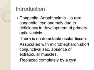
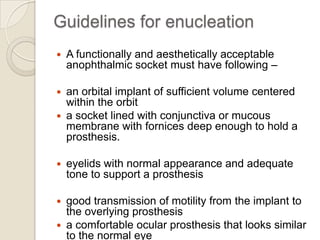
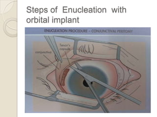
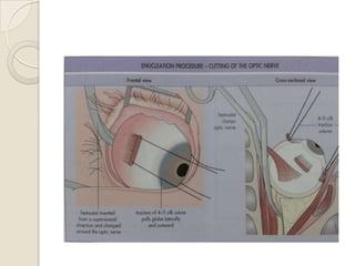
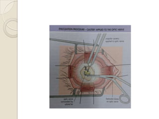
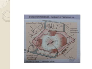
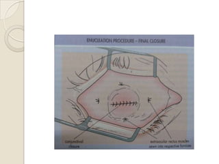
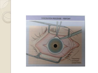
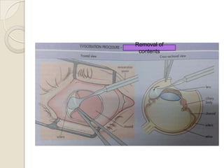
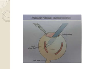
Congenital Anophthalmia:
Current concepts in
management
Current Opinion in Ophthalmology 2011,22:380384
Introduction


Congenital Anophthalmia – a rare
congenital eye anomaly due to
deficiency in development of primary
optic vesicle.
There is no detectable ocular tissue.
Associated with microblepharon,short
conjunctival sac, absence of
extraocular muscles.
Replaced completely by a cyst.
Purpose of review


The introduction of hydrogel socket &
orbital expanders has modified
approach towards rehabilitation of
congenital anophthalmia.
Recent advances
Hydrogel socket expander as an
outpatient procedure.
 Increased orbital volume confirmed by
CT ,MRI.
 Inflatable orbital tissue expander new
design

Clinical evaluation


Associate findings –

Coloboma,dermoids,sclerocornea,glaucoma,le
ns & optic nerve abnormalities.
-Canalicular stenosis
Systemic abnormalitiesAbnormalities of ears,palate,lower face
Cardiac,renal & genital anomalies,brain
abnormalities
- Anophthalmia-plus syndrome.
CT
Ultrasonography
Treatment


Goals:
-Simultaneous expansion of lids,soft
tissues,orbital bones /replace lost
volume
-maintain structure of orbit
-impart motility to prosthesis
1.Positioning of progressively enlarging
static acrylic conformers asap after
birth
- Orbital cyst –dynamic expander like
conformer
Drainage or Excision – Rapid growth
- Uncomfortable
to wear
MRI to exclude connection to brain
Orbital implant
Spherical implants:
 Inert material:


glass,silicone,methylmethacrylate
 Biointegrated:
Hydroxyapatite, porous
polyethylene
Inert spherical implants
Advantages
 Provide comfort and low rates of
extrusion.
 Cost-effective choice in patients.
Disadvantages
 decreased motility and implant migration.
Buried motility implants
 anterior surface projections push the
overlying
prosthesis with direct force and can
improve prosthetic motility.
 may pinch the conjunctiva between the
implant and the prosthesis - painful
Hydroxyapatite and porous
polyethylene implants allow for drilling
and placement of a peg to integrate
the prosthesis directly with the moving
implant.
 Pegging is usually carried out 6-12
months after enucleation. Pegged
porous implants offer excellent
motility,



Locations for implants -within the Tenon capsule
/behind the posteri or Tenon capsule in the
muscle cone.



Spheres may be covered with other materials
such as



sclera (homologous or cadaveric) or autogenous
fascia,



Secure closure of Tenon fasci a over the anterior
surface of an anophthalmic implant is an
important barrier to later extrusion.
Types
Hard spherical implant
 Inflatable soft tissue expander
 Hydrogel osmotic expander

Hard silicone spheres
-

Need of series of surgeries
Multiple general anaesthesias
Repeated trauma to soft tissues
Inflatable soft tissue
expanders
-

Better orbital bone stimulation &
socket enlargement.
Difficult to control direction ,maintain
expansion pressure.
Chance of displacing
conformer,extrusion
Hydrogel expander implant
To stimulate growth of conjunctival sac
& eyelids followed by serial
implantation for Orbital volume with
temporary tarsorraphy.
 Methylmethacrylate & Nvinylpyrrolidone
materials.
 Small Soft tissue incision – quick
surgery , recovery

Injectable pellet expanders through
trocar tru skin at inferior orbital rim to
deep orbit.
 Safe & minimally invasive technique
 Easy to insert , biocompatible.
 Migration & extrusion.

Positioning with cyanoacrylate glue
- No suture related complications
- Avoids multiple general anaesthesias
- Outpatient procedure with topical
anaesthesia.


Dermis –fat grafts
Outcomes:
-Good orbital volume ,adequate
fornices
-No excessive growth / need of
surgery
-Allows lid & socket expansion
 Problems:
-second surgical site,unpleasant scar
-delay in healing,chronic discharge



A study on evaluation of an integrated
orbital tissue expander in congenital
anophthalmos . Am J Ophthalmol
2011

-

An inflatable silicone globe sliding on
titanium T- plate secured to lateral
orbital rim with screws.
Inflating with transconjunctival inj of
normal saline ,30 G needle.

-
Outcomes:
- ease of insertion
- Absence of displacement
- Uniform pressure
- Reduced trauma


Guidelines for enucleation


A functionally and aesthetically acceptable
anophthalmic socket must have following –

an orbital implant of sufficient volume centered
within the orbit
 a socket lined with conjunctiva or mucous
membrane with fornices deep enough to hold a
prosthesis.




eyelids with normal appearance and adequate
tone to support a prosthesis

good transmission of motility from the implant to
the overlying prosthesis
 a comfortable ocular prosthesis that looks similar
to the normal eye

Steps of Enucleation with
orbital implant
Removal of
contents
Advantages of Evisceration in
orbital implant
Less disruption of orbital anatomy.
 Good motility of prosthesis
 Lower rate of
migration,extrusion,reoperation.

Anophthalmic Socket
Complications and Treatment
Deep superior sulcus
 Contracture of fornices
 Exposure & extrusion of implant
 Contracted socket
 Anophthalmic ectropion
 Anophthalmic ptosis
 Lash margin entropion
 Cosmetic Optics


Congenital anophthalmia

  • 1.
    Congenital Anophthalmia: Current conceptsin management Current Opinion in Ophthalmology 2011,22:380384
  • 2.
    Introduction  Congenital Anophthalmia –a rare congenital eye anomaly due to deficiency in development of primary optic vesicle. There is no detectable ocular tissue. Associated with microblepharon,short conjunctival sac, absence of extraocular muscles. Replaced completely by a cyst.
  • 4.
    Purpose of review  Theintroduction of hydrogel socket & orbital expanders has modified approach towards rehabilitation of congenital anophthalmia.
  • 5.
    Recent advances Hydrogel socketexpander as an outpatient procedure.  Increased orbital volume confirmed by CT ,MRI.  Inflatable orbital tissue expander new design 
  • 6.
    Clinical evaluation  Associate findings– Coloboma,dermoids,sclerocornea,glaucoma,le ns & optic nerve abnormalities. -Canalicular stenosis Systemic abnormalitiesAbnormalities of ears,palate,lower face Cardiac,renal & genital anomalies,brain abnormalities - Anophthalmia-plus syndrome. CT Ultrasonography
  • 7.
    Treatment  Goals: -Simultaneous expansion oflids,soft tissues,orbital bones /replace lost volume -maintain structure of orbit -impart motility to prosthesis
  • 8.
    1.Positioning of progressivelyenlarging static acrylic conformers asap after birth - Orbital cyst –dynamic expander like conformer Drainage or Excision – Rapid growth - Uncomfortable to wear MRI to exclude connection to brain
  • 9.
    Orbital implant Spherical implants: Inert material:  glass,silicone,methylmethacrylate  Biointegrated: Hydroxyapatite, porous polyethylene
  • 10.
    Inert spherical implants Advantages Provide comfort and low rates of extrusion.  Cost-effective choice in patients. Disadvantages  decreased motility and implant migration. Buried motility implants  anterior surface projections push the overlying prosthesis with direct force and can improve prosthetic motility.  may pinch the conjunctiva between the implant and the prosthesis - painful
  • 11.
    Hydroxyapatite and porous polyethyleneimplants allow for drilling and placement of a peg to integrate the prosthesis directly with the moving implant.  Pegging is usually carried out 6-12 months after enucleation. Pegged porous implants offer excellent motility, 
  • 12.
     Locations for implants-within the Tenon capsule /behind the posteri or Tenon capsule in the muscle cone.  Spheres may be covered with other materials such as  sclera (homologous or cadaveric) or autogenous fascia,  Secure closure of Tenon fasci a over the anterior surface of an anophthalmic implant is an important barrier to later extrusion.
  • 13.
    Types Hard spherical implant Inflatable soft tissue expander  Hydrogel osmotic expander 
  • 14.
    Hard silicone spheres - Needof series of surgeries Multiple general anaesthesias Repeated trauma to soft tissues
  • 15.
    Inflatable soft tissue expanders - Betterorbital bone stimulation & socket enlargement. Difficult to control direction ,maintain expansion pressure. Chance of displacing conformer,extrusion
  • 16.
    Hydrogel expander implant Tostimulate growth of conjunctival sac & eyelids followed by serial implantation for Orbital volume with temporary tarsorraphy.  Methylmethacrylate & Nvinylpyrrolidone materials.  Small Soft tissue incision – quick surgery , recovery 
  • 17.
    Injectable pellet expandersthrough trocar tru skin at inferior orbital rim to deep orbit.  Safe & minimally invasive technique  Easy to insert , biocompatible.  Migration & extrusion. 
  • 18.
    Positioning with cyanoacrylateglue - No suture related complications - Avoids multiple general anaesthesias - Outpatient procedure with topical anaesthesia. 
  • 19.
    Dermis –fat grafts Outcomes: -Goodorbital volume ,adequate fornices -No excessive growth / need of surgery -Allows lid & socket expansion  Problems: -second surgical site,unpleasant scar -delay in healing,chronic discharge 
  • 20.
     A study onevaluation of an integrated orbital tissue expander in congenital anophthalmos . Am J Ophthalmol 2011 - An inflatable silicone globe sliding on titanium T- plate secured to lateral orbital rim with screws. Inflating with transconjunctival inj of normal saline ,30 G needle. -
  • 21.
    Outcomes: - ease ofinsertion - Absence of displacement - Uniform pressure - Reduced trauma 
  • 22.
    Guidelines for enucleation  Afunctionally and aesthetically acceptable anophthalmic socket must have following – an orbital implant of sufficient volume centered within the orbit  a socket lined with conjunctiva or mucous membrane with fornices deep enough to hold a prosthesis.   eyelids with normal appearance and adequate tone to support a prosthesis good transmission of motility from the implant to the overlying prosthesis  a comfortable ocular prosthesis that looks similar to the normal eye 
  • 23.
    Steps of Enucleationwith orbital implant
  • 29.
  • 33.
    Advantages of Eviscerationin orbital implant Less disruption of orbital anatomy.  Good motility of prosthesis  Lower rate of migration,extrusion,reoperation. 
  • 36.
    Anophthalmic Socket Complications andTreatment Deep superior sulcus  Contracture of fornices  Exposure & extrusion of implant  Contracted socket  Anophthalmic ectropion  Anophthalmic ptosis  Lash margin entropion  Cosmetic Optics 