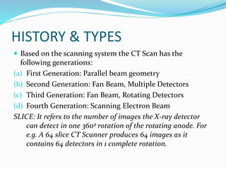
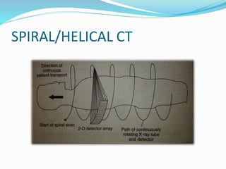
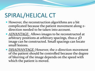
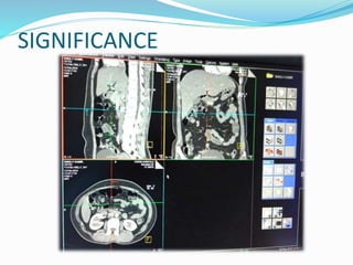
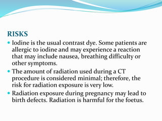
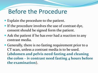
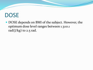
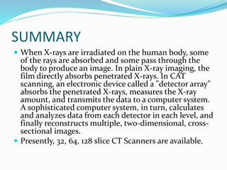
Presented by:
SHOUNAK NANDI
BME 3rd Year, 6th semester
Roll-130206014
Reg.n0. 131230110645
TABLE OF CONTENTS
 Introduction
 History and Types
 Components
 Working principle and circuitry
 Significance
 Risks and Precautions
 Dose
 Summary
 Conclusion
INTRODUCTION
 It is evident that X-Rays are used to examine the
internal structures of the human body. X-ray
absorption relative to their surroundings cause
sufficient contrast differences on the film to be
distinguished by the eye. Thus whilst the bone
structures are distinct, the shape and composition of
the soft tissues and organs are hard to be discerned.
INTRODUCTION(contd..)
 However, the limitations of X-Ray are-
(a) The superimposition of the three dimensional
information onto a single plane are too difficult and
confusing to be diagnosed.
(b) The radiographic plate has a limited dynamic range,
hence the large contrast differences are
distinguishable by the eye.
INTRODUCTION(contd..)
 Hence Computed Tomography scan, in short the C.T.
Scan has been designed which uses the X-Ray
radiation technique but with a different mechanism so
that the limitations could be bridged.
 The term ‘tomos’ is a Greek word meaning ‘to write a
slice or section’.
Head firstFeet first
Computed tomography
HISTORY & TYPES
 CT Scan was invented and developed by
G.N.Hounsfield at the Central Research Laboratories
of EMI Ltd, UK and introduced on a commercial scale
in 1972.
 As X-Ray CT is the most common CT in medicine and
various contexts, the other types include PET (Positron
Emission Tomography), SPECT(Single Positron
Emission Computed Tomography), CAT(Computed
Axial Tomography) Scan.
HISTORY & TYPES
 The early CT Scanners were specifically designed for
neuro-radiological investigations. But with the new
age CT Scan machines the image resolution has
increased, moreover it has enabled the doctors to
distinguish between different brain tissue, or even
normal and coagulated blood. Moreover, the ventricles
if the brain and the repositories of the CSF are clearly
observable from CT Scan.
 Whole body scanners are now commercially available.
HISTORY & TYPES
 Based on the scanning system the CT Scan has the
following generations:
(a) First Generation: Parallel beam geometry
(b) Second Generation: Fan Beam, Multiple Detectors
(c) Third Generation: Fan Beam, Rotating Detectors
(d) Fourth Generation: Scanning Electron Beam
SLICE: It refers to the number of images the X-ray detector
can detect in one 360º rotation of the rotating anode. For
e.g. A 64 slice CT Scanner produces 64 images as it
contains 64 detectors in 1 complete rotation.
WORKING PRINCIPLE & CIRCUITRY
 CT Scan reconstructs the image from a large number
of absorption profiles taken at regular intervals around
a slice, each profile being made up from a parallel set
of absorption values through the object.
 The X-Rays from a finely collimated source are made to
pass through a slice of the object or patient from a
variety of directions. Less X-Rays are transmitted
through a longer tissue path-length, while more for
shorter path length thus the radiation intensity of the
regions in the image varies accordingly.
WORKING PRINCIPLE & CIRCUITRY
 For a monochromatic X-Ray beam, the tissue
attenuation can be described by-
It=Ioe-µx
It= Transmitted intensity
Io =Incident radiation intensity
µ = Characteristic attenuation co-efficient of tissue
X = thickness of tissue
WORKING PRINCIPLE & CIRCUITRY
 The image formed by the CT Scan is considered as per
slice. This slice is considered to be as the sum of the
volume elements or voxels, each voxel having separate
attenuation coefficients. If a slice of heterogeneous
tissue is irradiated, the sum of the attenuation co-
efficients for each X-Ray beam is calculated by the
measured intensities for a given voxel width.
Computed Tomography uses the knowledge of
attenuation co-efficient sums, thus derived, from all
the irradiation directions to calculate the attenuation
co-efficients to form the CT-image.
WORKING PRINCIPLE & CIRCUITRY
 The X-Ray source and detectors are mounted opposite
to each other in a rigid gantry with patient lying in
between, by moving or both of these across relevant
sections which is how measurements are made.
 The X-Ray transmission readings are taken and stored
by the computer. In one system, there are 18 traverses
in 20s scanning cycle by 30 detectors with
approximately 600 position pulses that constitute the
image.
WORKING PRINCIPLE & CIRCUITRY
COMPONENTS
 The CT system consists of 4 major subsystems:
(a) Scanning system- Takes suitable readings for a
picture to be reconstructed, includes X-Ray source
and detectors.
(b) Processing unit- Converts the readings into
intelligible picture information
(c) Viewing part- Displays the image and includes
manipulative aids to assist diagnosis.
(d) Storage unit- This enables data storage.
COMPONENTS
(a) Scanning system
1. X-ray tube:
 Rotating anode tubes produce pulsed X-Ray
radiations having exposure time in the range of 2-
20seconds.
 Power requirement: 100-160kV; optimum at 120kV,
200-500mA.
 Energy spectrum ranges from 30-120keV.
COMPONENTS
(a) Scanning system
2. X-Ray detectors
 Xenon gas ionization detectors : Contains xenon gas
inside the gas chamber. The X-ray falls on the
chamber through a 1mm width slit and interacts with
xenon, thus producing positive ions and negative
electrons. The +ve voltage accelerates the ions to the
collector plate and produces an electric current in
the amplifier. The resulting current through the
electrode is a measure of the x-ray intensity.
COMPONENTS
(a) Scanning system
2. X-Ray detectors
 Scintillation PM detectors: Scintillation photo
multiplier detectors comprise scintillation crystals
and photodiodes.
 SOMATOM SCINTILLARC: 520 CsI crystals,
assembled with photodiodes arranged on a 42 degree
arc.
 Solid state detectors
COMPONENTS
(b) Processing system
Data Acquisition
*The dynamic range of the detection is 1:4,00,000. In
other words it can detect the smallest and the largest
signal whether the subject is obese or thin and thereby
produce an optimal image.
**A data acquisition systems consists of a precision pre-
amplifier, current to voltage converter, analog integrators,
multiplexers and ADC.
***The patient slice is divided up into several voxels, the
2D image of the slice contains several pixels which
corresponds to the attenuation coefficient of voxel in the
object slice.
COMPONENTS
(b) Processing system
Data Acquisition
Detector elements
Current to voltage
converter
Integrator Multiplexer
Analog to digital
converter computer
COMPONENTS
(c)Viewing system
Nowadays the CT image is obtained from a software
known as DICOM which processes the data to
reconstruct both cross-sectional as well as 3D image.
The images are interpreted by the radiologist from the
console room in front of a computer.
(d) Storing and Documentation
The data are stored in DVD or hard disk memory of
the computer.
GANTRY GEOMETRY
 The gantry looks like a doughnut which contains the
X-ray tube and the detector and other associated
mechanism.
 60-72cm diameter hole
 Can be tilted ±20degrees
 Provided with a light which indicate the CT slice
position
 Intercom available for communication between the
patient and the physician from the console room.
GANTRY CONSOLE (For intercom)
SPIRAL/HELICAL CT
 This is a scanning technique in which the X-Ray
rotates continuously around the patient while the
patient is continuously translated through the fan
beam. The focal spot traces a helix around the patient.
The projection data is used for the reconstruction of
multiple contiguous images.
 Spiral CT is used for 3D image- images taken are in
smooth continuous motion. The projection data
should be acquired in a single breath hold, at rates of
one slice per second.
SPIRAL/HELICAL CT
SPIRAL/HELICAL CT
 However, the reconstruction algorithms are a bit
complicated because the patient movement along z-
direction needed to be taken into account.
 ADVANTAGE: Allows images to be reconstructed at
arbitrary positions at arbitrary spacings, thus a 3D
image can be constructed. Small spacings can locate
small lesions.
 DISADVANTAGE: However, the z-direction movement
of the patient should be controlled because the degree
of blurring of the image depends on the speed with
which the patient is moved.
3D
RECONSTRUCT
IMAGE OF
THORACIC
CAVITY
SIGNIFICANCE
 Detection of tumors- benign and malignant at various
parts of the body.
(PET Positron Emission Tomography is a specialized
technique to detect cancer by allowing an isotope to
the body which highlights the cancerous cells.)
 Angioplasty
 Detection of aneurysm
 Brain : vessel coagulation findings, stroke
 Abdominal injury findings
SIGNIFICANCE
RISKS
 Iodine is the usual contrast dye. Some patients are
allergic to iodine and may experience a reaction
that may include nausea, breathing difficulty or
other symptoms.
 The amount of radiation used during a CT
procedure is considered minimal; therefore, the
risk for radiation exposure is very low.
 Radiation exposure during pregnancy may lead to
birth defects. Radiation is harmful for the foetus.
Before the Procedure
 Explain the procedure to the patient.
 If the procedure involves the use of contrast dye,
consent should be signed form the patient.
 Ask the patient if he has ever had a reaction to any
contrast media.
 Generally, there is no fasting requirement prior to a
CT scan, unless a contrast media is to be used.
(abdomen and pelvis need fasting and cleaning
the colon – iv contrast need fasting 4 hours before
the examination).
DOSE
 DOSE depends on BMI of the subject. However, the
optimum dose level ranges between 1.3±0.1
rad(J/kg) to 2.5 rad.
SUMMARY
 When X-rays are irradiated on the human body, some
of the rays are absorbed and some pass through the
body to produce an image. In plain X-ray imaging, the
film directly absorbs penetrated X-rays. In CAT
scanning, an electronic device called a "detector array"
absorbs the penetrated X-rays, measures the X-ray
amount, and transmits the data to a computer system.
A sophisticated computer system, in turn, calculates
and analyzes data from each detector in each level, and
finally reconstructs multiple, two-dimensional, cross-
sectional images.
 Presently, 32, 64, 128 slice CT Scanners are available.
REFERENCES
 R.S.Khandpur, “HANDBOOK OF BIO-MEDICAL
INSTRUMENTATION”, 2nd edition, Tata McGraw Hill
QUESTIONS??
THANK YOU FOR YOUR PATIENCE AND
CO-OPERATION!!!

COMPUTED TOMOGRAPHY SCAN

  • 1.
    Presented by: SHOUNAK NANDI BME3rd Year, 6th semester Roll-130206014 Reg.n0. 131230110645
  • 2.
    TABLE OF CONTENTS Introduction  History and Types  Components  Working principle and circuitry  Significance  Risks and Precautions  Dose  Summary  Conclusion
  • 3.
    INTRODUCTION  It isevident that X-Rays are used to examine the internal structures of the human body. X-ray absorption relative to their surroundings cause sufficient contrast differences on the film to be distinguished by the eye. Thus whilst the bone structures are distinct, the shape and composition of the soft tissues and organs are hard to be discerned.
  • 4.
    INTRODUCTION(contd..)  However, thelimitations of X-Ray are- (a) The superimposition of the three dimensional information onto a single plane are too difficult and confusing to be diagnosed. (b) The radiographic plate has a limited dynamic range, hence the large contrast differences are distinguishable by the eye.
  • 5.
    INTRODUCTION(contd..)  Hence ComputedTomography scan, in short the C.T. Scan has been designed which uses the X-Ray radiation technique but with a different mechanism so that the limitations could be bridged.  The term ‘tomos’ is a Greek word meaning ‘to write a slice or section’.
  • 6.
  • 7.
    HISTORY & TYPES CT Scan was invented and developed by G.N.Hounsfield at the Central Research Laboratories of EMI Ltd, UK and introduced on a commercial scale in 1972.  As X-Ray CT is the most common CT in medicine and various contexts, the other types include PET (Positron Emission Tomography), SPECT(Single Positron Emission Computed Tomography), CAT(Computed Axial Tomography) Scan.
  • 8.
    HISTORY & TYPES The early CT Scanners were specifically designed for neuro-radiological investigations. But with the new age CT Scan machines the image resolution has increased, moreover it has enabled the doctors to distinguish between different brain tissue, or even normal and coagulated blood. Moreover, the ventricles if the brain and the repositories of the CSF are clearly observable from CT Scan.  Whole body scanners are now commercially available.
  • 9.
    HISTORY & TYPES Based on the scanning system the CT Scan has the following generations: (a) First Generation: Parallel beam geometry (b) Second Generation: Fan Beam, Multiple Detectors (c) Third Generation: Fan Beam, Rotating Detectors (d) Fourth Generation: Scanning Electron Beam SLICE: It refers to the number of images the X-ray detector can detect in one 360º rotation of the rotating anode. For e.g. A 64 slice CT Scanner produces 64 images as it contains 64 detectors in 1 complete rotation.
  • 10.
    WORKING PRINCIPLE &CIRCUITRY  CT Scan reconstructs the image from a large number of absorption profiles taken at regular intervals around a slice, each profile being made up from a parallel set of absorption values through the object.  The X-Rays from a finely collimated source are made to pass through a slice of the object or patient from a variety of directions. Less X-Rays are transmitted through a longer tissue path-length, while more for shorter path length thus the radiation intensity of the regions in the image varies accordingly.
  • 11.
    WORKING PRINCIPLE &CIRCUITRY  For a monochromatic X-Ray beam, the tissue attenuation can be described by- It=Ioe-µx It= Transmitted intensity Io =Incident radiation intensity µ = Characteristic attenuation co-efficient of tissue X = thickness of tissue
  • 12.
  • 13.
    WORKING PRINCIPLE &CIRCUITRY  The image formed by the CT Scan is considered as per slice. This slice is considered to be as the sum of the volume elements or voxels, each voxel having separate attenuation coefficients. If a slice of heterogeneous tissue is irradiated, the sum of the attenuation co- efficients for each X-Ray beam is calculated by the measured intensities for a given voxel width. Computed Tomography uses the knowledge of attenuation co-efficient sums, thus derived, from all the irradiation directions to calculate the attenuation co-efficients to form the CT-image.
  • 14.
    WORKING PRINCIPLE &CIRCUITRY  The X-Ray source and detectors are mounted opposite to each other in a rigid gantry with patient lying in between, by moving or both of these across relevant sections which is how measurements are made.  The X-Ray transmission readings are taken and stored by the computer. In one system, there are 18 traverses in 20s scanning cycle by 30 detectors with approximately 600 position pulses that constitute the image.
  • 15.
  • 16.
    COMPONENTS  The CTsystem consists of 4 major subsystems: (a) Scanning system- Takes suitable readings for a picture to be reconstructed, includes X-Ray source and detectors. (b) Processing unit- Converts the readings into intelligible picture information (c) Viewing part- Displays the image and includes manipulative aids to assist diagnosis. (d) Storage unit- This enables data storage.
  • 17.
    COMPONENTS (a) Scanning system 1.X-ray tube:  Rotating anode tubes produce pulsed X-Ray radiations having exposure time in the range of 2- 20seconds.  Power requirement: 100-160kV; optimum at 120kV, 200-500mA.  Energy spectrum ranges from 30-120keV.
  • 18.
    COMPONENTS (a) Scanning system 2.X-Ray detectors  Xenon gas ionization detectors : Contains xenon gas inside the gas chamber. The X-ray falls on the chamber through a 1mm width slit and interacts with xenon, thus producing positive ions and negative electrons. The +ve voltage accelerates the ions to the collector plate and produces an electric current in the amplifier. The resulting current through the electrode is a measure of the x-ray intensity.
  • 19.
    COMPONENTS (a) Scanning system 2.X-Ray detectors  Scintillation PM detectors: Scintillation photo multiplier detectors comprise scintillation crystals and photodiodes.  SOMATOM SCINTILLARC: 520 CsI crystals, assembled with photodiodes arranged on a 42 degree arc.  Solid state detectors
  • 20.
    COMPONENTS (b) Processing system DataAcquisition *The dynamic range of the detection is 1:4,00,000. In other words it can detect the smallest and the largest signal whether the subject is obese or thin and thereby produce an optimal image. **A data acquisition systems consists of a precision pre- amplifier, current to voltage converter, analog integrators, multiplexers and ADC. ***The patient slice is divided up into several voxels, the 2D image of the slice contains several pixels which corresponds to the attenuation coefficient of voxel in the object slice.
  • 21.
    COMPONENTS (b) Processing system DataAcquisition Detector elements Current to voltage converter Integrator Multiplexer Analog to digital converter computer
  • 22.
    COMPONENTS (c)Viewing system Nowadays theCT image is obtained from a software known as DICOM which processes the data to reconstruct both cross-sectional as well as 3D image. The images are interpreted by the radiologist from the console room in front of a computer. (d) Storing and Documentation The data are stored in DVD or hard disk memory of the computer.
  • 23.
    GANTRY GEOMETRY  Thegantry looks like a doughnut which contains the X-ray tube and the detector and other associated mechanism.  60-72cm diameter hole  Can be tilted ±20degrees  Provided with a light which indicate the CT slice position  Intercom available for communication between the patient and the physician from the console room.
  • 24.
  • 25.
    SPIRAL/HELICAL CT  Thisis a scanning technique in which the X-Ray rotates continuously around the patient while the patient is continuously translated through the fan beam. The focal spot traces a helix around the patient. The projection data is used for the reconstruction of multiple contiguous images.  Spiral CT is used for 3D image- images taken are in smooth continuous motion. The projection data should be acquired in a single breath hold, at rates of one slice per second.
  • 26.
  • 27.
    SPIRAL/HELICAL CT  However,the reconstruction algorithms are a bit complicated because the patient movement along z- direction needed to be taken into account.  ADVANTAGE: Allows images to be reconstructed at arbitrary positions at arbitrary spacings, thus a 3D image can be constructed. Small spacings can locate small lesions.  DISADVANTAGE: However, the z-direction movement of the patient should be controlled because the degree of blurring of the image depends on the speed with which the patient is moved.
  • 28.
  • 29.
    SIGNIFICANCE  Detection oftumors- benign and malignant at various parts of the body. (PET Positron Emission Tomography is a specialized technique to detect cancer by allowing an isotope to the body which highlights the cancerous cells.)  Angioplasty  Detection of aneurysm  Brain : vessel coagulation findings, stroke  Abdominal injury findings
  • 30.
  • 31.
    RISKS  Iodine isthe usual contrast dye. Some patients are allergic to iodine and may experience a reaction that may include nausea, breathing difficulty or other symptoms.  The amount of radiation used during a CT procedure is considered minimal; therefore, the risk for radiation exposure is very low.  Radiation exposure during pregnancy may lead to birth defects. Radiation is harmful for the foetus.
  • 32.
    Before the Procedure Explain the procedure to the patient.  If the procedure involves the use of contrast dye, consent should be signed form the patient.  Ask the patient if he has ever had a reaction to any contrast media.  Generally, there is no fasting requirement prior to a CT scan, unless a contrast media is to be used. (abdomen and pelvis need fasting and cleaning the colon – iv contrast need fasting 4 hours before the examination).
  • 33.
    DOSE  DOSE dependson BMI of the subject. However, the optimum dose level ranges between 1.3±0.1 rad(J/kg) to 2.5 rad.
  • 34.
    SUMMARY  When X-raysare irradiated on the human body, some of the rays are absorbed and some pass through the body to produce an image. In plain X-ray imaging, the film directly absorbs penetrated X-rays. In CAT scanning, an electronic device called a "detector array" absorbs the penetrated X-rays, measures the X-ray amount, and transmits the data to a computer system. A sophisticated computer system, in turn, calculates and analyzes data from each detector in each level, and finally reconstructs multiple, two-dimensional, cross- sectional images.  Presently, 32, 64, 128 slice CT Scanners are available.
  • 35.
    REFERENCES  R.S.Khandpur, “HANDBOOKOF BIO-MEDICAL INSTRUMENTATION”, 2nd edition, Tata McGraw Hill
  • 36.
  • 37.
    THANK YOU FORYOUR PATIENCE AND CO-OPERATION!!!