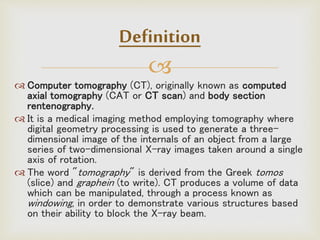
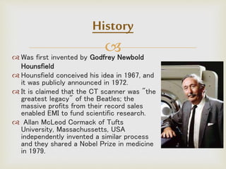
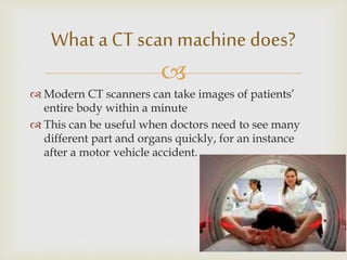
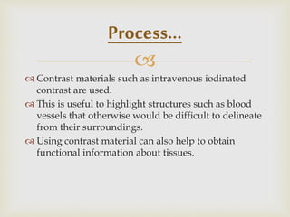
By
Jekadeshnaidu Panirselvam BPT
MSc Human Anatomy
Facultyof Medicine
AIMST University

Definition
History
Body CT
Generations
Components
Diagnostic Usage
Benefits Vs. Risks
Contents

Definition
 Computer tomography (CT), originally known as computed
axial tomography (CAT or CT scan) and body section
rentenography.
 It is a medical imaging method employing tomography where
digital geometry processing is used to generate a three-
dimensional image of the internals of an object from a large
series of two-dimensional X-ray images taken around a single
axis of rotation.
 The word "tomography" is derived from the Greek tomos
(slice) and graphein (to write). CT produces a volume of data
which can be manipulated, through a process known as
windowing, in order to demonstrate various structures based
on their ability to block the X-ray beam.

 Is a non-invasive medical test that helps physicians
diagnose and treat medical conditions.
 - Combines special x-ray equipment with
sophisticated computers to produce multiple images
or pictures of the inside of the body.
 - These cross-sectional images of the area being
studied can then be examined on a computer
monitor, printed or transferred to a CD.
Definition
 Was first invented by Godfrey Newbold
Hounsfield
 Hounsfield conceived his idea in 1967, and
it was publicly announced in 1972.
 It is claimed that the CT scanner was "the
greatest legacy" of the Beatles; the
massive profits from their record sales
enabled EMI to fund scientific research.
 Allan McLeod Cormack of Tufts
University, Massachussetts, USA
independently invented a similar process
and they shared a Nobel Prize in medicine
in 1979.
History

 Simple motion of a tube and Detector was used before
CT to create images at a given depth.
 All anatomy not at the target level was blurred. This
gave a somewhat crude image and was quickly replaced
by CT.
 With the advent of digital detectors and the ability to
post process this imaging method is making a
comeback.
Tomosynthesis

 Modern CT scanners can take images of patients’
entire body within a minute
 This can be useful when doctors need to see many
different part and organs quickly, for an instance
after a motor vehicle accident.
What a CT scan machine does?

One of the best and fastest tools for examining
the chest, abdomen and pelvis
It provides detailed, cross-sectional views of all
types of tissue.
Used to examine patients with severe injuries
from incidents such as a motor vehicle accident.
Performed on patients with acute symptoms
such as abdominal pain or difficulty breathing.
Body CT

 Often the best method for detecting many different cancers,
including lung, liver, kidney and pancreatic cancer, since the
image allows a physician to confirm the presence of a tumor
and measure its size, precise location and the extent of the
tumor's involvement with other nearby tissue.
 An examination that plays a significant role in the detection,
diagnosis and treatment of vascular diseases that can lead to
stroke, kidney failure or even death.
 CT is commonly used to assess for pulmonary embolism as
well as for abdominal aortic aneurysms (AAA).
Body CT

Broken bones
Blood clots in brain and chest
Abnormalities of lung; pneumonia
Inflammation in the abdomen; appendicitis or
gallbladder disease
Stone formation; kidney or gallbladder
Blocked bowel passage or twisted bowel
Cancers in various parts of the body
What a Body CT can see?

 Quickly identify injuries to the lungs, heart and
vessels, liver, spleen, kidneys, bowel or other
internal organs in cases of trauma.
 Guide biopsies and other procedures such as abscess
drainages and minimally invasive tumor treatments
 Plan for and assess the results of surgery, such as
organ transplants or gastric bypass.
Physician often use CT for

Generation Configuration Detector Beam Min. Scan
Time
First Translate -
rotate
1 -2 Pencil thin 2.5 min
Second Translate -
rotate
3 -52 Narrow fan 10sec
Third Rotate -
rotate
256 –
1000
Wide fan 0.5 sec
Forth Rotate - fixed 600 -
4800
Wide fan 1 sec
Fifth Electron beam 1284 Wide fan
electron
beam
33nsec
Generations

The X-ray beam was not wide enough to cover the
entire width of the 'slice' of interest.
A mechanical arrangement was required to move the X-
ray source and detector horizontally across the field of
view.
After a sweep, the source/detector assembly would be
rotated a few degrees, and another sweep performed.
This process would be repeated until 360 degrees (or
180 degrees) had been covered. The complex motion
placed a limit on the minimum scan time at
approximately 20 seconds per image.
1st & 2nd Generation

The X-ray beam is able to cover the entire field
of view of the scanner.
This avoids the need for any horizontal motion;
an entire 'line' can be captured in an instant.
This allowed simplification of the motion to
rotation of the X-ray source.
3rd & 4th Generation

Third and fourth generation designs differ in the
arrangement of the detectors.
In 3rd generation, the detector array is as wide
as the beam, and must therefore rotate as the
source rotates.
In 4th generation, an entire ring of stationary
detectors are used.
3rd & 4th Generation

Electron beam tomography (EBCT) was
introduced in the early 1980s, by medical
physicist Andrew Castagnini.
 It is a method of improving the temporal
resolution of CT scanners.
 Because the X-ray source has to rotate by
over 180 degrees in order to capture an image
the technique is inherently unable to capture
dynamic events or movements that are quicker
than the rotation time.
Electron Beam CT

Instead of rotating a conventional X-ray tube
around the patient, the EBCT machine houses a
huge vacuum tube in which an electron beam is
electro-magnetically steered towards an array
of tungsten X-ray anodes arranged circularly
around the patient.
Each anode is hit in turn by the electron beam
and emits X-rays that are collimated and
detected as in conventional CT.
Electron Beam CT

The lack of moving parts allows very quick
scanning, with single slice making the technique
ideal for capturing images of the heart.
 EBCT has found particular use for assessment
of coronary artery calcium, a means of
predicting risk of coronary artery disease.
Electron Beam CT

 Main components of CT includes;
 CT Scanner
 Gantry
 Data Acquisition System
 Operating Console
Components

The gantry assembly is the largest of these
systems. - - It is made up of all the equipment
related to the patient, including the patient
support, the positioning couch, the mechanical
supports, and the scanner housing. It also
contains the heart of the CAT scanner, the x-
ray tube, as well as detectors that generate
and detect x rays.
Gantry
High
Voltage
Generator
X-ray tube
Pre
Patient
Collimator
Patient
Post
Patient
Collimator
Detector
Gantry
X ray source
Pre Patient Collimation
Post Patient
Collimation
X-ray Detector

Two types of detectors are used
Scintillation Detectors
Gas Filled Detectors
Scintillation Detectors
 Materials Used
 Sodium Iodide
 Bismuth Germanium Oxide
 Cesium Iodide
 Cadmium Tungstate
Detectors
Scintillator
Crystal
Photo
Multiplier
Detector
Rings

Gas Filled Detectors
 Materials Used
 Xenon
 Krypton
 Xenon + Krypton
Detectors

Data Acquisition System
(DAS)
 The DAS consists of the following parts
 X-ray photons come on the detector.
 The detector detects the intensity in form of current.
 The current is converted into voltage.
 The analog integrator removes spikes.
 The analog signal is converted into digital form.
 This signal can now be processed and reconstructed in
the computer.
Detector
Current to
Voltage
Convertor
Pre
Amplifier
Analog
Integrator
Analog to
Digital
Convertor
Computer

After enough transmission measurements
(detector) it will be sent to the computer for
processing
A software called Fourier Slice Transform is
used.
More than 250,000 reconstruction algorithms
are used to compute the image.
Image Reconstruction

 The operating console is the master control center of the
CAT scanner.
 It is used to input all of the factors related to taking a scan.
 Typically, this console is made up of a computer, a keyboard,
and multiple monitors.
 Often there are two different control consoles, one used by
the CAT scanner operator, and the other used by the
physician.
 The operator's console controls such variables as the
thickness of the imaged tissue slice, mechanical movement of
the patient couch, and other radiographic technique factors.
Operating Console

Helical, also called spiral, CT was introduced in
the early 1990s, with much of the development
led by Willi Kalender and Kazuhiro Katada.
 In older CT scanners, the X-ray source would
move in a circular fashion to acquire a single
'slice', once the slice had been completed, the
scanner table would move to position the
patient for the next slice; meanwhile the X-ray
source/detectors would reverse direction to
avoid tangling their cables.
Helical or Spiral CT

In helical CT the X-ray source are attached to a
freely rotating gantry.
During a scan, the table moves the patient smoothly
through the scanner; the name derives from the
helical path traced out by the X-ray beam.
Helical or Spiral CT

Multislice CT scanners are similar in concept to
the helical or spiral CT but there are more than
one detector ring.
 It began with two rings in mid nineties, with a 2
solid state ring model designed and built by
Elscint (Haifa) called CT TWIN, with one second
rotation.
Later, it was presented 4, 8, 16, 32, 40 and 64
detector rings, with increasing rotation speeds.
Multislice CT

The major benefit of multi-slice CT is the increased
speed of volume coverage. This allows large
volumes to be scanned at the optimal time .
The ability of multi-slice scanners to achieve
isotropic resolution even on routine studies means
that maximum image quality is not restricted to
images in the axial plane - and studies can be freely
viewed in any desired plane.
Multislice CT

First ,CT completely eliminates the superimposition
of images of structures outside the area of interest.
Second, because of the inherent high-contrast
resolution of CT, differences between tissues that
differ in physical density by less than 1% can be
distinguished.
Third, data from a single CT imaging procedure
consisting of either multiple contiguous or one helical
scan can be viewed as images in the axial, coronal, or
sagittal planes, depending on the diagnostic task.
This is referred to as multiplanar reformatted
imaging.
Advantages

Because CT scans rely on intravenously administered
contrast agents in order to provide superior image quality,
there is a low but non-negligible level of risk associated
with the contrast agents themselves.
Certain patients may experience severe and potentially
life-threatening allergic reactions to the contrast dye.
The contrast agent may also induce kidney damage. The
risk of this is increased with patients who have
preexisting renal insufficiency, preexisting diabetes, or
reduced intravascular volume.
Hazards :Adverse reactions to contrast
agents

In general, if a patient has normal kidney function,
then the risks of contrast nephropathy are
negligible.
Patients with mild kidney impairment are usually
advised to ensure full hydration for several hours
before and after the injection.
For moderate kidney failure, the use of iodinated
contrast should be avoided; this may mean using an
alternative technique instead of CT e.g. MRI.
Hazards :Adverse reactions to contrast
agents

Process…
CT scan
reveals
mummy
inside
Buddha
Statue

X-ray slice data is generated using an X-ray
source that rotates around the object; X-ray
sensors are positioned on the opposite side of the
circle from the X-ray source.
 Many data scans are progressively taken as the
object is gradually passed through the gantry.
They are combined together by the mathematical
procedure known as tomographic reconstruction.
Process…

 Contrast materials such as intravenous iodinated
contrast are used.
 This is useful to highlight structures such as blood
vessels that otherwise would be difficult to delineate
from their surroundings.
 Using contrast material can also help to obtain
functional information about tissues.
Process…

 The technologist begins by positioning patient on the
CT examination table, usually lying flat on back or
less commonly, on one side or on stomach.
 Straps and pillows may be used to help you maintain
the correct position and to hold still during the exam.
 Depending on the part of the body being scanned,
patient may be asked to keep hands over head.
How is the CAT scan performed?

 If contrast material is used, it will be swallowed, injected
through an intravenous line (IV) or administered by
enema, depending on the type of examination.
 Next, the table will move quickly through the scanner to
determine the correct starting position for the scans.
 Then, the table will move slowly through the machine as
the actual CT scanning is performed.
 Depending on the type of CT scan, the machine may
make several passes.
 Patient may be asked to hold breath during the scanning.
How is the CAT scan performed?

 Any motion, whether breathing or body movements, can
lead to artefacts on the images.
 This is similar to the blurring seen on a photograph taken
of a moving object.
 When the examination is completed, you will be asked to
wait until the technologist verifies that the images are of
high enough quality for accurate interpretation.
 The CT examination is usually completed within 30
minutes. The portion requiring intravenous contrast
injection usually lasts only 10 to 30 seconds.

Windowing is the process of using the calculated
Hounsfield units (HU) to make an image.
The various radio density amplitudes are mapped to 256
shades of gray. These shades of gray can be distributed
over a wide range of HU values to get an overview of
structures.
Alternatively, these shades of gray can be distributed
over a narrow range of HU values (called a "narrow
window"). In this way, variations in the internal makeup
of the structure can be discerned. This is a commonly
used image processing technique known as contrast
compression.
Windowing

Although CT is a relatively accurate test, it is liable
to produce artifacts, such as the following:
Aliasing Artifact or Streaks
• These appear as dark lines which radiate away
from sharp corners.
• It occurs because it is impossible for the
scanner to 'sample' or take enough projections of
the object, which is usually metallic. It can also
occur when an insufficient penetration of the x-
ray occurs.
Artefacts

Ring Artifact
• Probably the most common mechanical artifact, the
image of one or many 'rings' appears within an image.
This is due to a detector fault.
Noise Artifact
• This appears as gaining on the image and is caused
by a low signal to noise ratio. This occurs more
commonly when a thin slice thickness is used. It can
also occur when the kV or mA is too low.
Artefacts

Motion Artifact
• This is seen as blurring which is caused by patient
movement. This is not so much a problem these
days with faster scanning times in the use of MDCT.
Beam Hardening
• This can give a 'cupped appearance'. It occurs when
there is more attenuation in the center of the object
than around the edge. This is easily corrected by
filtration .
Artefacts
 CT scanning is painless, non- invasive and accurate.
 A major advantage of CT is its ability to image bone, soft
tissue and blood vessels all at the same time.
 Unlike conventional x-rays, CT scanning provides very detailed
images of many types of tissue as well as the lungs, bones, and
blood vessels.
 CT examinations are fast and simple; in emergency cases, they
can reveal internal injuries and bleeding quickly enough to help
save lives.
 CT has been shown to be a cost-effective imaging tool for a
wide range of clinical problems.
 CT is less sensitive to patient movement than MRI.
Benefits Vs. Risks
 CT can be performed if you have an implanted medical device of
any kind, unlike MRI.
 CT imaging provides real-time imaging, making it a good tool for
guiding minimally invasive procedures such as needle biopsies and
needle aspirations of many areas of the body, particularly the
lungs, abdomen, pelvis and bones.
 A diagnosis determined by CT scanning may eliminate the need for
exploratory surgery and surgical biopsy.
 No radiation remains in a patient's body after a CT examination.
 X-rays used in CT scans usually have no immediate side effects.
Benefits…

There is no conclusive evidence that radiation
at amounts delivered by a CT scan causes
cancer.
Large population studies have shown a slight
increase in cancer from larger amounts of
radiation, such as from radiation therapy.
Thus, there is always concern that this risk may
also apply to the lower amounts of radiation
delivered by a CT exam.
Risks

 The effective radiation dose for this procedure varies.
 CT scanning is, in general, not recommended for
pregnant women unless medically necessary because of
potential risk to the baby.
 Manufacturers of intravenous contrast indicate mothers
should not breastfeed their babies for 24-48 hours after
contrast medium is given.
 The risk of serious allergic reaction to contrast materials
that contain iodine is extremely rare, and radiology
departments are well-equipped to deal with them.
Risks

Soft-tissue details in areas such as the brain, internal
pelvic organs, and joints (such as knees and shoulders) can
often be better evaluated with magnetic resonance imaging
(MRI).
In pregnant women, while CT can be performed safely,
other imaging exams not involving radiation, such as
ultrasound or MRI, is preferred if they are likely to be as
good as CT in diagnosing condition.
A person who is very large may not fit into the opening of a
conventional CT scanner or may be over the weight limit—
usually 450 pounds—for the moving table.
Limitations
Computerised tomography scan

Computerised tomography scan

  • 1.
    By Jekadeshnaidu Panirselvam BPT MScHuman Anatomy Facultyof Medicine AIMST University
  • 2.
  • 3.
     Definition  Computer tomography(CT), originally known as computed axial tomography (CAT or CT scan) and body section rentenography.  It is a medical imaging method employing tomography where digital geometry processing is used to generate a three- dimensional image of the internals of an object from a large series of two-dimensional X-ray images taken around a single axis of rotation.  The word "tomography" is derived from the Greek tomos (slice) and graphein (to write). CT produces a volume of data which can be manipulated, through a process known as windowing, in order to demonstrate various structures based on their ability to block the X-ray beam.
  • 4.
      Is anon-invasive medical test that helps physicians diagnose and treat medical conditions.  - Combines special x-ray equipment with sophisticated computers to produce multiple images or pictures of the inside of the body.  - These cross-sectional images of the area being studied can then be examined on a computer monitor, printed or transferred to a CD. Definition
  • 5.
     Was firstinvented by Godfrey Newbold Hounsfield  Hounsfield conceived his idea in 1967, and it was publicly announced in 1972.  It is claimed that the CT scanner was "the greatest legacy" of the Beatles; the massive profits from their record sales enabled EMI to fund scientific research.  Allan McLeod Cormack of Tufts University, Massachussetts, USA independently invented a similar process and they shared a Nobel Prize in medicine in 1979. History
  • 6.
      Simple motionof a tube and Detector was used before CT to create images at a given depth.  All anatomy not at the target level was blurred. This gave a somewhat crude image and was quickly replaced by CT.  With the advent of digital detectors and the ability to post process this imaging method is making a comeback. Tomosynthesis
  • 7.
      Modern CTscanners can take images of patients’ entire body within a minute  This can be useful when doctors need to see many different part and organs quickly, for an instance after a motor vehicle accident. What a CT scan machine does?
  • 8.
     One of thebest and fastest tools for examining the chest, abdomen and pelvis It provides detailed, cross-sectional views of all types of tissue. Used to examine patients with severe injuries from incidents such as a motor vehicle accident. Performed on patients with acute symptoms such as abdominal pain or difficulty breathing. Body CT
  • 9.
      Often thebest method for detecting many different cancers, including lung, liver, kidney and pancreatic cancer, since the image allows a physician to confirm the presence of a tumor and measure its size, precise location and the extent of the tumor's involvement with other nearby tissue.  An examination that plays a significant role in the detection, diagnosis and treatment of vascular diseases that can lead to stroke, kidney failure or even death.  CT is commonly used to assess for pulmonary embolism as well as for abdominal aortic aneurysms (AAA). Body CT
  • 10.
     Broken bones Blood clotsin brain and chest Abnormalities of lung; pneumonia Inflammation in the abdomen; appendicitis or gallbladder disease Stone formation; kidney or gallbladder Blocked bowel passage or twisted bowel Cancers in various parts of the body What a Body CT can see?
  • 11.
      Quickly identifyinjuries to the lungs, heart and vessels, liver, spleen, kidneys, bowel or other internal organs in cases of trauma.  Guide biopsies and other procedures such as abscess drainages and minimally invasive tumor treatments  Plan for and assess the results of surgery, such as organ transplants or gastric bypass. Physician often use CT for
  • 12.
     Generation Configuration DetectorBeam Min. Scan Time First Translate - rotate 1 -2 Pencil thin 2.5 min Second Translate - rotate 3 -52 Narrow fan 10sec Third Rotate - rotate 256 – 1000 Wide fan 0.5 sec Forth Rotate - fixed 600 - 4800 Wide fan 1 sec Fifth Electron beam 1284 Wide fan electron beam 33nsec Generations
  • 13.
     The X-ray beamwas not wide enough to cover the entire width of the 'slice' of interest. A mechanical arrangement was required to move the X- ray source and detector horizontally across the field of view. After a sweep, the source/detector assembly would be rotated a few degrees, and another sweep performed. This process would be repeated until 360 degrees (or 180 degrees) had been covered. The complex motion placed a limit on the minimum scan time at approximately 20 seconds per image. 1st & 2nd Generation
  • 14.
     The X-ray beamis able to cover the entire field of view of the scanner. This avoids the need for any horizontal motion; an entire 'line' can be captured in an instant. This allowed simplification of the motion to rotation of the X-ray source. 3rd & 4th Generation
  • 15.
     Third and fourthgeneration designs differ in the arrangement of the detectors. In 3rd generation, the detector array is as wide as the beam, and must therefore rotate as the source rotates. In 4th generation, an entire ring of stationary detectors are used. 3rd & 4th Generation
  • 16.
     Electron beam tomography(EBCT) was introduced in the early 1980s, by medical physicist Andrew Castagnini.  It is a method of improving the temporal resolution of CT scanners.  Because the X-ray source has to rotate by over 180 degrees in order to capture an image the technique is inherently unable to capture dynamic events or movements that are quicker than the rotation time. Electron Beam CT
  • 17.
     Instead of rotatinga conventional X-ray tube around the patient, the EBCT machine houses a huge vacuum tube in which an electron beam is electro-magnetically steered towards an array of tungsten X-ray anodes arranged circularly around the patient. Each anode is hit in turn by the electron beam and emits X-rays that are collimated and detected as in conventional CT. Electron Beam CT
  • 18.
     The lack ofmoving parts allows very quick scanning, with single slice making the technique ideal for capturing images of the heart.  EBCT has found particular use for assessment of coronary artery calcium, a means of predicting risk of coronary artery disease. Electron Beam CT
  • 19.
      Main componentsof CT includes;  CT Scanner  Gantry  Data Acquisition System  Operating Console Components
  • 20.
     The gantry assemblyis the largest of these systems. - - It is made up of all the equipment related to the patient, including the patient support, the positioning couch, the mechanical supports, and the scanner housing. It also contains the heart of the CAT scanner, the x- ray tube, as well as detectors that generate and detect x rays. Gantry
  • 21.
  • 22.
     Two types ofdetectors are used Scintillation Detectors Gas Filled Detectors Scintillation Detectors  Materials Used  Sodium Iodide  Bismuth Germanium Oxide  Cesium Iodide  Cadmium Tungstate Detectors Scintillator Crystal Photo Multiplier Detector Rings
  • 23.
     Gas Filled Detectors Materials Used  Xenon  Krypton  Xenon + Krypton Detectors
  • 24.
     Data Acquisition System (DAS) The DAS consists of the following parts  X-ray photons come on the detector.  The detector detects the intensity in form of current.  The current is converted into voltage.  The analog integrator removes spikes.  The analog signal is converted into digital form.  This signal can now be processed and reconstructed in the computer. Detector Current to Voltage Convertor Pre Amplifier Analog Integrator Analog to Digital Convertor Computer
  • 25.
     After enough transmissionmeasurements (detector) it will be sent to the computer for processing A software called Fourier Slice Transform is used. More than 250,000 reconstruction algorithms are used to compute the image. Image Reconstruction
  • 26.
      The operatingconsole is the master control center of the CAT scanner.  It is used to input all of the factors related to taking a scan.  Typically, this console is made up of a computer, a keyboard, and multiple monitors.  Often there are two different control consoles, one used by the CAT scanner operator, and the other used by the physician.  The operator's console controls such variables as the thickness of the imaged tissue slice, mechanical movement of the patient couch, and other radiographic technique factors. Operating Console
  • 27.
     Helical, also calledspiral, CT was introduced in the early 1990s, with much of the development led by Willi Kalender and Kazuhiro Katada.  In older CT scanners, the X-ray source would move in a circular fashion to acquire a single 'slice', once the slice had been completed, the scanner table would move to position the patient for the next slice; meanwhile the X-ray source/detectors would reverse direction to avoid tangling their cables. Helical or Spiral CT
  • 28.
     In helical CTthe X-ray source are attached to a freely rotating gantry. During a scan, the table moves the patient smoothly through the scanner; the name derives from the helical path traced out by the X-ray beam. Helical or Spiral CT
  • 29.
     Multislice CT scannersare similar in concept to the helical or spiral CT but there are more than one detector ring.  It began with two rings in mid nineties, with a 2 solid state ring model designed and built by Elscint (Haifa) called CT TWIN, with one second rotation. Later, it was presented 4, 8, 16, 32, 40 and 64 detector rings, with increasing rotation speeds. Multislice CT
  • 30.
     The major benefitof multi-slice CT is the increased speed of volume coverage. This allows large volumes to be scanned at the optimal time . The ability of multi-slice scanners to achieve isotropic resolution even on routine studies means that maximum image quality is not restricted to images in the axial plane - and studies can be freely viewed in any desired plane. Multislice CT
  • 31.
     First ,CT completelyeliminates the superimposition of images of structures outside the area of interest. Second, because of the inherent high-contrast resolution of CT, differences between tissues that differ in physical density by less than 1% can be distinguished. Third, data from a single CT imaging procedure consisting of either multiple contiguous or one helical scan can be viewed as images in the axial, coronal, or sagittal planes, depending on the diagnostic task. This is referred to as multiplanar reformatted imaging. Advantages
  • 32.
     Because CT scansrely on intravenously administered contrast agents in order to provide superior image quality, there is a low but non-negligible level of risk associated with the contrast agents themselves. Certain patients may experience severe and potentially life-threatening allergic reactions to the contrast dye. The contrast agent may also induce kidney damage. The risk of this is increased with patients who have preexisting renal insufficiency, preexisting diabetes, or reduced intravascular volume. Hazards :Adverse reactions to contrast agents
  • 33.
     In general, ifa patient has normal kidney function, then the risks of contrast nephropathy are negligible. Patients with mild kidney impairment are usually advised to ensure full hydration for several hours before and after the injection. For moderate kidney failure, the use of iodinated contrast should be avoided; this may mean using an alternative technique instead of CT e.g. MRI. Hazards :Adverse reactions to contrast agents
  • 34.
  • 35.
     X-ray slice datais generated using an X-ray source that rotates around the object; X-ray sensors are positioned on the opposite side of the circle from the X-ray source.  Many data scans are progressively taken as the object is gradually passed through the gantry. They are combined together by the mathematical procedure known as tomographic reconstruction. Process…
  • 36.
      Contrast materialssuch as intravenous iodinated contrast are used.  This is useful to highlight structures such as blood vessels that otherwise would be difficult to delineate from their surroundings.  Using contrast material can also help to obtain functional information about tissues. Process…
  • 37.
      The technologistbegins by positioning patient on the CT examination table, usually lying flat on back or less commonly, on one side or on stomach.  Straps and pillows may be used to help you maintain the correct position and to hold still during the exam.  Depending on the part of the body being scanned, patient may be asked to keep hands over head. How is the CAT scan performed?
  • 38.
      If contrastmaterial is used, it will be swallowed, injected through an intravenous line (IV) or administered by enema, depending on the type of examination.  Next, the table will move quickly through the scanner to determine the correct starting position for the scans.  Then, the table will move slowly through the machine as the actual CT scanning is performed.  Depending on the type of CT scan, the machine may make several passes.  Patient may be asked to hold breath during the scanning. How is the CAT scan performed?
  • 39.
      Any motion,whether breathing or body movements, can lead to artefacts on the images.  This is similar to the blurring seen on a photograph taken of a moving object.  When the examination is completed, you will be asked to wait until the technologist verifies that the images are of high enough quality for accurate interpretation.  The CT examination is usually completed within 30 minutes. The portion requiring intravenous contrast injection usually lasts only 10 to 30 seconds.
  • 40.
     Windowing is theprocess of using the calculated Hounsfield units (HU) to make an image. The various radio density amplitudes are mapped to 256 shades of gray. These shades of gray can be distributed over a wide range of HU values to get an overview of structures. Alternatively, these shades of gray can be distributed over a narrow range of HU values (called a "narrow window"). In this way, variations in the internal makeup of the structure can be discerned. This is a commonly used image processing technique known as contrast compression. Windowing
  • 41.
     Although CT isa relatively accurate test, it is liable to produce artifacts, such as the following: Aliasing Artifact or Streaks • These appear as dark lines which radiate away from sharp corners. • It occurs because it is impossible for the scanner to 'sample' or take enough projections of the object, which is usually metallic. It can also occur when an insufficient penetration of the x- ray occurs. Artefacts
  • 42.
     Ring Artifact • Probablythe most common mechanical artifact, the image of one or many 'rings' appears within an image. This is due to a detector fault. Noise Artifact • This appears as gaining on the image and is caused by a low signal to noise ratio. This occurs more commonly when a thin slice thickness is used. It can also occur when the kV or mA is too low. Artefacts
  • 43.
     Motion Artifact • Thisis seen as blurring which is caused by patient movement. This is not so much a problem these days with faster scanning times in the use of MDCT. Beam Hardening • This can give a 'cupped appearance'. It occurs when there is more attenuation in the center of the object than around the edge. This is easily corrected by filtration . Artefacts
  • 44.
     CT scanningis painless, non- invasive and accurate.  A major advantage of CT is its ability to image bone, soft tissue and blood vessels all at the same time.  Unlike conventional x-rays, CT scanning provides very detailed images of many types of tissue as well as the lungs, bones, and blood vessels.  CT examinations are fast and simple; in emergency cases, they can reveal internal injuries and bleeding quickly enough to help save lives.  CT has been shown to be a cost-effective imaging tool for a wide range of clinical problems.  CT is less sensitive to patient movement than MRI. Benefits Vs. Risks
  • 45.
     CT canbe performed if you have an implanted medical device of any kind, unlike MRI.  CT imaging provides real-time imaging, making it a good tool for guiding minimally invasive procedures such as needle biopsies and needle aspirations of many areas of the body, particularly the lungs, abdomen, pelvis and bones.  A diagnosis determined by CT scanning may eliminate the need for exploratory surgery and surgical biopsy.  No radiation remains in a patient's body after a CT examination.  X-rays used in CT scans usually have no immediate side effects. Benefits…
  • 46.
     There is noconclusive evidence that radiation at amounts delivered by a CT scan causes cancer. Large population studies have shown a slight increase in cancer from larger amounts of radiation, such as from radiation therapy. Thus, there is always concern that this risk may also apply to the lower amounts of radiation delivered by a CT exam. Risks
  • 47.
      The effectiveradiation dose for this procedure varies.  CT scanning is, in general, not recommended for pregnant women unless medically necessary because of potential risk to the baby.  Manufacturers of intravenous contrast indicate mothers should not breastfeed their babies for 24-48 hours after contrast medium is given.  The risk of serious allergic reaction to contrast materials that contain iodine is extremely rare, and radiology departments are well-equipped to deal with them. Risks
  • 48.
     Soft-tissue details inareas such as the brain, internal pelvic organs, and joints (such as knees and shoulders) can often be better evaluated with magnetic resonance imaging (MRI). In pregnant women, while CT can be performed safely, other imaging exams not involving radiation, such as ultrasound or MRI, is preferred if they are likely to be as good as CT in diagnosing condition. A person who is very large may not fit into the opening of a conventional CT scanner or may be over the weight limit— usually 450 pounds—for the moving table. Limitations