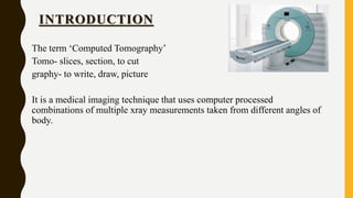
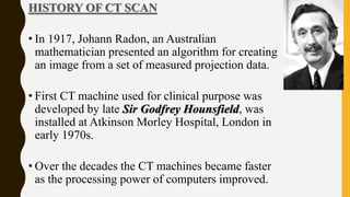
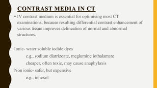
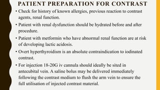
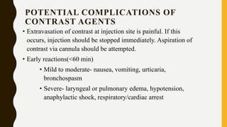
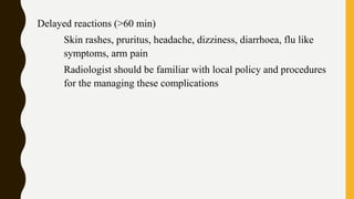
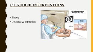
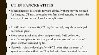
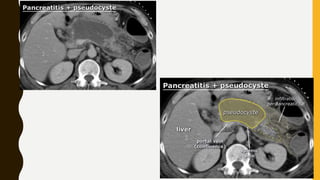
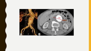
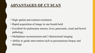
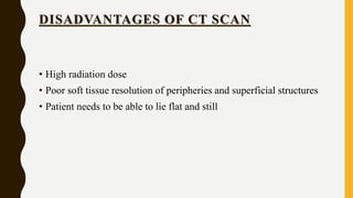
CT scan uses X-rays and computers to create detailed images of the inside of the body. It has replaced conventional angiography as the primary method for examining blood vessels. The document discusses the history, components, principles, types (including spiral, multidetector, angiography), advantages, and risks of CT scanning. CT guided procedures such as biopsy and drainage are also summarized.
Computed tomography (CT scan) is a medical imaging procedure that uses computer-processed X-rays to produce tomographic images or 'slices' of specific areas of the body. These cross-sectional images are used for diagnostic and therapeutic purposes in various medical disciplines.
this slide sharer contents are basic principle of CT fluoroscopy , software and hardware parts of equipment and image aqua cation and radiation dose comparison and videos related to equipment .
principle of ct scanner
generations
scanning motion
EMI unit
xray beam
x ray tube
advantages
disadvantages
in this you PPT got clear idea about generation of ct
if you have any doubt text me
insta ID - ___sadham_____
principle of ct scanner
generations
scanning motion
EMI unit
xray beam
x ray tube
advantages
disadvantages
in this you PPT got clear idea about generation of ct
if you have any doubt text me
insta ID - ___sadham_____
Neck angiography cect neck angiography carotid angiography
CT scan neck angiography
Carotid angiography useful for medical radiology students thank you process explain in simple language for more content like this presentation
generations of CT, explains each generations of CT, muti detector computer tomography, slip ring technology, main terminologies such as FOV , pitch, voxel and matrix, pixel size equation. EBCT, Basic configuration of CT, Data acquisition systems DAS, multi-slice CT
The Indian Dental Academy is the Leader in continuing dental education , training dentists in all aspects of dentistry and
offering a wide range of dental certified courses in different formats.
- Video recording of this lecture in English language: https://youtu.be/lK81BzxMqdo
- Video recording of this lecture in Arabic language: https://youtu.be/Ve4P0COk9OI
- Link to download the book free: https://nephrotube.blogspot.com/p/nephrotube-nephrology-books.html
- Link to NephroTube website: www.NephroTube.com
- Link to NephroTube social media accounts: https://nephrotube.blogspot.com/p/join-nephrotube-on-social-media.html
Lung Cancer: Artificial Intelligence, Synergetics, Complex System Analysis, S...Oleg Kshivets
RESULTS: Overall life span (LS) was 2252.1±1742.5 days and cumulative 5-year survival (5YS) reached 73.2%, 10 years – 64.8%, 20 years – 42.5%. 513 LCP lived more than 5 years (LS=3124.6±1525.6 days), 148 LCP – more than 10 years (LS=5054.4±1504.1 days).199 LCP died because of LC (LS=562.7±374.5 days). 5YS of LCP after bi/lobectomies was significantly superior in comparison with LCP after pneumonectomies (78.1% vs.63.7%, P=0.00001 by log-rank test). AT significantly improved 5YS (66.3% vs. 34.8%) (P=0.00000 by log-rank test) only for LCP with N1-2. Cox modeling displayed that 5YS of LCP significantly depended on: phase transition (PT) early-invasive LC in terms of synergetics, PT N0—N12, cell ratio factors (ratio between cancer cells- CC and blood cells subpopulations), G1-3, histology, glucose, AT, blood cell circuit, prothrombin index, heparin tolerance, recalcification time (P=0.000-0.038). Neural networks, genetic algorithm selection and bootstrap simulation revealed relationships between 5YS and PT early-invasive LC (rank=1), PT N0—N12 (rank=2), thrombocytes/CC (3), erythrocytes/CC (4), eosinophils/CC (5), healthy cells/CC (6), lymphocytes/CC (7), segmented neutrophils/CC (8), stick neutrophils/CC (9), monocytes/CC (10); leucocytes/CC (11). Correct prediction of 5YS was 100% by neural networks computing (area under ROC curve=1.0; error=0.0).
CONCLUSIONS: 5YS of LCP after radical procedures significantly depended on: 1) PT early-invasive cancer; 2) PT N0--N12; 3) cell ratio factors; 4) blood cell circuit; 5) biochemical factors; 6) hemostasis system; 7) AT; 8) LC characteristics; 9) LC cell dynamics; 10) surgery type: lobectomy/pneumonectomy; 11) anthropometric data. Optimal diagnosis and treatment strategies for LC are: 1) screening and early detection of LC; 2) availability of experienced thoracic surgeons because of complexity of radical procedures; 3) aggressive en block surgery and adequate lymph node dissection for completeness; 4) precise prediction; 5) adjuvant chemoimmunoradiotherapy for LCP with unfavorable prognosis.
Prix Galien International 2024 Forum ProgramLevi Shapiro
June 20, 2024, Prix Galien International and Jerusalem Ethics Forum in ROME. Detailed agenda including panels:
- ADVANCES IN CARDIOLOGY: A NEW PARADIGM IS COMING
- WOMEN’S HEALTH: FERTILITY PRESERVATION
- WHAT’S NEW IN THE TREATMENT OF INFECTIOUS,
ONCOLOGICAL AND INFLAMMATORY SKIN DISEASES?
- ARTIFICIAL INTELLIGENCE AND ETHICS
- GENE THERAPY
- BEYOND BORDERS: GLOBAL INITIATIVES FOR DEMOCRATIZING LIFE SCIENCE TECHNOLOGIES AND PROMOTING ACCESS TO HEALTHCARE
- ETHICAL CHALLENGES IN LIFE SCIENCES
- Prix Galien International Awards Ceremony
Report Back from SGO 2024: What’s the Latest in Cervical Cancer?bkling
Are you curious about what’s new in cervical cancer research or unsure what the findings mean? Join Dr. Emily Ko, a gynecologic oncologist at Penn Medicine, to learn about the latest updates from the Society of Gynecologic Oncology (SGO) 2024 Annual Meeting on Women’s Cancer. Dr. Ko will discuss what the research presented at the conference means for you and answer your questions about the new developments.
Title: Sense of Smell
Presenter: Dr. Faiza, Assistant Professor of Physiology
Qualifications:
MBBS (Best Graduate, AIMC Lahore)
FCPS Physiology
ICMT, CHPE, DHPE (STMU)
MPH (GC University, Faisalabad)
MBA (Virtual University of Pakistan)
Learning Objectives:
Describe the primary categories of smells and the concept of odor blindness.
Explain the structure and location of the olfactory membrane and mucosa, including the types and roles of cells involved in olfaction.
Describe the pathway and mechanisms of olfactory signal transmission from the olfactory receptors to the brain.
Illustrate the biochemical cascade triggered by odorant binding to olfactory receptors, including the role of G-proteins and second messengers in generating an action potential.
Identify different types of olfactory disorders such as anosmia, hyposmia, hyperosmia, and dysosmia, including their potential causes.
Key Topics:
Olfactory Genes:
3% of the human genome accounts for olfactory genes.
400 genes for odorant receptors.
Olfactory Membrane:
Located in the superior part of the nasal cavity.
Medially: Folds downward along the superior septum.
Laterally: Folds over the superior turbinate and upper surface of the middle turbinate.
Total surface area: 5-10 square centimeters.
Olfactory Mucosa:
Olfactory Cells: Bipolar nerve cells derived from the CNS (100 million), with 4-25 olfactory cilia per cell.
Sustentacular Cells: Produce mucus and maintain ionic and molecular environment.
Basal Cells: Replace worn-out olfactory cells with an average lifespan of 1-2 months.
Bowman’s Gland: Secretes mucus.
Stimulation of Olfactory Cells:
Odorant dissolves in mucus and attaches to receptors on olfactory cilia.
Involves a cascade effect through G-proteins and second messengers, leading to depolarization and action potential generation in the olfactory nerve.
Quality of a Good Odorant:
Small (3-20 Carbon atoms), volatile, water-soluble, and lipid-soluble.
Facilitated by odorant-binding proteins in mucus.
Membrane Potential and Action Potential:
Resting membrane potential: -55mV.
Action potential frequency in the olfactory nerve increases with odorant strength.
Adaptation Towards the Sense of Smell:
Rapid adaptation within the first second, with further slow adaptation.
Psychological adaptation greater than receptor adaptation, involving feedback inhibition from the central nervous system.
Primary Sensations of Smell:
Camphoraceous, Musky, Floral, Pepperminty, Ethereal, Pungent, Putrid.
Odor Detection Threshold:
Examples: Hydrogen sulfide (0.0005 ppm), Methyl-mercaptan (0.002 ppm).
Some toxic substances are odorless at lethal concentrations.
Characteristics of Smell:
Odor blindness for single substances due to lack of appropriate receptor protein.
Behavioral and emotional influences of smell.
Transmission of Olfactory Signals:
From olfactory cells to glomeruli in the olfactory bulb, involving lateral inhibition.
Primitive, less old, and new olfactory systems with different path
Flu Vaccine Alert in Bangalore Karnatakaaddon Scans
As flu season approaches, health officials in Bangalore, Karnataka, are urging residents to get their flu vaccinations. The seasonal flu, while common, can lead to severe health complications, particularly for vulnerable populations such as young children, the elderly, and those with underlying health conditions.
Dr. Vidisha Kumari, a leading epidemiologist in Bangalore, emphasizes the importance of getting vaccinated. "The flu vaccine is our best defense against the influenza virus. It not only protects individuals but also helps prevent the spread of the virus in our communities," he says.
This year, the flu season is expected to coincide with a potential increase in other respiratory illnesses. The Karnataka Health Department has launched an awareness campaign highlighting the significance of flu vaccinations. They have set up multiple vaccination centers across Bangalore, making it convenient for residents to receive their shots.
To encourage widespread vaccination, the government is also collaborating with local schools, workplaces, and community centers to facilitate vaccination drives. Special attention is being given to ensuring that the vaccine is accessible to all, including marginalized communities who may have limited access to healthcare.
Residents are reminded that the flu vaccine is safe and effective. Common side effects are mild and may include soreness at the injection site, mild fever, or muscle aches. These side effects are generally short-lived and far less severe than the flu itself.
Healthcare providers are also stressing the importance of continuing COVID-19 precautions. Wearing masks, practicing good hand hygiene, and maintaining social distancing are still crucial, especially in crowded places.
Protect yourself and your loved ones by getting vaccinated. Together, we can help keep Bangalore healthy and safe this flu season. For more information on vaccination centers and schedules, residents can visit the Karnataka Health Department’s official website or follow their social media pages.
Stay informed, stay safe, and get your flu shot today!
ARTIFICIAL INTELLIGENCE IN HEALTHCARE.pdfAnujkumaranit
Artificial intelligence (AI) refers to the simulation of human intelligence processes by machines, especially computer systems. It encompasses tasks such as learning, reasoning, problem-solving, perception, and language understanding. AI technologies are revolutionizing various fields, from healthcare to finance, by enabling machines to perform tasks that typically require human intelligence.
MANAGEMENT OF ATRIOVENTRICULAR CONDUCTION BLOCK.pdfJim Jacob Roy
Cardiac conduction defects can occur due to various causes.
Atrioventricular conduction blocks ( AV blocks ) are classified into 3 types.
This document describes the acute management of AV block.
New Directions in Targeted Therapeutic Approaches for Older Adults With Mantl...i3 Health
i3 Health is pleased to make the speaker slides from this activity available for use as a non-accredited self-study or teaching resource.
This slide deck presented by Dr. Kami Maddocks, Professor-Clinical in the Division of Hematology and
Associate Division Director for Ambulatory Operations
The Ohio State University Comprehensive Cancer Center, will provide insight into new directions in targeted therapeutic approaches for older adults with mantle cell lymphoma.
STATEMENT OF NEED
Mantle cell lymphoma (MCL) is a rare, aggressive B-cell non-Hodgkin lymphoma (NHL) accounting for 5% to 7% of all lymphomas. Its prognosis ranges from indolent disease that does not require treatment for years to very aggressive disease, which is associated with poor survival (Silkenstedt et al, 2021). Typically, MCL is diagnosed at advanced stage and in older patients who cannot tolerate intensive therapy (NCCN, 2022). Although recent advances have slightly increased remission rates, recurrence and relapse remain very common, leading to a median overall survival between 3 and 6 years (LLS, 2021). Though there are several effective options, progress is still needed towards establishing an accepted frontline approach for MCL (Castellino et al, 2022). Treatment selection and management of MCL are complicated by the heterogeneity of prognosis, advanced age and comorbidities of patients, and lack of an established standard approach for treatment, making it vital that clinicians be familiar with the latest research and advances in this area. In this activity chaired by Michael Wang, MD, Professor in the Department of Lymphoma & Myeloma at MD Anderson Cancer Center, expert faculty will discuss prognostic factors informing treatment, the promising results of recent trials in new therapeutic approaches, and the implications of treatment resistance in therapeutic selection for MCL.
Target Audience
Hematology/oncology fellows, attending faculty, and other health care professionals involved in the treatment of patients with mantle cell lymphoma (MCL).
Learning Objectives
1.) Identify clinical and biological prognostic factors that can guide treatment decision making for older adults with MCL
2.) Evaluate emerging data on targeted therapeutic approaches for treatment-naive and relapsed/refractory MCL and their applicability to older adults
3.) Assess mechanisms of resistance to targeted therapies for MCL and their implications for treatment selection
micro teaching on communication m.sc nursing.pdfAnurag Sharma
Microteaching is a unique model of practice teaching. It is a viable instrument for the. desired change in the teaching behavior or the behavior potential which, in specified types of real. classroom situations, tends to facilitate the achievement of specified types of objectives.
2. • INTRODUCTION
• HISTORY
• PARTS OF CT SCAN MACHINE
• PRINCIPLE OF CT SCAN
• TYPES OF CT SCAN-
CONVETIONAL CT
SPIRAL CT
MULTIDETECTOR CT
• CONTRAST MEDIA IN CT
• CT GUIDED INTERVENTIONS
• SPECIAL SCANS-
CT ANGIOGRAPHY
CARDIAC CT
• ADVANTAGES OF CT
• DISADVANTAGES OF CT
3. INTRODUCTION
The term ‘Computed Tomography’
Tomo- slices, section, to cut
graphy- to write, draw, picture
It is a medical imaging technique that uses computer processed
combinations of multiple xray measurements taken from different angles of
body.
4. HISTORY OF CT SCAN
• In 1917, Johann Radon, an Australian
mathematician presented an algorithm for creating
an image from a set of measured projection data.
• First CT machine used for clinical purpose was
developed by late Sir Godfrey Hounsfield, was
installed at Atkinson Morley Hospital, London in
early 1970s.
• Over the decades the CT machines became faster
as the processing power of computers improved.
5. • In late 1980s , the development of slip ring technology enables
continuous revolution of Xray unit , which not only reduce the
acquisition time per axial image to 1s but also allowed helical data to be
acquired. This revolunised CT because the whole organ systems could be
now examined continuously during a single breath hold.
• The next step of development occurred in late 1990s, when detectors
were split into multiple thin rows along z axis to permit acquisition of
multiple sections simultaneously, MULTIDETECTOR CT. this decreased
acquisition time further and made it possible to use thin sections.
6. • In 2000s introduction of cardiac CT, based on ECG synchronisation to
freeze cardiac motion. The number of detector increased from 4 to
somewhere between 64 and 320 depending on manufacture. Rapid tube
rotation (<0.3s) and dual source system with two Xray tube were
developed to optimise cardiac imaging.
7. PARTS OF CT SCAN MACHINE
• Gantry
• X-Ray Tubes
• X-Ray Detectors
• Filter
• Collimator
8. GANTRY
• It is the major component of CT system
• It’s a movable frame that contains x ray tube, filter, detector.
Revolve around the patient. Acquire information at different angles
and projections.
• Slipring technology eliminated the need of cables and allow
continuous rotation of gantry components.
• The opening through which patient passes is called gantry
aperature. Diameter ranges from 50 to 85cms
9. XRAY TUBE
• 3 main parts of Xray tube anode, cathode, filament.
• When filament is heated , electrons are ejected from its surface and
move towards anode
10. COLLIMATOR
• It’s the important component for reducing radiation dose and
improving image quality by reducing scatter radiation
• These determine the section thickness that will be utilised in CT
scan procedure
• Narrowing and widening of beam
11. FILTER
• Made of aluminium or Teflon
• Provide equal photon distribution across the Xray beam.
• Reduce overall patient dose by removing softer radiation
12. DETECTORS
• They gather information by measuring the Xray attenuation through
objects
• Scintillation detectors- use solid material in which the energy of
Xray is converted to light photons. E.g., sodium iodide, cadmium
tungstate
• Gas ionisation detectors- based on ionisation of gas inside closed
chamber when Xray energy is absorbed into gas. 100% effective
utilisation of energy. E.g., xenon
13. PRINCIPLE OF CT SCAN
The Xray pass through body and are detected by a detector
positioned on opposite side of body. The projection data must be
acquired from multiple angle around the body. From these data
the computer reconstructs an image
14. CT NUMBER
• CT image is composed of thousands of tiny squares (pixels) each of
which computer assigned a CT number from -1000 to +1000,
measured in Hounsfield unit, named after sir Godfrey Hounsfield.
• CT number vary according to the density of tissue. It is the measure
of how much Xray beam is absorbed.
• Dense substance absorbs more Xray, high CT number, increased
attenuation, displayed as whiter densities (Hyperdense)
• Less dense substance absorbs few Xray, has low CT number,
decreased attenuation. Displayed as black densities (Hypodense)
15. CT IMAGE QUALITY
• Spatial resolution- ability to differentiate small objects that are
adjacent to one another
• Contrast resolution- ability of CT scan to differentiate small
attenuation .
• Image noise- one of the limiting factor of CT image quality. It’s a
portion of signal that contains no information. It is characterized by
grainy appearance of image
• Image artefact- any distortion or error in the image that is unrelated
to subject. Motion, metallic objects, equipment malfunction may
cause artifacts.
16. MEASURE OF RADIATION
• Absorbed radiation is measured by Absorbed dose.
• The unit for absorbed dose is Gray
• Energy absorbed per unit of mass.
• Absorbed dose does not take into account the biological effect of that
radiation.
• Effective dose attempt to corelate the absorbed dose with potential
biological effect.
• Unit for these dose is Sievert(Sv)
17. BIOLOGICAL EFFECT OF CT RADIATION
• Deterministic effect- dose dependent
eg, hail loss, skin erythema, sterility
• Stochastic effect- dose independent. By chance
eg, cancer
18. CONVENTIONAL CT
• Traditionally operated in step and shoot method, data acquisition
and patient positioning phase.
• During data acquisition phase , the Xray tube rotates around the
patient, who is maintained in stationary position. Latter patient is
transported to the next prescribed scanning location
• Power to Xray tube via cord
• More scan time (interscan delay)
19. SPIRAL/ HELICAL CT
• First spiral CT was introduced for clinical application in eary 1990s.
• This is characterized by continuous patient transport through the
gantry with Xray tube rotates around patient simultaneously
acquiring data.
• Power to Xray tube via slip ring
• Advantage is reduced scan time (no interscan delay)
20. MULTIDETECTOR CT
• Combination of spiral CT with multiple detector.
• Current models are capable of acquiring 64, 128 or 256 channels of
helical data simultaneously.
• MDCT can reduce scan time, permit imaging with thinner
collimation. The use of thinner collimation in conjunction with high
resolution reconstruction algorithms yields images of higher spatial
resolution.
• Single breath hold
21. CONTRAST MEDIA IN CT
• IV contrast medium is essential for optimising most CT
examinations, because resulting differential contrast enhancement of
various tissue improves delineation of normal and abnormal
structures.
Ionic- water soluble iodide dyes
e.g., sodium diatrizoate, meglumine iothalamate
cheaper, often toxic, may cause anaphylaxis
Non ionic- safer, but expensive
e.g., iohexol
22. PATIENT PREPARATION FOR CONTRAST
• Check for history of known allergies, previous reaction to contrast
agents, renal function.
• Patient with renal dysfunction should be hydrated before and after
procedure.
• Patient with metformin who have abnormal renal function are at risk
of developing lactic acidosis.
• Overt hyperthyroidism is an absolute contraindication to iodinated
contrast.
• For injection 18-20G iv cannula should ideally be sited in
antecubital vein. A saline bolus may be delivered immediately
following the contrast medium to flush the arm vein to ensure the
full utilisation of injected contrast material.
23. POTENTIAL COMPLICATIONS OF
CONTRAST AGENTS
• Extravasation of contrast at injection site is painful. If this
occurs, injection should be stopped immediately. Aspiration of
contrast via cannula should be attempted.
• Early reactions(<60 min)
• Mild to moderate- nausea, vomiting, urticaria,
bronchospasm
• Severe- laryngeal or pulmonary edema, hypotension,
anaphylactic shock, respiratory/cardiac arrest
24. Delayed reactions (>60 min)
Skin rashes, pruritus, headache, dizziness, diarrhoea, flu like
symptoms, arm pain
Radiologist should be familiar with local policy and procedures
for the managing these complications
26. PLANNING AND PATIENT PREPARATION
FOR CT GUIDED INTERVENTION
• Informed consent should be acquired before undertaking any
interventional procedure, with the advised of all relative risk
• Patient should often be positioned slightly off-centre so that there is
adequate space between gantry and patient.
• The skin entry site should be localised and marked. All procedure
should be performed under aseptic condition.
• Local anaesthesia should be used as required
27. PROCEDURE CT GUIDED
INTERVENTION
• Following suitable anaesthesia, skin should be punctured and needle
to be advanced.
• Low dose images may be acquired at intervals to assess the position
of needle in relation to target.
• However single shot acquisition is preferred over CT fluoroscopy
because of low radiation exposure
28. PATIENT FOLLOW UP AFTER CT GUIDED
INTERVENTION
• Following CT guided procedure , regular routine observation (pulse,
BP, spo2) should be performed for 4-6 hrs
• Following pulmonary procedure, chest radiograph to be performed
(1-4 hr) to exclude pneumothorax
29. CT ANGIOGRAPHY
• Protocols combine with high resolution helical CT acquisition with
iv iodinated contrast
• Image acquisition during arterial, venous and/or equilibrium phases
• Both intraluminal and extraluminal structure is revealed using this
technique, with detection of intimal calcification and mural
thrombosis.
30. CARDIAC CT
• Based on synchronisation of data acquisition with the cardiac cycle.
• Coronary CT angiography- assess coronary arteries and occlusions
• Coronary CT calcium scan- look for calcium deposits in coronary
arteries (increased risk of MI)
31. VIRTUAL COLONOSCOPY /
CT COLONOGRAPHY
• Introduced in 1994.
• It is a relative new non invasive method of imaging of colon in
which thin section helical CT data are used to generate image of
colon.
• Detects colonic polyps. Also display internal density of detected
lesion as well as extra colonic abdominal pathology
32. CT IN SURGICAL PRACTICES
With current multidetector scanner the examination of head, spine, chest,
abdomen, pelvis can be completed in less than 5 minutes. However much
more time is taken up in transferring the patient and the associated
monitoring equipment. Thus total time may exceeds 30 minutes. Hence
CT should be reserved for individuals whose condition is stable.
• Head & Chest trauma Diverticulitis
• Pancreatitis Appendicitis
• Aortic aneurysm Bowel obstruction
• Ischemia/infarction of bowel wall
33. CT HEAD
• Typically used to detect infarction, tumor, calcifications,
haemorrhage
34. CT CHEST
• Haemothorax, pneumothorax
• Detect both acute and chronic changes in lung parenchyma
35.
36.
37. CT IN BOWEL WALL ISCHEMIA
• Iv contrast may be used to look for thrombus/emboli in mesenteric
vessel.
• Ischemia is difficult to diagnose radiologically, but may suspected
clinically- bowel wall thickening, submucosal edema, free fluid
(haemorrhagic) between the folds of mesentery. Ischemia is strongly
suspected if these findings are seen associated with loop obstruction
or strangulated herina
• Bowel wall ischemia leads to transmural infarction , evidence of
pneumatosis (air in bowel wall) . The air in bowel wall may track
into mesenteric vein and to portal vein.
38. CT IN APPENDICITIS
• In past clinical diagnosis of appendicitis obviated any need for imaging,
but with proven accuracy of available imaging modalities has become
increasingly popular to reduce negative appendicectomy rates.
• In children , pregnant female ultrasound is the best, to avoid radiation
exposure.
• Retrocecal appendicitis can readily escape detection with ultrasound.
Hence CT is the next modality of choice in most adults.
• The diagnosis of appendicitis in CT requires identification of thickened
appendix (>7mm), with peri appendiceal inflammatory changes. Other
sign may include free fluid, thickening caecal pole
• Both CT and ultrasound can also identify collections if an inflamed
appendix ruptures.
39.
40. CT IN DIVERTICULITIS
• Typically presents with left iliac fossa pain and pyrexia
• Typical CT appearance is of pericolic inflammatory change around
diverticulum, most commonly in sigmoid colon.
• Complications of diverticulitis include perforation, abscess
formation. CT is modality of choice to identify these and can be
used to guide percutaneous abscess drainage as a bridge to definite
surgery.
41.
42. CT IN PANCREATITIS
• When diagnosis is straight forward clinically there may be no need
for imaging. CT may be used to confirm the diagnosis, to assess the
severity of process and look for complication.
• In mild acute pancreatitis, CT may be normal, may show enlarged
edematous gland.
• More sever attack may show peripancreatic fluid collection,
vascular complication such as pseudo-aneurysm and necrosis of
gland itself or surrounding fat.
• Necrosis typically develop after 48-72 hours after the onset of
symptoms and manifest on CT as lack of enhancement of the area.
43.
44.
45. CT IN AORTIC ANEURYSM
• If a pulsatile mass is felt in abdomen, diagnosis of possible
abdominal aortic aneurysm is suspected.
• Ultrasound is modality of choice , provided aorta is not obscured by
bowel gas.
• If aneurysm is identified and information regarding extend and size
is required ( for surgical or endovascular repair planning), CT
angiography is indicated from arch to pubic symphysis in arterial
phase after iv contrast
• In case of suspected aneurysm rupture , urgent CT angiography to
performed , provided patient is hemodynamically stable
46.
47. CT IN ONCOLOGY
• TNM Staging
• Tumour – MRI
• Nodes- pet CT
• Mets- CT (lung mets, liver mets)
• PET Scan
48. PET SCAN
• It’s a functional imaging technique that uses radiotracers to
visualise and measure change in metabolic processes.
• Technique with FDG(fluorodeoxyglucose) an analogue of glucose.
Reflects tumor cell metabolism.
• Pet imaging with oxygen 15 indirectly measures blood flow to the
brain
49.
50. ADVANTAGES OF CT SCAN
• High spatial and contrast resolution
• Rapid acquisition of image in one breath hold
• Excellent for pulmonary masses, liver, pancreatic, renal and bowel
pathology
• Multiplanar reconstruction and 3 dimensional imaging
• Ability to guide intervention such as percutaneous biopsy and
drainage
51. DISADVANTAGES OF CT SCAN
• High radiation dose
• Poor soft tissue resolution of peripheries and superficial structures
• Patient needs to be able to lie flat and still
52. REFERENCES
• Grainger & Allison’s Diagnostic Radiology – 6th edition
• Learning Radiology , Recognising the Basics – 3rd edition
• Basic Radiology – 2nd edition
• Bailey & Love’s Short Practice of Surgery – 27th edition