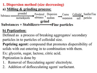
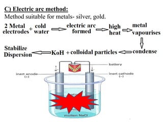
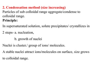
Downloaded 583 times

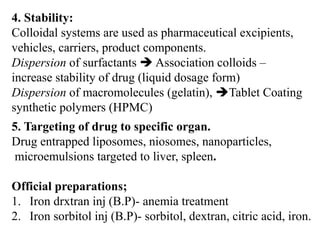
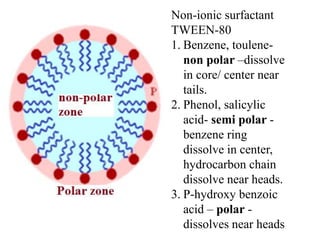
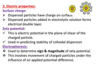
![Relative viscosity (ɳ rel) = ɳ/ ɳ0 = 1+2.5ɸ
Specific viscocity (ɳ sp) = ɳ/ ɳ0 -1= 2.5ɸ
(ɳ sp) = 2.5ɸ
(ɳ sp)/ɸ = 2.5 (ɸ = concentration of particles)
(ɳ sp)/C = 2.5 = K (K = Intrinsic viscosity factor)
Molecular weight
determination:
[ɳ] = Kma
ɳ =intrinsic viscosity
(viscometer)
K, a = constants of polymer,
M= molecular weight of
polymer.](https://image.slidesharecdn.com/colloids-180817061437/85/Colloids-39-320.jpg)
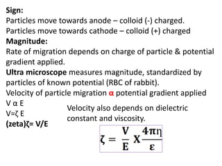
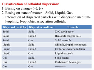
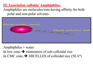
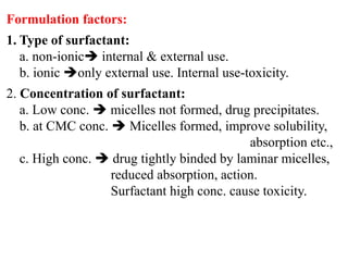
![At Equilibrium Charge balance Electro neutrality
Out side [Na+]o = [Cl-]o
In side [Na+]I = [Cl-]I + [R-]I
According to principle of escaping tendency of the
electrolytes concentration on both sides of the membrane
should be same. (outside = inside)
[Na+]o [Cl-]o = [Na+]I [Cl-]I
Converting to [Cl-] concentrations.
[Cl-]o [Cl-]o = ([Cl-]I + [R-]I) [Cl-]I
[Cl-]o2 = [Cl-]I](https://image.slidesharecdn.com/colloids-180817061437/85/Colloids-43-320.jpg)
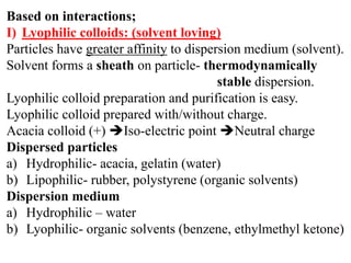
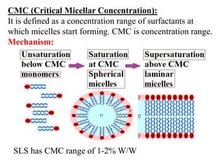
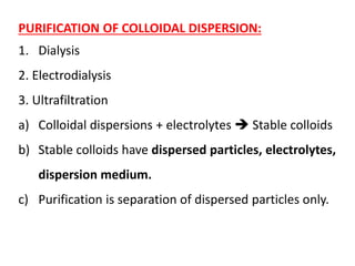
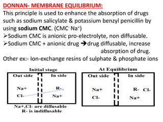
![[R-] = CMC-
[Cl-] = Drug = [D-]
Equation helps in selecting appropriate concentration of
components.
CASE- 1
If [R-]I/[D-]I= 8; then [D-]o /[D-]I = 3 D out= 3 D in
CASE- 2
If [R-]I/[D-]I= 99; then [D-]o /[D-]I = 10 D out = 10 D in
(GIT) (Blood)](https://image.slidesharecdn.com/colloids-180817061437/85/Colloids-44-320.jpg)
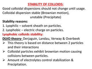
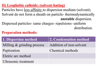
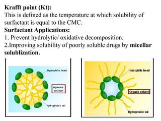
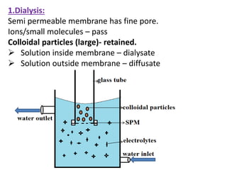
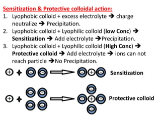
![ Addition of oppositely charged colloid
Gelatin Colloid [+] + Acacia Colloid [-] Electrostatic
attractive forces Solvent sheath break Particles
aggregate.
Addition of non-solvent.
Colloidal Dispersion + Alcohol/Acetone
Water(solvent) + Alcohol/Acetone(non-solvent) Solution.
No water, No solvent Sheath Unstable colloid.](https://image.slidesharecdn.com/colloids-180817061437/85/Colloids-51-320.jpg)


This document discusses colloids, which are uniform dispersions of small particles within a medium, detailing their definitions, characteristics, pharmaceutical applications, and various methods of preparation and purification. Characteristics such as particle size, shape, surface charge, and interactions with dispersion mediums are explored, alongside their implications in drug absorption, stability, and targeting. The document also outlines the physical properties of colloids, including optical, kinetic, and electrical properties, along with the stability and purification techniques of colloidal systems.