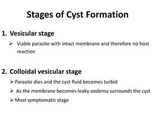
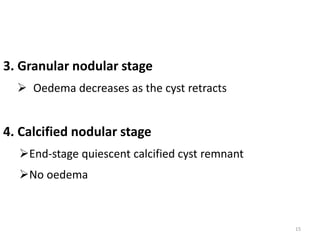
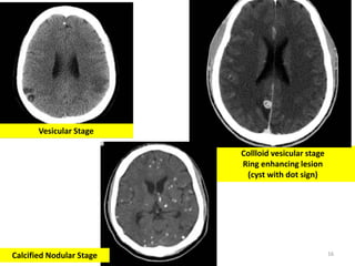
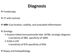
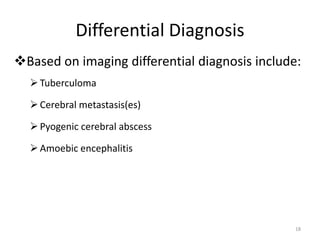
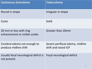
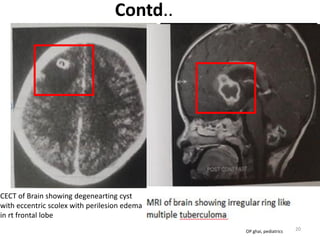
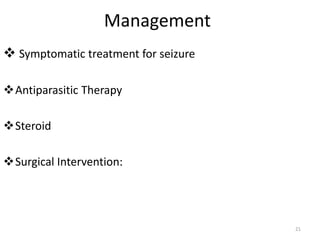
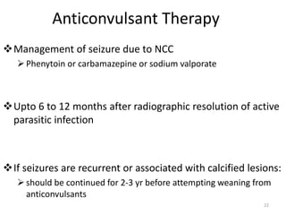
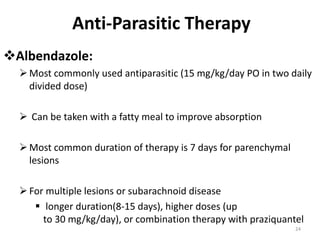
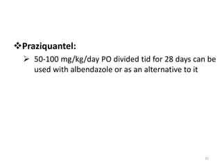
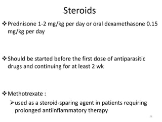
NEUROCYSTICERCOSIS
Dr. Prashant Raj Bhatt
1
Contents
 Introduction to Taeniasis
 Lifecycle of Taenia solium
 Anatomical Classification of NCC
 Stages of cyst formation
 Diagnosis
 NCC vs Tuberculoma based on imaging
 Management of parenchymal NCC
 Prevention and control
2
Neurocysticercosis
3
Definitive host:
Human
Intermediate host:
Cow/Cattle
• Definitive
host: Human
• Intermediate
host: Pig
Taeniasis
 Taeniasis is the infection of humans with the adult tapeworm
of Taenia saginata, T. solium or T. asiatica
 Taenia solium (pork tapeworm) is the main cause of human
cysticercosis
 Cysticercosis
 Caused by the presence, of the larval forms of
 Taenina solium i.e Cysticercus cellulosae and Cysticercus racemose
 T. saginata i.e Cysticercus bovis occurs very rarely
4
5
Target Tissues
Eyes
CNS
Striated muscles
Probably due to high glycogen and glucose content of these
tissues
CNS and Eye involvement is termed as Neurocysticercosis
Clinical features
Diverse clinical presentation of NCC is determined by:
Location of cysts
Size of cysts
Cyst load (number of cysts)
Host’s immune response
Anatomical Classification of NCC
 Parenchymal
 Intraventricular
 Meningeal
 Spinal
 Ocular Nelson
Parenchymal NCC
 Seizures (87%)
Simple partial with secondary generalization
 Generalized tonic-clonic
 Complex partial or complex partial with secondary generalization
 Headache, nausea and vomiting
Stroke
Hemiparesis
Focal neurologic deficits
Frontal lobe involvement
Psychosis, dementia, intellectual impairment
Intraventricular NCC
 5- 10% of all cases
 4th ventricle :Most common site for obstruction
 Up to 20% of cases is associated with obstructive
hydrocephalus and signs of raised ICP
Meningeal NCC
Meningeal irritation resembling Tubercular meningitis
 Raised ICP from inflammation, oedema and presence of
cyst obstructing flow of CSF
 Spinal NCC
– Spinal cord compression
– Nerve root pain
– Transverse myelitis
Ocular NCC
– Visual impairment (decreased visual acquity)
– Retinal detachment, iridocyclitis
iridocyclitis - inflammation of the iris and ciliary body of the eye
1. Vesicular stage
 Viable parasite with intact membrane and therefore no host
reaction
2. Colloidal vesicular stage
Parasite dies and the cyst fluid becomes turbid
 As the membrane becomes leaky oedema surrounds the cyst
Most symptomatic stage
Stages of Cyst Formation
3. Granular nodular stage
 Oedema decreases as the cyst retracts
4. Calcified nodular stage
End-stage quiescent calcified cyst remnant
No oedema
15
16
Collloid vesicular stage
Ring enhancing lesion
(cyst with dot sign)
Calcified Nodular Stage
Vesicular Stage
Diagnosis
 Fundoscopy
 CT with contrast
 MRI: Cyst location, viability, and associated inflammation
 Serology
Enzyme-linked immunotransfer blot (EITB): serologic diagnosis
sensitivity of 98% specificity of 100%
ELISA in CSF
sensitivity of 87% specificity of 95%
 Biopsy and histopathology
Differential Diagnosis
Based on imaging differential diagnosis include:
Tuberculoma
Cerebral metastasis(es)
Pyogenic cerebral abscess
Amoebic encephalitis
18
Cysticercus Granuloma Tuberculoma
Round in shape Irregular in shape
Cystic Solid
20 mm or less with ring
enhancement or visible scolex
Greater than 20mm
Cerebral edema not enough to
produce midline shift
Severe perifocal edema, midline
shift and raised ICP
Usually focal neurological deficit is
not present
Focal neurological deficit
19
Contd..
CECT of Brain showing degenearting cyst
with eccentric scolex with perilesion edema
in rt frontal lobe
Caseating granulomatous inflammation
associated with a fibrous type capsule,
20OP ghai, pediatrics
Management
 Symptomatic treatment for seizure
Antiparasitic Therapy
Steroid
Surgical Intervention:
21
Anticonvulsant Therapy
Management of seizure due to NCC
Phenytoin or carbamazepine or sodium valporate
Upto 6 to 12 months after radiographic resolution of active
parasitic infection
If seizures are recurrent or associated with calcified lesions:
should be continued for 2-3 yr before attempting weaning from
anticonvulsants
22
Prior to Therapy
These condition should be ruled out prior to initiating
anti parasitic and steroid therapy
Tuberculosis
Ocular cysticercosis
Strongyloidiasis
23
Anti-Parasitic Therapy
Albendazole:
Most commonly used antiparasitic (15 mg/kg/day PO in two daily
divided dose)
 Can be taken with a fatty meal to improve absorption
Most common duration of therapy is 7 days for parenchymal
lesions
For multiple lesions or subarachnoid disease
 longer duration(8-15 days), higher doses (up
to 30 mg/kg/day), or combination therapy with praziquantel
24
Praziquantel:
 50-100 mg/kg/day PO divided tid for 28 days can be
used with albendazole or as an alternative to it
25
Steroids
Prednisone 1-2 mg/kg per day or oral dexamethasone 0.15
mg/kg per day
Should be started before the first dose of antiparasitic
drugs and continuing for at least 2 wk
Methotrexate :
used as a steroid-sparing agent in patients requiring
prolonged antiinflammatory therapy
26
Indication of Surgery
 Symptomatic hydrocephalus due to NCC
 NCC of ventricle
 Giant cysticerci with life-threatening mass effect
 cysticerci adjacent to vascular structures
 Ocular and spinal NCC
Giant cysticerci : Rare condition defined as size in its largest dimension 27
Follow up and monitoring
 Intermittent radiographic surveillance to evaluate for resolution of
the cysticerci and development of calcifications
 Imaging at 1 to 2 month and 6 month
 Imaging should be repeated prior to discontinuing antiepileptic
drugs
 Antiparasitic therapy should be considered for patients with growing
cysts off therapy 28
Prevention and Controll
 Wash hands with soap and warm water after using the toilet,
changing diapers, and before handling food
 Wash and peel all raw vegetables and fruits before eating
 Adequate cooking of meat products
 Storage of meet in freezing condition*
 Veterinary vaccines for several cestode infections
Note *: (Cysticerci do not survive temperatures below -10o C and above 50o C)
THANK YOU
30

Neurocysticercosis

  • 1.
  • 2.
    Contents  Introduction toTaeniasis  Lifecycle of Taenia solium  Anatomical Classification of NCC  Stages of cyst formation  Diagnosis  NCC vs Tuberculoma based on imaging  Management of parenchymal NCC  Prevention and control 2
  • 3.
  • 4.
    Taeniasis  Taeniasis isthe infection of humans with the adult tapeworm of Taenia saginata, T. solium or T. asiatica  Taenia solium (pork tapeworm) is the main cause of human cysticercosis  Cysticercosis  Caused by the presence, of the larval forms of  Taenina solium i.e Cysticercus cellulosae and Cysticercus racemose  T. saginata i.e Cysticercus bovis occurs very rarely 4
  • 5.
  • 6.
    Target Tissues Eyes CNS Striated muscles Probablydue to high glycogen and glucose content of these tissues CNS and Eye involvement is termed as Neurocysticercosis
  • 7.
    Clinical features Diverse clinicalpresentation of NCC is determined by: Location of cysts Size of cysts Cyst load (number of cysts) Host’s immune response
  • 8.
    Anatomical Classification ofNCC  Parenchymal  Intraventricular  Meningeal  Spinal  Ocular Nelson
  • 9.
    Parenchymal NCC  Seizures(87%) Simple partial with secondary generalization  Generalized tonic-clonic  Complex partial or complex partial with secondary generalization  Headache, nausea and vomiting Stroke Hemiparesis Focal neurologic deficits Frontal lobe involvement Psychosis, dementia, intellectual impairment
  • 10.
    Intraventricular NCC  5-10% of all cases  4th ventricle :Most common site for obstruction  Up to 20% of cases is associated with obstructive hydrocephalus and signs of raised ICP
  • 11.
    Meningeal NCC Meningeal irritationresembling Tubercular meningitis  Raised ICP from inflammation, oedema and presence of cyst obstructing flow of CSF
  • 12.
     Spinal NCC –Spinal cord compression – Nerve root pain – Transverse myelitis Ocular NCC – Visual impairment (decreased visual acquity) – Retinal detachment, iridocyclitis iridocyclitis - inflammation of the iris and ciliary body of the eye
  • 13.
    1. Vesicular stage Viable parasite with intact membrane and therefore no host reaction 2. Colloidal vesicular stage Parasite dies and the cyst fluid becomes turbid  As the membrane becomes leaky oedema surrounds the cyst Most symptomatic stage Stages of Cyst Formation
  • 14.
    3. Granular nodularstage  Oedema decreases as the cyst retracts 4. Calcified nodular stage End-stage quiescent calcified cyst remnant No oedema 15
  • 15.
    16 Collloid vesicular stage Ringenhancing lesion (cyst with dot sign) Calcified Nodular Stage Vesicular Stage
  • 16.
    Diagnosis  Fundoscopy  CTwith contrast  MRI: Cyst location, viability, and associated inflammation  Serology Enzyme-linked immunotransfer blot (EITB): serologic diagnosis sensitivity of 98% specificity of 100% ELISA in CSF sensitivity of 87% specificity of 95%  Biopsy and histopathology
  • 17.
    Differential Diagnosis Based onimaging differential diagnosis include: Tuberculoma Cerebral metastasis(es) Pyogenic cerebral abscess Amoebic encephalitis 18
  • 18.
    Cysticercus Granuloma Tuberculoma Roundin shape Irregular in shape Cystic Solid 20 mm or less with ring enhancement or visible scolex Greater than 20mm Cerebral edema not enough to produce midline shift Severe perifocal edema, midline shift and raised ICP Usually focal neurological deficit is not present Focal neurological deficit 19
  • 19.
    Contd.. CECT of Brainshowing degenearting cyst with eccentric scolex with perilesion edema in rt frontal lobe Caseating granulomatous inflammation associated with a fibrous type capsule, 20OP ghai, pediatrics
  • 20.
    Management  Symptomatic treatmentfor seizure Antiparasitic Therapy Steroid Surgical Intervention: 21
  • 21.
    Anticonvulsant Therapy Management ofseizure due to NCC Phenytoin or carbamazepine or sodium valporate Upto 6 to 12 months after radiographic resolution of active parasitic infection If seizures are recurrent or associated with calcified lesions: should be continued for 2-3 yr before attempting weaning from anticonvulsants 22
  • 22.
    Prior to Therapy Thesecondition should be ruled out prior to initiating anti parasitic and steroid therapy Tuberculosis Ocular cysticercosis Strongyloidiasis 23
  • 23.
    Anti-Parasitic Therapy Albendazole: Most commonlyused antiparasitic (15 mg/kg/day PO in two daily divided dose)  Can be taken with a fatty meal to improve absorption Most common duration of therapy is 7 days for parenchymal lesions For multiple lesions or subarachnoid disease  longer duration(8-15 days), higher doses (up to 30 mg/kg/day), or combination therapy with praziquantel 24
  • 24.
    Praziquantel:  50-100 mg/kg/dayPO divided tid for 28 days can be used with albendazole or as an alternative to it 25
  • 25.
    Steroids Prednisone 1-2 mg/kgper day or oral dexamethasone 0.15 mg/kg per day Should be started before the first dose of antiparasitic drugs and continuing for at least 2 wk Methotrexate : used as a steroid-sparing agent in patients requiring prolonged antiinflammatory therapy 26
  • 26.
    Indication of Surgery Symptomatic hydrocephalus due to NCC  NCC of ventricle  Giant cysticerci with life-threatening mass effect  cysticerci adjacent to vascular structures  Ocular and spinal NCC Giant cysticerci : Rare condition defined as size in its largest dimension 27
  • 27.
    Follow up andmonitoring  Intermittent radiographic surveillance to evaluate for resolution of the cysticerci and development of calcifications  Imaging at 1 to 2 month and 6 month  Imaging should be repeated prior to discontinuing antiepileptic drugs  Antiparasitic therapy should be considered for patients with growing cysts off therapy 28
  • 28.
    Prevention and Controll Wash hands with soap and warm water after using the toilet, changing diapers, and before handling food  Wash and peel all raw vegetables and fruits before eating  Adequate cooking of meat products  Storage of meet in freezing condition*  Veterinary vaccines for several cestode infections Note *: (Cysticerci do not survive temperatures below -10o C and above 50o C)
  • 29.