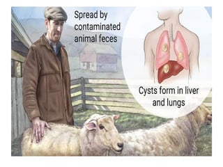
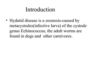
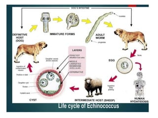
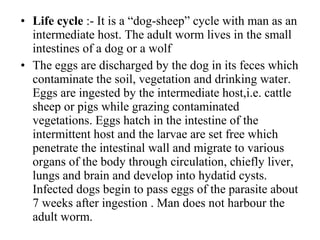
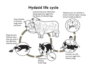
HYDATID DISEASE
Prepared by:-
MR. VANINDARKUMAR B.C
GOVERNMENT COLLEGE OF NURSING
FORT, BENGALURU-02
Introduction
• Hydatid disease is a zoonosis-caused by
metacystodes(infective larva) of the cystode
genus Echinococcus, the adult worms are
found in dogs and other carnivores.
Problem
• Hydatidosis is a world wide public health
problem. It is prevalent in all sheep-rearing
countries, e.g. Australia, Newzealand,
Tasmania, Middle East countries, Turkey,
Latin America and the Far East, Etc. Its foci
are also prevalent in india, with highest
prevalence in, i,e. Andhra pradesh and Tamil
nadu.
Epidemiological Factors
• Agent Factors:-
Echinococcus species are small tape-worms, rarely
more than 7mm length. The scolex(head) has four
suckers, two rows of hooks, one small and one large
on the rostellum.
They have 4-6 proglottids. At present there are four
species which are considered valid to cause
hydatidosis, namely E. granulosus, E.multilocularis,
E. oligarthus and E.vogeli.
• Reservoir of infection :-
Carnivores infected with adult worms, especially
dogs and wolves.
• Sources of infection :-
Feces of dogs containing eggs of Echinococcus.
• Life cycle :- It is a “dog-sheep” cycle with man as an
intermediate host. The adult worm lives in the small
intestines of a dog or a wolf
• The eggs are discharged by the dog in its feces which
contaminate the soil, vegetation and drinking water.
Eggs are ingested by the intermediate host,i.e. cattle
sheep or pigs while grazing contaminated
vegetations. Eggs hatch in the intestine of the
intermittent host and the larvae are set free which
penetrate the intestinal wall and migrate to various
organs of the body through circulation, chiefly liver,
lungs and brain and develop into hydatid cysts.
Infected dogs begin to pass eggs of the parasite about
7 weeks after ingestion . Man does not harbour the
adult worm.
Host factors
• Human behavior,i,e. close association
between infected dogs and man especially
during child-hood and contamination of hands
with the feces of dogs.
• Uncontrolled slaughter of herbivorous
animals,
• Hydatid disease is an occupational
disease.Shepherds and their families and shoe-
makers in the endemic areas are at increased
risk of getting the disease.
Mode of Transmission
• Ingestion of food, unwashed vegetables and
water contaminated with feces of infected dogs
containing eggs of the cystodes.
• Handling or playing with infected dogs, e.g.
hand to mouth transfer of eggs through feces
of dogs.
• Inhalation of dust contaminated with infected
eggs. Direct transmission from person to
person does not occur.
Incubation period
• Varies from months to years. Depends upon
the number and location of cysts and how
rapidly they grow.
Clinical Features
• Symptoms appear usually after several years of
exposure. Symptoms depend upon the size and the
anatomical size of the cysts.
• The patient may have a single cysts or multiple
cysts. The cysts are filled with watery fluid and
contain a large number of tapeworm heads.The
cysts are located mostly in the right lobe of the
liver followed by the kidney,
brain,lungs,peritoneum and long bones.
• Liver enlargment, jaundice in the liver.
• Death may occur due to vital organs involvement.
• Spontaneous recovery may occur when there is no
growth of cysts.
• Large cysts may rupture releasing volumes of liquid
and thousands of the worm protoscolices. This can
cause allergic reactions.
Diagnosis
• History of residence in the endemic area and close
contact with dog.
• Presence of slowly growing cystic tumour.
• X-ray shows the location of the cyst.
• Ultrasonography and CAT scan-modern techniques.
• Serological :- Indirect immunofluorescent test is quite
sensitive and specific. ELISA is also very sensitive
and superior to some serological procedure.
Prevention
• Preventing dogs from ingestion of sheep offal at
slaughter houses and farm houses and to dead animals.
• proper meat inspection and destruction of infected
viscera.
• Control of dogs, i.e. elimination of stray dogs, registration
of dogs, surveillance of dogs based on periodic stool
examination.
• Health education regarding prevention, particularly to the
butchers, dog owners, animal breeders and shepherds.
• Children and other general public should also be educated
about dangers of close association with dogs.
Control
• No isolation, concurrent disinfection, quarantine or
terminal disinfection needed.
• There is no specific treatment.Surgical removal of
cysts is done but it is not without risk since accidental
penetration of one of the cysts can leads to
anaphylatic shock which may lead to death.
• Mebendazole(veermox) has been found very useful in
mice. It may become the drug of choice.
Hydatid disease

Hydatid disease

  • 1.
    HYDATID DISEASE Prepared by:- MR.VANINDARKUMAR B.C GOVERNMENT COLLEGE OF NURSING FORT, BENGALURU-02
  • 3.
    Introduction • Hydatid diseaseis a zoonosis-caused by metacystodes(infective larva) of the cystode genus Echinococcus, the adult worms are found in dogs and other carnivores.
  • 5.
    Problem • Hydatidosis isa world wide public health problem. It is prevalent in all sheep-rearing countries, e.g. Australia, Newzealand, Tasmania, Middle East countries, Turkey, Latin America and the Far East, Etc. Its foci are also prevalent in india, with highest prevalence in, i,e. Andhra pradesh and Tamil nadu.
  • 6.
    Epidemiological Factors • AgentFactors:- Echinococcus species are small tape-worms, rarely more than 7mm length. The scolex(head) has four suckers, two rows of hooks, one small and one large on the rostellum. They have 4-6 proglottids. At present there are four species which are considered valid to cause hydatidosis, namely E. granulosus, E.multilocularis, E. oligarthus and E.vogeli.
  • 7.
    • Reservoir ofinfection :- Carnivores infected with adult worms, especially dogs and wolves. • Sources of infection :- Feces of dogs containing eggs of Echinococcus.
  • 9.
    • Life cycle:- It is a “dog-sheep” cycle with man as an intermediate host. The adult worm lives in the small intestines of a dog or a wolf • The eggs are discharged by the dog in its feces which contaminate the soil, vegetation and drinking water. Eggs are ingested by the intermediate host,i.e. cattle sheep or pigs while grazing contaminated vegetations. Eggs hatch in the intestine of the intermittent host and the larvae are set free which penetrate the intestinal wall and migrate to various organs of the body through circulation, chiefly liver, lungs and brain and develop into hydatid cysts. Infected dogs begin to pass eggs of the parasite about 7 weeks after ingestion . Man does not harbour the adult worm.
  • 11.
    Host factors • Humanbehavior,i,e. close association between infected dogs and man especially during child-hood and contamination of hands with the feces of dogs. • Uncontrolled slaughter of herbivorous animals, • Hydatid disease is an occupational disease.Shepherds and their families and shoe- makers in the endemic areas are at increased risk of getting the disease.
  • 12.
    Mode of Transmission •Ingestion of food, unwashed vegetables and water contaminated with feces of infected dogs containing eggs of the cystodes. • Handling or playing with infected dogs, e.g. hand to mouth transfer of eggs through feces of dogs. • Inhalation of dust contaminated with infected eggs. Direct transmission from person to person does not occur.
  • 13.
    Incubation period • Variesfrom months to years. Depends upon the number and location of cysts and how rapidly they grow.
  • 14.
    Clinical Features • Symptomsappear usually after several years of exposure. Symptoms depend upon the size and the anatomical size of the cysts. • The patient may have a single cysts or multiple cysts. The cysts are filled with watery fluid and contain a large number of tapeworm heads.The cysts are located mostly in the right lobe of the liver followed by the kidney, brain,lungs,peritoneum and long bones.
  • 15.
    • Liver enlargment,jaundice in the liver. • Death may occur due to vital organs involvement. • Spontaneous recovery may occur when there is no growth of cysts. • Large cysts may rupture releasing volumes of liquid and thousands of the worm protoscolices. This can cause allergic reactions.
  • 16.
    Diagnosis • History ofresidence in the endemic area and close contact with dog. • Presence of slowly growing cystic tumour. • X-ray shows the location of the cyst. • Ultrasonography and CAT scan-modern techniques. • Serological :- Indirect immunofluorescent test is quite sensitive and specific. ELISA is also very sensitive and superior to some serological procedure.
  • 17.
    Prevention • Preventing dogsfrom ingestion of sheep offal at slaughter houses and farm houses and to dead animals. • proper meat inspection and destruction of infected viscera. • Control of dogs, i.e. elimination of stray dogs, registration of dogs, surveillance of dogs based on periodic stool examination. • Health education regarding prevention, particularly to the butchers, dog owners, animal breeders and shepherds. • Children and other general public should also be educated about dangers of close association with dogs.
  • 18.
    Control • No isolation,concurrent disinfection, quarantine or terminal disinfection needed. • There is no specific treatment.Surgical removal of cysts is done but it is not without risk since accidental penetration of one of the cysts can leads to anaphylatic shock which may lead to death. • Mebendazole(veermox) has been found very useful in mice. It may become the drug of choice.