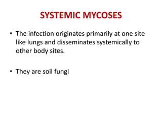
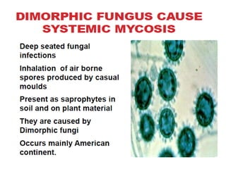
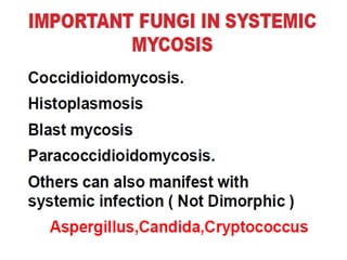
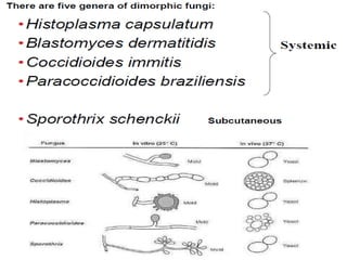
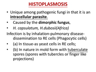
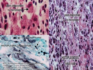
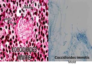
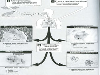
SYSTEMIC FUNGAL
INFECTIONS
SYSTEMIC MYCOSES
• The infection originates primarily at one site
like lungs and disseminates systemically to
other body sites.
• They are soil fungi
HISTOPLASMOSIS
• Unique among pathogenic fungi in that it is an
intracellular parasite.
• Caused by the dimorphic fungus,
• H. capsulatum, H.dubosii(Africa)
Infection is by inhalation-pulmonary disease-
dissemination to RE cells (Phagocytic cells)
• (a) In tissue-as yeast cells in RE cells;
• (b) In nature in mold form with tuberculate
spores (spores with tubercles or finger like
projections)
Pathogenesis
• asymptomatic infection,
• ac influenza like illness,
• Chr. Form-resemble TB
• Later it progressive with widespread infection
of RE system
Disseminated infection occurs most often in
old age, infancy, impaired immune response
(due to neutropenia, haematological
malignancy, AIDS and high doses of steroids)
(i) In tissue –yeast form
cells found as oval budding
cells; 2-4um; also yeast
forms seen on blood agar
at 370C;
(ii) On Sabouraud’s agar at
room temp-white cottony
mycelial growth with
tuberculate spores (large
thick walled spherical
spores with tubercles or
finger like projections).
Worldwide in distribution,
most common in USA.
Lab diagnosis
• Microscopy after Giemsa/,
Wright-of sputum, pus, blood
(AIDS),
• culture on Sabouraud’s (25-300C)
• histoplasmin skin testing-
delayed hypersensitivity test
• Liver,lung biopsy-PAS,
Methenamine silver
• Culture at 370C used for
conversion from mold to yeast-
for confirmation
• Serological test-CFT, Latex
agglutination
BLASTOMYCOSIS
(North America Blastomycosis)
• Dimorphic fungus Blastomyces
dermatitidis ;
• causes suppurative granulomatous
lesions of skin and lungs.
• Seen in North America & Africa;
• Source–soil-inhalation-lungs-
dissemination.
• In tissue and culture at 370C budding
yeasts with double contoured walls
and a single broad based bud, from
a single pole on the mother cell.
• On culture at room temp it gives rise
to filamentous septate hyphae.
BLASTOMYCOSIS/Pathogenesis
• Slowly progressive disease, chest radiography
resembles TB, Malignancy
• Disseminated disease seen in
immunosuppressed
• Skin lesions are raised and well demarcated
Lab diagnosis
• Direct microscopy of pus, scrappings from skin
lesions or sputum-thick walled yeast cells with
buds on a broad base,
• The buds remain attached until they are
almost the size of parent cell forming chain of
3 or 4 cells
PARACOCCIDIOIDOMYCOSIS/South American
blastomycosis
• Is caused by a dimorphic fungus
Paracoccidioides brasiliensis - Chr
granulomatous disease of skin, mucosa,
lymphnodes and internal organs.
• Disease confined to South America.
• The yeast phase is found in tissue and
culture at 370C which is spherical, thick
walled cell, with multiple buddings, ship
steering wheel appearance;
• at room temp mycelial form develops.
Paracoccidioides
brasiliensis
Lab diagnosis
• Yeast-multiple
buddings- ship steering
wheel appearance
• Skin test
• Serological test-
precipitin, CFT
Paracoccidioides brasiliensis
COCCIDIOIDOMYCOSIS
• Is caused by dimorphic fungus
Coccidioides immitis ,
• Seen in South Western USA-dry
region; Infection is acquired by
inhalation.
• Disease resembles clinically and
histologically disseminated
tuberculosis.
• Tissue form (Yeast phase) is a
spherule with thick doubly
refractile wall and is filled with
endospores, occurs at-370C.
Coccidioides immitis
Mold
OPPORTUNISTIC MYCOSES
• immuno-compromised individuals and are rare in
normal individuals
• Incidence of opportunistic mycoses increasing
due to-
(a) Use of immunosuppressive drugs in organ
transplantation
(b) Chemotherapy in cancer treatment
(c) Immunodeficiency due to HIV infection/AIDS
• Constitute 15% of all nosocomial infections in
ICUs
SN Opportunistic
mycoses
Causative
agent
Group
1 Candidiasis Candida
albicans
Yeast like
fungus
2 Cryptococco
sis
Cryptococc
us
neoformans
True Yeast
3 Pneumocyst
is carinii
Pneumonia
(PCP)
Pneumocys
tis carinii
Taxonomic
status?(mole
culr-fungus)
4 Aspergillosis Aspergillus
fumigatus
Filamentous
mould
• .
•
5 Mucormycosis
(zygomycoses,
phycomycose
s)
Rhizopus,
Absidia &
Mucor spp
Filamentous
mould (non
septate)
6 Penicillosis Penicillium
marneffei
Dimorphic
fungus
7 Histoplasmosi
s
Histoplasma
capsulatum
Dimorphic
fungus
CRYPTOCOCCOSIS
• Cryptococcus neoformans- subacute / chr.
infection. Soil saprophytes; abundant in faeces
of pigeons / birds.
• Infection : is by inhalation; Worldwide in
distribution.
• Four serological types of cryptococcal capsular
polysaccharide–A,B,C,& D
• Cryptococcal meningitis is the most serious
type of infection – often seen in AIDS.
CRYPTOCOCCOSIS
• Birds do not get infected due to high body
temperature
• C. neoformans var neoformans-(A,D) &C
• C. neoformans var gatti-(B,C)
• Pathogenesis-
(a)Commonest form-self limiting pulmonary form,
Causing, Acute pneumonic type,
(d) Meningeal form.
• *Caused by true yeast-Cryptococcus
neoformans. Found worldwide.
• *Round or oval budding yeast cell with
thick prominent polysaccharide capsule.
• *Organism is especially abundant in soil
containing bird (especially pigeon)
droppings.
• *Cryptococcal meningitis is the most
serious form of disseminated infection.
Laboratory diagnosis-CRYPTOCOCCOSIS
• India ink / Nigrosine – Capsule +
• Ability to grow at 370C : smooth, mucoid cream coloured
colonies
• Hydrolyse urea
• Budding yeast – Grams stain, wet mount
• Tissue section – PAS
• Serological test for capsular antigen
Cryptococcus gram stain Capsulated yeast in India Ink
preperation
Cryptococcus
neoformans
on Niger
seed agar
CANDIDIASIS
• Caused by yeast like fungus Candida albicans
(resembles like pseudohyphae). Part of normal body
flora.
• (a) Oral candidiasis (Thrush)-raised white paques
over oral mucosa, tongue and gums. Presence of
oesophagial candidiasis is a hallmark of progression
of HIV infection to AIDS.
• (b) Vaginal candidiasis- Itching and burning pain of
vulva & vagina with whitish discharge. Infection is
recurrent in HIV +ve women
CANDIDIASIS (CANDIDOSIS, MONILIASIS)
• (c) Systemic candidiasis- Potentially life threatening.
May involve GIT, brain, liver, kidney & spleen etc.
Common sign of deep candidiasis-
• Candida endocarditis in surgery/ in drug addicts.
• Cadidaemia in postoperative/immunosuppressed pts
MOST COMMONLY ISOLATED
CANDIDA SPECIES
• C. albicans
• C. tropicalis
• C. parapsilosis
• C. kefyr
• C. glabrata
• C. krusei
• C. guillermondii
• C. lusitaniae
Lab diagnosis
(a) Microscopy-wet mount and Grams stain;
(b) Culture on Sabouraud’s/blood agar;
(c) Germ tube formation in serum at 370C and
(d) Chlamydospore fomation if cultured on
corn meal agar.
(e) Serology-immunodiffusion,CIE,ELISA
(f) PCR
ASPERGILLOSIS
Caused primarily by Aspergillus
fumigatus, ubiquitous, filamentous
septate mould. Rarely pathogenic
in normal host
• Disease manifests as
(a) Pulmonary aspergillosis-
(i) Aspergillus asthma
(ii) Bronchopulmonary
aspergillosis
(iii) Aspergilloma (fungus
ball)
aspergillosis
(b) Disseminating aspergillosis- Invasive
aspergillosis of lung then dissemination to
brain, kidney other organs etc.
Diagnosis -Aspergillus hyphae
characteristically form ‘V’ shaped branches
(septate hyphae that branch at 450 angle)
Aspergillus
Lab diagnosis
• Direct microscopy
• Culture-SDA
• A fumigatus grows at 450C
• Skin test
• Serological test
• PCR
MUCORMYCOSIS
• Ubiquitous in nature. Infection
occurs worldwide-they are
saprophytic
• Most common form of disease
which can be fatal within a week
is Rhinocerebral mucormycosis in
which infection begins in the
nasal mucosa or sinuses and
progresses to the orbits, the
palate and the brain. The disease
is aggressive and fatal.
• Broad aseptate hyphae, and form
angled branches
• Species are rhizopus, mucor and
absidia.
• Can be cultured in Sabouraud’s
agar without cycloheximide.
• Penicillium marneffei is an
important opportunistic
pathogen in HIV infected.
• Penicillium marneffei cause
serious disseminated
disease with characteristic
papular skin lesions in AIDS
pts of South East Asia
• Diagnosis-brush like
arrangement of conidia-
(penicillium=brush)-chains
of spores from the ends of
short branches of
conidiophores
conidiophores
PENICILLIOSIS
Other opportunistic mycoses
• Sporotrichosis- Sporothrix schenkii (brain &
skin)
• Dermatophytoses (Nail & skin)
(a) Trichophyton spp.
(b) Epidermophyton floccosum

Systemic mycology

  • 1.
  • 2.
    SYSTEMIC MYCOSES • Theinfection originates primarily at one site like lungs and disseminates systemically to other body sites. • They are soil fungi
  • 6.
    HISTOPLASMOSIS • Unique amongpathogenic fungi in that it is an intracellular parasite. • Caused by the dimorphic fungus, • H. capsulatum, H.dubosii(Africa) Infection is by inhalation-pulmonary disease- dissemination to RE cells (Phagocytic cells) • (a) In tissue-as yeast cells in RE cells; • (b) In nature in mold form with tuberculate spores (spores with tubercles or finger like projections)
  • 7.
    Pathogenesis • asymptomatic infection, •ac influenza like illness, • Chr. Form-resemble TB • Later it progressive with widespread infection of RE system Disseminated infection occurs most often in old age, infancy, impaired immune response (due to neutropenia, haematological malignancy, AIDS and high doses of steroids)
  • 8.
    (i) In tissue–yeast form cells found as oval budding cells; 2-4um; also yeast forms seen on blood agar at 370C; (ii) On Sabouraud’s agar at room temp-white cottony mycelial growth with tuberculate spores (large thick walled spherical spores with tubercles or finger like projections). Worldwide in distribution, most common in USA.
  • 9.
    Lab diagnosis • Microscopyafter Giemsa/, Wright-of sputum, pus, blood (AIDS), • culture on Sabouraud’s (25-300C) • histoplasmin skin testing- delayed hypersensitivity test • Liver,lung biopsy-PAS, Methenamine silver • Culture at 370C used for conversion from mold to yeast- for confirmation • Serological test-CFT, Latex agglutination
  • 11.
    BLASTOMYCOSIS (North America Blastomycosis) •Dimorphic fungus Blastomyces dermatitidis ; • causes suppurative granulomatous lesions of skin and lungs. • Seen in North America & Africa; • Source–soil-inhalation-lungs- dissemination. • In tissue and culture at 370C budding yeasts with double contoured walls and a single broad based bud, from a single pole on the mother cell. • On culture at room temp it gives rise to filamentous septate hyphae.
  • 12.
    BLASTOMYCOSIS/Pathogenesis • Slowly progressivedisease, chest radiography resembles TB, Malignancy • Disseminated disease seen in immunosuppressed • Skin lesions are raised and well demarcated
  • 13.
    Lab diagnosis • Directmicroscopy of pus, scrappings from skin lesions or sputum-thick walled yeast cells with buds on a broad base, • The buds remain attached until they are almost the size of parent cell forming chain of 3 or 4 cells
  • 15.
    PARACOCCIDIOIDOMYCOSIS/South American blastomycosis • Iscaused by a dimorphic fungus Paracoccidioides brasiliensis - Chr granulomatous disease of skin, mucosa, lymphnodes and internal organs. • Disease confined to South America. • The yeast phase is found in tissue and culture at 370C which is spherical, thick walled cell, with multiple buddings, ship steering wheel appearance; • at room temp mycelial form develops. Paracoccidioides brasiliensis
  • 16.
    Lab diagnosis • Yeast-multiple buddings-ship steering wheel appearance • Skin test • Serological test- precipitin, CFT Paracoccidioides brasiliensis
  • 17.
    COCCIDIOIDOMYCOSIS • Is causedby dimorphic fungus Coccidioides immitis , • Seen in South Western USA-dry region; Infection is acquired by inhalation. • Disease resembles clinically and histologically disseminated tuberculosis. • Tissue form (Yeast phase) is a spherule with thick doubly refractile wall and is filled with endospores, occurs at-370C. Coccidioides immitis
  • 18.
  • 22.
  • 23.
    OPPORTUNISTIC MYCOSES • immuno-compromisedindividuals and are rare in normal individuals • Incidence of opportunistic mycoses increasing due to- (a) Use of immunosuppressive drugs in organ transplantation (b) Chemotherapy in cancer treatment (c) Immunodeficiency due to HIV infection/AIDS • Constitute 15% of all nosocomial infections in ICUs
  • 24.
    SN Opportunistic mycoses Causative agent Group 1 CandidiasisCandida albicans Yeast like fungus 2 Cryptococco sis Cryptococc us neoformans True Yeast 3 Pneumocyst is carinii Pneumonia (PCP) Pneumocys tis carinii Taxonomic status?(mole culr-fungus) 4 Aspergillosis Aspergillus fumigatus Filamentous mould
  • 25.
    • . • 5 Mucormycosis (zygomycoses, phycomycose s) Rhizopus, Absidia& Mucor spp Filamentous mould (non septate) 6 Penicillosis Penicillium marneffei Dimorphic fungus 7 Histoplasmosi s Histoplasma capsulatum Dimorphic fungus
  • 26.
    CRYPTOCOCCOSIS • Cryptococcus neoformans-subacute / chr. infection. Soil saprophytes; abundant in faeces of pigeons / birds. • Infection : is by inhalation; Worldwide in distribution. • Four serological types of cryptococcal capsular polysaccharide–A,B,C,& D • Cryptococcal meningitis is the most serious type of infection – often seen in AIDS.
  • 27.
    CRYPTOCOCCOSIS • Birds donot get infected due to high body temperature • C. neoformans var neoformans-(A,D) &C • C. neoformans var gatti-(B,C) • Pathogenesis- (a)Commonest form-self limiting pulmonary form, Causing, Acute pneumonic type, (d) Meningeal form.
  • 28.
    • *Caused bytrue yeast-Cryptococcus neoformans. Found worldwide. • *Round or oval budding yeast cell with thick prominent polysaccharide capsule. • *Organism is especially abundant in soil containing bird (especially pigeon) droppings. • *Cryptococcal meningitis is the most serious form of disseminated infection.
  • 29.
    Laboratory diagnosis-CRYPTOCOCCOSIS • Indiaink / Nigrosine – Capsule + • Ability to grow at 370C : smooth, mucoid cream coloured colonies • Hydrolyse urea • Budding yeast – Grams stain, wet mount • Tissue section – PAS • Serological test for capsular antigen Cryptococcus gram stain Capsulated yeast in India Ink preperation Cryptococcus neoformans on Niger seed agar
  • 30.
    CANDIDIASIS • Caused byyeast like fungus Candida albicans (resembles like pseudohyphae). Part of normal body flora. • (a) Oral candidiasis (Thrush)-raised white paques over oral mucosa, tongue and gums. Presence of oesophagial candidiasis is a hallmark of progression of HIV infection to AIDS. • (b) Vaginal candidiasis- Itching and burning pain of vulva & vagina with whitish discharge. Infection is recurrent in HIV +ve women
  • 31.
    CANDIDIASIS (CANDIDOSIS, MONILIASIS) •(c) Systemic candidiasis- Potentially life threatening. May involve GIT, brain, liver, kidney & spleen etc. Common sign of deep candidiasis- • Candida endocarditis in surgery/ in drug addicts. • Cadidaemia in postoperative/immunosuppressed pts
  • 32.
    MOST COMMONLY ISOLATED CANDIDASPECIES • C. albicans • C. tropicalis • C. parapsilosis • C. kefyr • C. glabrata • C. krusei • C. guillermondii • C. lusitaniae
  • 33.
    Lab diagnosis (a) Microscopy-wetmount and Grams stain; (b) Culture on Sabouraud’s/blood agar; (c) Germ tube formation in serum at 370C and (d) Chlamydospore fomation if cultured on corn meal agar. (e) Serology-immunodiffusion,CIE,ELISA (f) PCR
  • 34.
    ASPERGILLOSIS Caused primarily byAspergillus fumigatus, ubiquitous, filamentous septate mould. Rarely pathogenic in normal host • Disease manifests as (a) Pulmonary aspergillosis- (i) Aspergillus asthma (ii) Bronchopulmonary aspergillosis (iii) Aspergilloma (fungus ball)
  • 35.
    aspergillosis (b) Disseminating aspergillosis-Invasive aspergillosis of lung then dissemination to brain, kidney other organs etc. Diagnosis -Aspergillus hyphae characteristically form ‘V’ shaped branches (septate hyphae that branch at 450 angle)
  • 36.
  • 37.
    Lab diagnosis • Directmicroscopy • Culture-SDA • A fumigatus grows at 450C • Skin test • Serological test • PCR
  • 38.
    MUCORMYCOSIS • Ubiquitous innature. Infection occurs worldwide-they are saprophytic • Most common form of disease which can be fatal within a week is Rhinocerebral mucormycosis in which infection begins in the nasal mucosa or sinuses and progresses to the orbits, the palate and the brain. The disease is aggressive and fatal. • Broad aseptate hyphae, and form angled branches • Species are rhizopus, mucor and absidia. • Can be cultured in Sabouraud’s agar without cycloheximide.
  • 39.
    • Penicillium marneffeiis an important opportunistic pathogen in HIV infected. • Penicillium marneffei cause serious disseminated disease with characteristic papular skin lesions in AIDS pts of South East Asia • Diagnosis-brush like arrangement of conidia- (penicillium=brush)-chains of spores from the ends of short branches of conidiophores conidiophores PENICILLIOSIS
  • 40.
    Other opportunistic mycoses •Sporotrichosis- Sporothrix schenkii (brain & skin) • Dermatophytoses (Nail & skin) (a) Trichophyton spp. (b) Epidermophyton floccosum