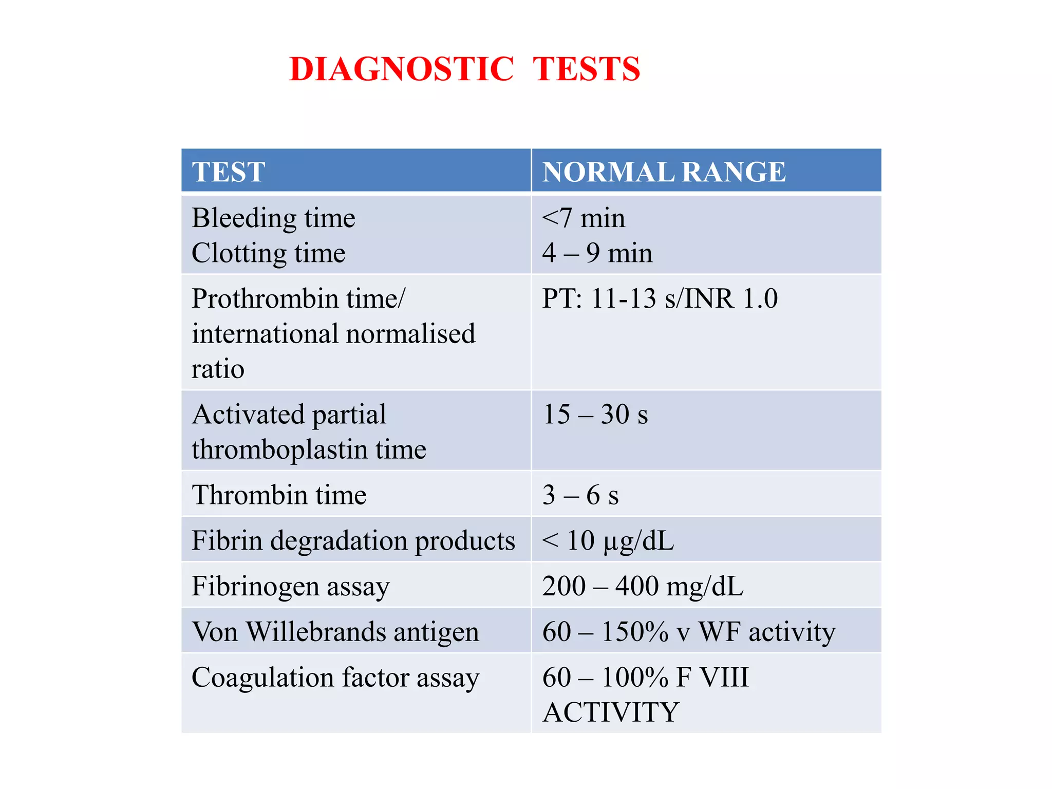
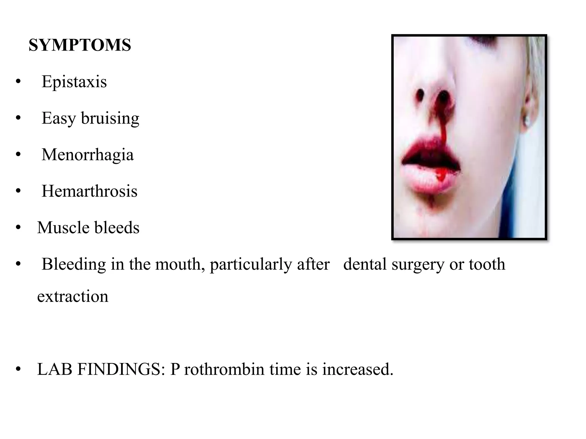
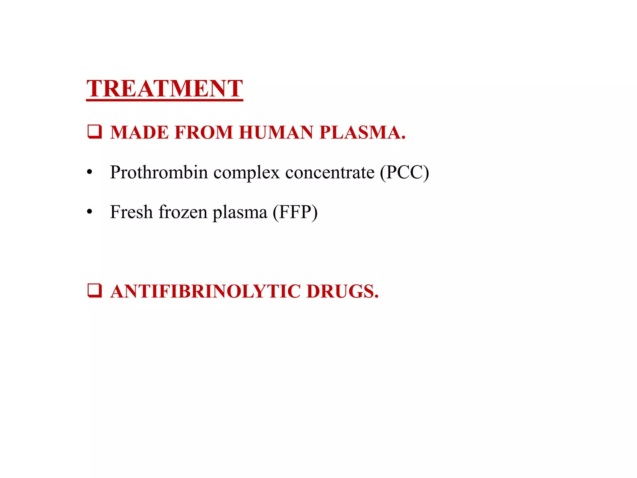
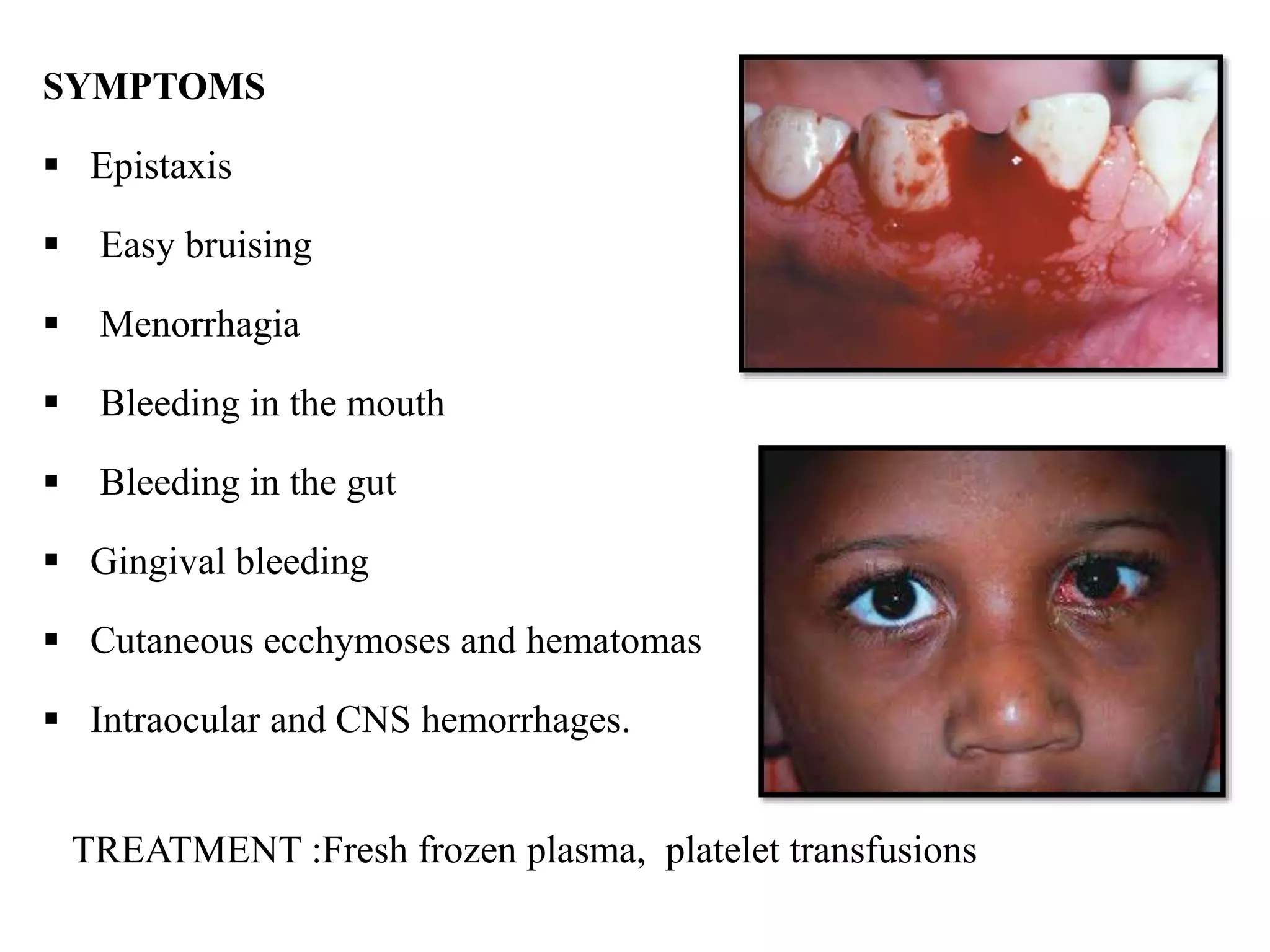
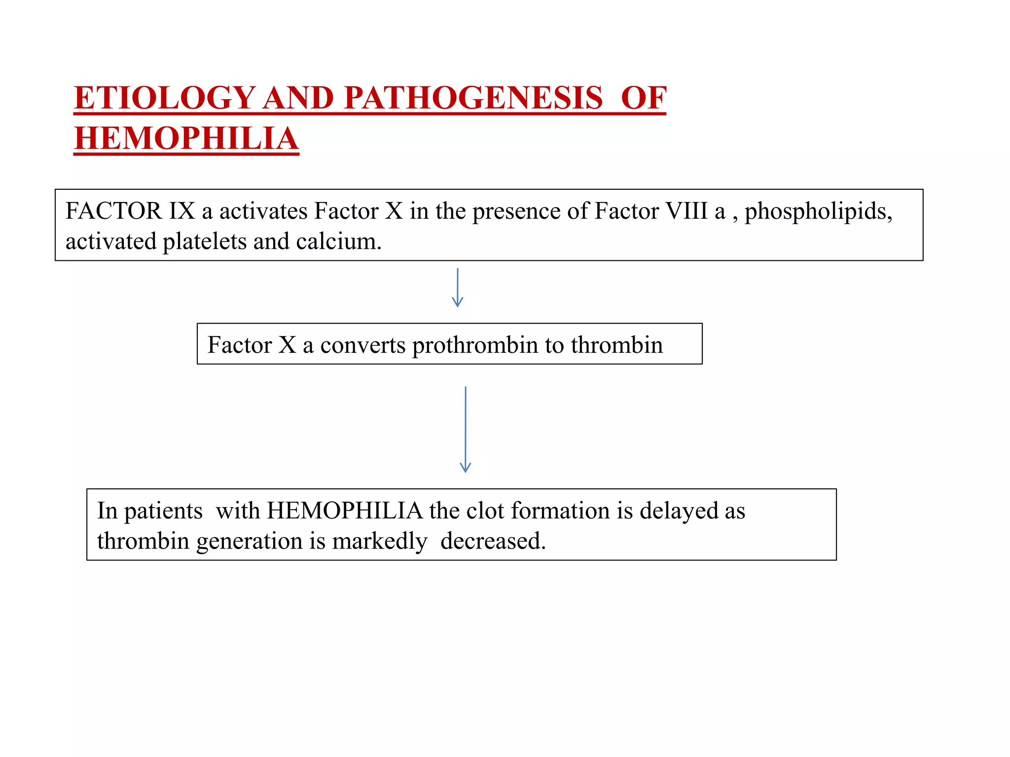
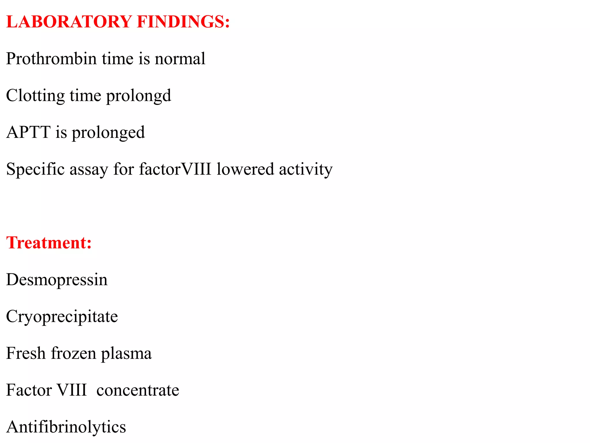
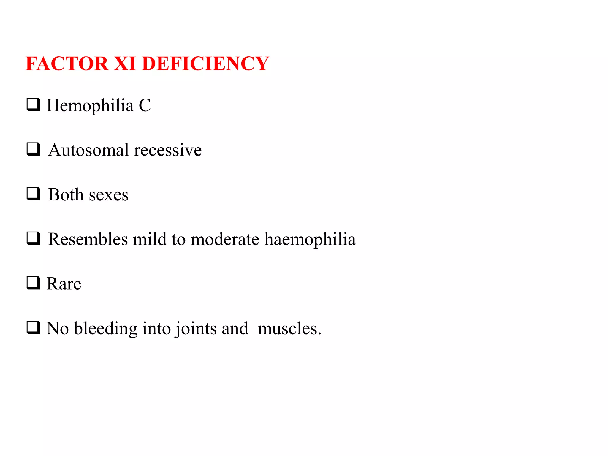
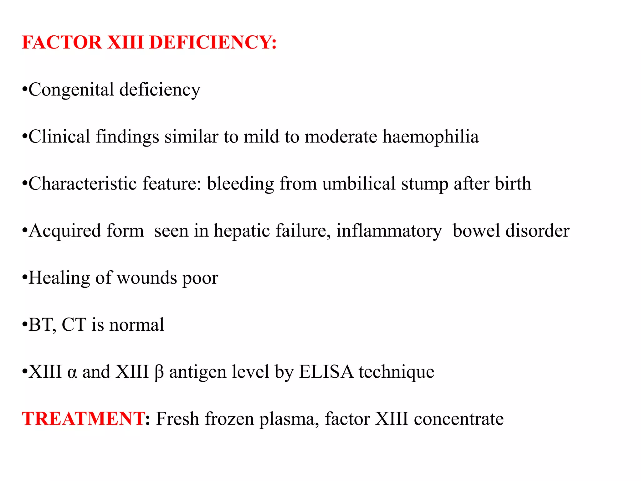
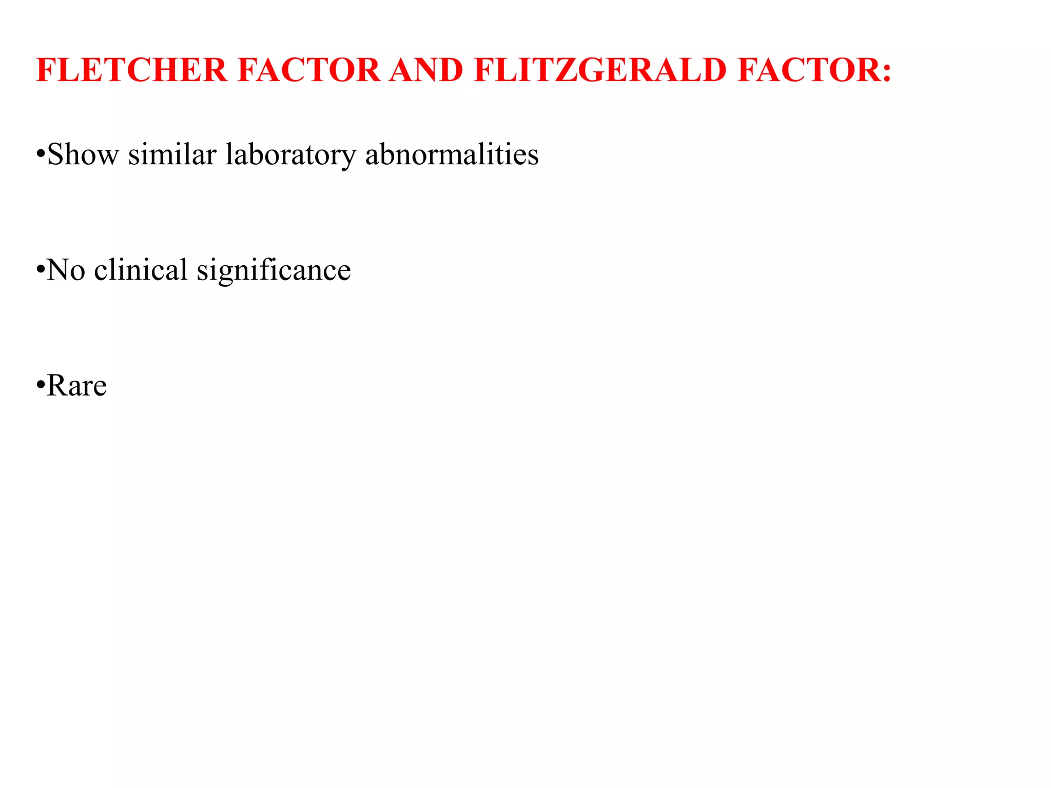
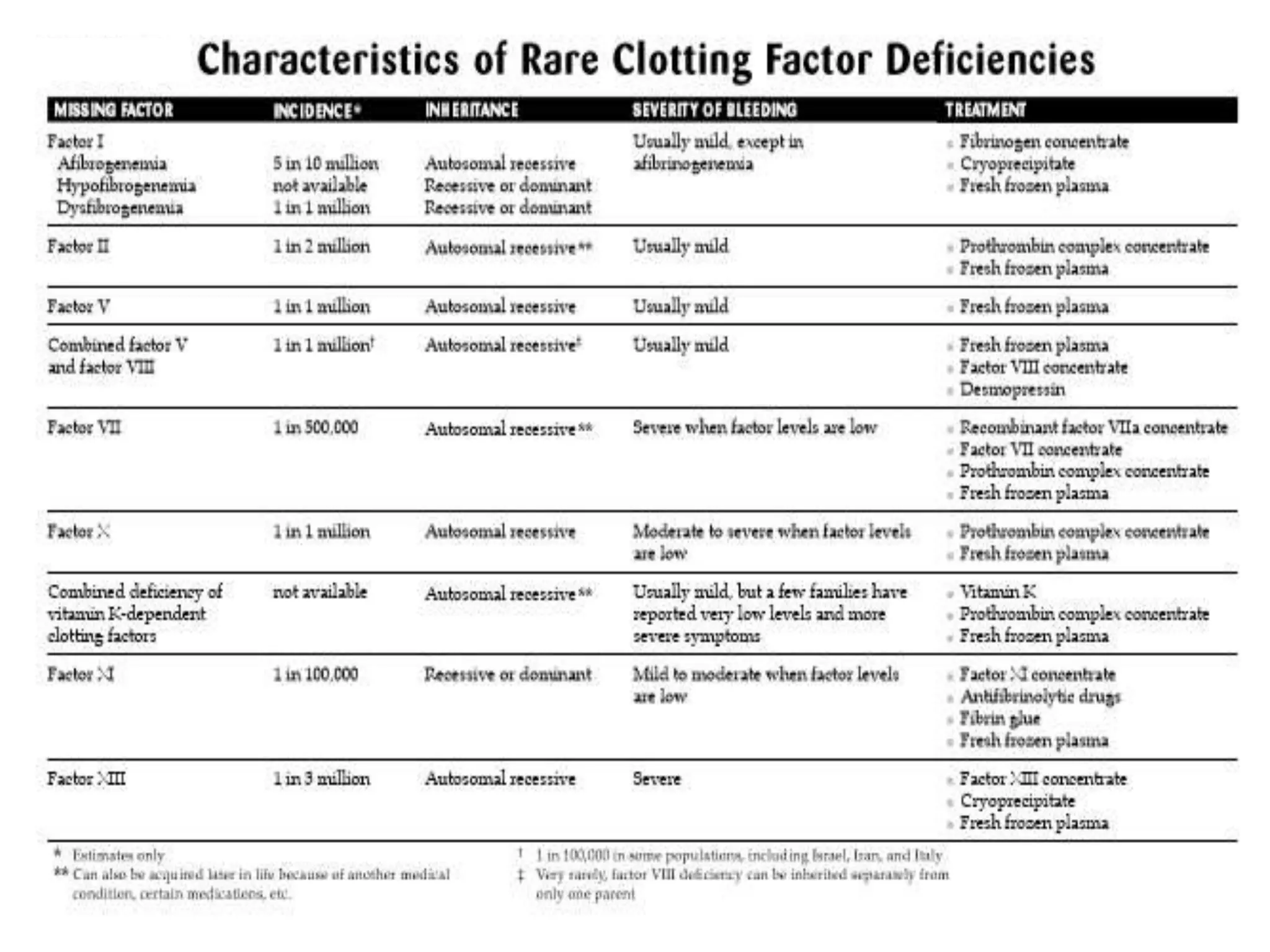
SlideShare a Scribd company logo
DISEASES INVOLVING BLOOD FACTORS
PRIYANKA SHETTY
CONTENTS
• INTRODUCTION
• BLOOD COAGULATION
• HISTORY
• ENZYME CASCADE
• CLOTTING FACTORS
• INHIBITORS
• CLASSIFICATION OF CLOTTING FACTORS
• DIAGNOSTIC TESTS
• VARIOUS CLOTTING FACTOR DISORDERS
• DENTAL CONSIDERATIONS
• RECENT ADVANCES
• REFERENCES
INTRODUCTION
Hemostasis is the process of forming clots in the walls of
damaged blood vessels and preventing blood loss while
maintaining blood in a fluid state within the vascular system.
MECHANISMS
 Vasoconstriction
 Platelet plug formation
 Blood coagulation or clotting
 Clot retraction
BLOOD COAGULATION /CLOTTING
Coagulation or clotting is the process in which blood looses its fluidity and
becomes a jelly like mass in a few minutes after it is shed.
HISTORY:
1720 JEAN LOUIS PETIT : Hemostasis was caused by blood clots.
1860 RUDOLF VIRCHOW described blot clots and their tendency to embolise.
1905 PAUL MORAWITZ assembled 4 coagulation factors .
1940 PAUL OWREN :Recognized the other factors.
FACTORS INVOLVED IN BLOOD COAGULATION
Coagulation of blood occurs through a series of reactions due to activation
of a group of substances. The substances necessary for clotting are called
CLOTTING FACTORS. They are:
Factor VI Presence has not been proved
Factor VII Stable factor
Factor VIII Antihemophilic factor
Factor IX Christmas factor
Factor X Stuart Prower factor
Factor XI Plasma thromboplastin
antecedent
Factor XII Hegman factor
Factor XIII Fibrin stabilisng factor
Factor I Fibrinogen
Factor II Prothrombin
Factor III Thromboplastin
(tissue factor)
Factor IV Calcium
Factor V Labile factor
(Proaccelerin or
accelerator globulin)
FACTORS INVOLVED IN BLOOD COAGULATION
 Clotting factors are proteins in the form of enzymes.
Proenzymes enzymes clot formation.
 Reactions involved in the conversion of proenzymes to active enzymes is
explained by ENZYME CASCADE THEORY .
3 STAGES:
 Formation of prothrombin activator
 Conversion of prothrombin into thrombin
 Conversion of fibrinogen into fibrin.
FEATURES INTRINSIC
MECHANISM
EXTRINSIC
MECHANISM
SOURCE OF
THROMBOPLASTIN
Damaged platelets,
Intrinsic source
Damaged tissue,
Extrinsic source
ROLE OF FACTOR
VII
Not required Required
NUMBER OF
REACTIONS
More Less
TIME Takes few minutes Few seconds
STARTS FROM
ACTIVATION OF
Factor XII Factor VII
COMPARISON BETWEEN INTRINSIC AND EXTRINSIC
MECHANISM OF BLOOD CLOTTING
Clot is a mesh of thin fibrils entangling the blood cells. These fibrils
consist of fibrin. The fibrin is formed from fibrinogen
FACTOR I /FIBRINOGEN
• 3 polypeptide chains - alpha, beta and gamma.
Fibrinogen Fibrin
Prothrombin
Fibrin forms a mesh around the wound leading - blood clot.
INHERITED DISORDERS (MUTATIONS IN FIBRINOGEN ) INCLUDE :
 Afibrinogenemia
 Hypofibrinogenemia
 Hyperfibrinogenemia
The gene for factor I is located on the 4th chromosome.
FACTOR II /PROTHROMBIN
Vitamin K-dependent serine protease.
Prothrombin FACTOR Xa Thrombin.
Soluble fibrinogen THROMBIN Insoluble fibrin.
• Activates factors V, VIII, XI and XIII.
• Thrombin along with thrombomodulin present on endothelial cell surfaces form
a complex that converts protein C to activated protein C (APC).
• Patients suffer from dysprothrombinemia or hypoprothrombinemia.
• The gene for thrombin is located on the eleventh chromosome (11p11).
FACTOR III / THROMBOPLASTIN /TISSUE FACTOR:
 Found in the tissue cell.
 Surface glycoprotein.
 This factor enables cells to initiate the blood coagulation cascades, and it
functions as the high-affinity receptor for the coagulation factor VII.
 Acts as a cofactor in the factor VIIa- catalysed activation of factor X to Xa.
 The gene for tissue factor is located on the first chromosome (1p22) .
FACTOR IV /CALCIUM
• Calcium ions are essential in low concentrations for normal blood coagulation
• Binding of coagulation factor complexes to phospholipid
• Acts as a co factor in the coagulation process
• Essential for formation of intrinsic and extrinsic thromboplastin
• Conversion of prothrombin to thrombin
• No evidence that coagulation disorder results from pathological reduction in
ionised calcium
FACTOR V / PROACCELERIN OR LABILE FACTOR
 Enzymatically inactive cofactor to the serine protease FXa
Ca + phosholipid activation of prothrombin to thrombin.
 Factor V mutation
 Factor V deficiency ( Parahemophilia)
 Myocardial Infarction
 Deep Vein Thrombosis.
 first chromosome (1q23).
FACTOR VII (STABLE OR PROCONVERTIN)
 Vitamin K-dependent Serine Protease.
 Activates factors IX and X simultaneously with tissue factor in the extrinsic
pathway.
 Deficiency is rare (congenital Proconvertin deficiency) and inherits recessively.
 The gene for factor VII is located on the thirteenth chromosome (13q34).
FACTOR VIII ANTI-HEMOPHILIC FACTOR (AHF).
 It is a cofactor in the activation of factor X to FXa, which is catalyzed
by factor IXa in the presence of calcium and phospholipids.
 Mutations in the factor VIII gene results in HEMOPHILIA A.
 CLASSICAL HEMOPHILIA, an X-linked recessive coagulation disorder.
 The gene for factor VIII is located on the long arm of X chromosome
(Xq28).
FACTOR IX ( CHRISTMAS FACTOR)
 It is a proenzyme serine protease, which in the presence of calcium activates
factor X.
 Deficiency cause Hemophilia B or Christmas disease.
 High antigen or activity levels of factor IX is associated with an increased risk of
thromboembolism.
 X chromosome (Xq27).
FACTOR X ( STUART-PROWER FACTOR)
 Both intrinsic and extrinsic pathway of blood coagulation.
 Factor X is activated to FXa by factors IX and VII.
 It is the first member of the common pathway of blood coagulation. FXa cleaves
prothrombin to thrombin.
 Deficiency :Bleeding diathesis and hemorrhages.
 The gene for factor X is located on the thirteenth chromosome (13q32).
FACTOR XI / PLASMA THROMBOPLASTIN ANTECEDENT
 It is a serine protease zymogen which is activated to factor XIa by factor XIIa.
 Hemophilia C.
 Individuals with severe deficiency do not show excessive bleeding conditions and
hemorrhage normally occurs after trauma or surgery.
 Gene in chromosone 4
FACTOR XII /HAGEMAN FACTOR .
 Zymogen form of factor XIIa, which activates factor XI and Prekallikrein.
 Deficiency does not cause excessive hemorrhage due to lack of involvement
of factor XIIa in thrombin formation.
 Increases the risk of thrombosis, due to inadequate activation of the
fibrinolytic pathway.
 Long arm of the fifth chromosome (5q33).
FACTOR XIII ( FIBRIN STABILIZING FACTOR)
 Composed of two subunits- Alpha (A) and Beta (B).
 Activated by thrombin into factor XIIIa in presence of calcium.
 Forms ε-(γ-glutamyl)-lysyl bonds between the fibrin chains & stabilizes the blood
clot.
 Reduces the sensitivity of the clot to degradation by proteases.
 Genetic defects in the factor XIII gene leads to life long bleeding diathesis.
 F13A is located on the sixth chromosome (6p24).
 F13B gene is located on the long arm of first chromosome (1q32).
VON WILLEBRAND FACTOR (vWF)
 Binding of platelets to the site of injury by forming a bridge between collagen
matrix and platelet-surface receptor complex.
 Hereditary or acquired defects of vWF lead to von Willebrand disease
 The gene for vWF is located on short arm of the twelfth chromosome.
Kallikrein is a serine protease.
Inactive form called prekallikrein kallikrein by factor XIIa.
High-molecular-weight kininogen (HMWK) is also called as the Williams-
Fitzgerald-Flaujeac factor.
It is enzymatically inactive and functions as a cofactor for the activation of
kallikrein and factor XII
Plasminogen is a glycoprotein which circulates as proenzyme. It is
converted to plasmin by tissue plasminogen activator in the presence of a
fibrin clot.
Dissolve the fibrin of blood clots.
Wound healing and the maintenance of liver homeostasis.
INHIBITORS:
ANTITHROMBIN III/ANTITHROMBIN
Natural inhibitor of the activated serine proteases of the coagulation
system.
Inhibits factors Xa, IXa and thrombin.
PROTEIN C is a serine protease enzyme.
Its function is to inactivate factors Va and VIIIa.
It is activated by thrombin to activated protein C (APC). APC along
with protein S degrades factors Va and VIIIa.
Protein S -Vitamin K-dependent plasma glycoprotein.
Cofactor to protein C, thus enhancing the inactivation of
factors Va and VIIIa .
Protein Z : Degradation of factor Xa.
Heparin cofactor II - Serine protease inhibitor.
Inhibits thrombin and factor X.
CLASSIFICATION OF
CLOTTING DISORDERS
TEST NORMAL RANGE
Bleeding time
Clotting time
<7 min
4 – 9 min
Prothrombin time/
international normalised
ratio
PT: 11-13 s/INR 1.0
Activated partial
thromboplastin time
15 – 30 s
Thrombin time 3 – 6 s
Fibrin degradation products < 10 µg/dL
Fibrinogen assay 200 – 400 mg/dL
Von Willebrands antigen 60 – 150% v WF activity
Coagulation factor assay 60 – 100% F VIII
ACTIVITY
DIAGNOSTIC TESTS
CONGENITAL DISORDERS
FACTOR I DEFICIENCY
Factor I deficiency is an umbrella term for several related disorders known
as congenital fibrinogen defects.
 Afibrinogenemia
 Hypofibrinogenemia
 Dysfibrinogenemia
Hypodysfibrinogenemia is a combined defect that involves both low levels
of fibrinogen and impaired function.
Afibrinogenemia : Autosomal recessive disorder
Hypofibrinogenemia, dysfibrinogenemia and hypodysfibrinogenemia
Recessive or dominant .
Males and females.
SYMPTOMS OF AFIBRINOGENEMIA
• Nosebleeds (epistaxis)
• Easy bruising
• Menorrhagia
• Muscle bleeds
• Cutaneous ecchymoses and hematomas
•Bleeding from the umbilical cord stump at birth
• Bleeding in the mouth, particularly after dental surgery or tooth
extraction
•Gingival bleeding
• Abnormal bleeding during or after injury, surgery
•Problems during pregnancy
HYPOFIBRINOGENEMIA Symptoms are similar to those seen in
afibrinogenemia.
DYSFIBRINOGENEMIA
• Symptoms depend on how the fibrinogen (which is present in normal
quantities) is functioning.
• Bleeding (similar to those seen in afibrinogenemia)
• Signs of thrombosis (abnormal blood clots in blood vessels) instead of
bleeding.
HYPODYSFIBRINOGENEMIA Symptoms are variable and depend on the
amount of fibrinogen that is produced and how it is functioning.
TREATMENT:
 Fibrinogen concentrate
 Cryoprecipitate
 Fresh frozen plasma (FFP)
FACTOR II (PROTHROMBIN) DEFICIENCY/
HYPOPROTHROMBINEMIA
• Autosomal recessive disorder
• Males and females.
• Very rare
• Inherited bleeding disorder
• It can also be acquired later in life as a result of liver disease, vitamin K
deficiency, or certain medications such as Coumadin.
• Acquired factor II deficiency is more common.
SYMPTOMS
• Epistaxis
• Easy bruising
• Menorrhagia
• Hemarthrosis
• Muscle bleeds
• Bleeding in the mouth, particularly after dental surgery or tooth
extraction
• LAB FINDINGS: P rothrombin time is increased.
TREATMENT
 MADE FROM HUMAN PLASMA.
• Prothrombin complex concentrate (PCC)
• Fresh frozen plasma (FFP)
 ANTIFIBRINOLYTIC DRUGS.
FACTOR V DEFICIENCY
 Inherited bleeding disorder
 PARAHEMOPHILIA
 Autosomal recessive disorder.
 Affects both males and females.
 Rare
LAB FINDINGS:
Clotting and Prothrombin time prolonged
SYMPTOMS
 Epistaxis
 Easy bruising
 Menorrhagia
 Bleeding in the mouth
 Bleeding in the gut
 Gingival bleeding
 Cutaneous ecchymoses and hematomas
 Intraocular and CNS hemorrhages.
TREATMENT :Fresh frozen plasma, platelet transfusions
COMBINED FACTOR V AND FACTOR VIII DEFICIENCY
 Combined factor V and factor VIII deficiency is an inherited bleeding
disorder.
 Autosomal recessive disorder.
 Affects both males and females.
 Rare
 Normally the disorder is caused by a single gene defect that affects the
body’s ability to transport factor V and factor VIII outside the cell and into
the bloodstream, and not by a problem with the gene for either factor.
SYMPTOMS
• Skin bleeding
• Menorrhagia
• Bleeding in the mouth
• Abnormal bleeding during or after injury, surgery, or childbirth
TREATMENT:
Desmopressin
Factor VIII concentrate
Fresh frozen plasma
FACTOR VII DEFICIENCY
• Inherited bleeding disorder .
• Produces less factor VII
• Autosomal recessive disorder
• Rare.
• It can also be acquired later in life as a result of liver disease, vitamin K
deficiency, or certain medications such as Coumadin.
SYMPTOMS
• Nosebleeds (epistaxis) .
• Easy bruising .
• Menorrhagia.
• Bleeding in the mouth, particularly after dental surgery or tooth extraction.
• Bleeding in the head (newborns) .
TREATMENT:
• Recombitant factor VII
• Prothrombin complex conc. factor VII
• Factor VII conc
• Fresh frozen plasma
FACTOR VIII DEFICIENCY (Bleeder s disease, disease of
Hapsburg,Disease of Kings)
 Hemophilia A - most common hereditary disease associated
with factor VIII
 Reduction in the amount or activity of factor VIII.
 This protein serves as a cofactor for factor IX in the activation of
factor X in the coagulation cascade
 Hemophilia A is inherited as an X-linked recessive trait
 Occurs in males.
Genetic abnormality includes:
• Deletion of variable size
• Abnormalities of stop codon
• Frame shift defects
• 45% of severe Hemophilia A results from inverse mutation
ETIOLOGY AND PATHOGENESIS OF
HEMOPHILIA
FACTOR IX a activates Factor X in the presence of Factor VIII a , phospholipids,
activated platelets and calcium.
Factor X a converts prothrombin to thrombin
In patients with HEMOPHILIA the clot formation is delayed as
thrombin generation is markedly decreased.
CLASSIFICATION OF HAEMOPHILIA:
CLINICAL FEATURES:
• Bleeding into muscle, degeneration & contracture of the muscle.
• Compression of the peripheral nerve by large haematoma peripheral nerve lesions.
• Haemophilic cysts & pseudotumour is common in pelvic & knee region enlarge &
sometime erode through skin & cause ulceration on the skin.
• Haematuria is common in severe haemphilia. Bilateral obstruction by clots
acuteoliguric renal pain.
• Spontaneous intracranial & intraspinal haemorrhage is common
Retroperitoneal and mesentric bleeding
Gastrointestinal bleeding
Epistaxis
Menorhhagia
Oral Manifestation:
Oral hematoma
Bleeding gums during tooth shedding
Bleeding after extraction
Palatal and tongue purpura
Ecchymosis
TMJ hemarthrosis
Mandibular pseudo tumour
Muscle hematoma (pseudotumor) Hemarthrosis (joint bleeding)
COMPLICATIONS OF HEMOPHILIA
Claw hand deformity of hand
Atrophy of muscles
LABORATORY FINDINGS:
Prothrombin time is normal
Clotting time prolongd
APTT is prolonged
Specific assay for factorVIII lowered activity
Treatment:
Desmopressin
Cryoprecipitate
Fresh frozen plasma
Factor VIII concentrate
Antifibrinolytics
HAEMOPHILIA – B 1X DEFICIENCY
(Christmas Disease/ Plasma thromboplastin component deficiency))
• Christmas disease is a congenital sex-linked disorder due to absence
reduction or functional abnormality of factor IX
• Incidence 1 per 50,000 male
• Clinical features are same as Haemophilia – A
• Factor IX coagulation activities is reduced or absent.
• X linked disorder
• Occurs one fifth as frequently as hemophilia A
• Prolonged APTT level
• Bioassay of Factor IX has lowered activity
FACTOR X DEFICIENCY
 Inherited bleeding disorder
 Autosomal recessive disorder
 Males and females.
 Rare
TREATMENT
Prothrombin complex concentrate containing factor X.
Fresh frozen plasma.
 Antifibrinolytic drugs.
FACTOR XI DEFICIENCY
 Hemophilia C
 Autosomal recessive
 Both sexes
 Resembles mild to moderate haemophilia
 Rare
 No bleeding into joints and muscles.
FACTOR XII DEFICIENCY:
•Absence of clinical haemorrhage
•Prolonged coagulation time
•Prolonged APTT
•Discovered on routine laboratory testing
•No treatment required
FACTOR XIII DEFICIENCY:
•Congenital deficiency
•Clinical findings similar to mild to moderate haemophilia
•Characteristic feature: bleeding from umbilical stump after birth
•Acquired form seen in hepatic failure, inflammatory bowel disorder
•Healing of wounds poor
•BT, CT is normal
•XIII α and XIII β antigen level by ELISA technique
TREATMENT: Fresh frozen plasma, factor XIII concentrate
FLETCHER FACTOR AND FLITZGERALD FACTOR:
•Show similar laboratory abnormalities
•No clinical significance
•Rare
von Willebrand’s Disease (vWD)(Pseudohemophilia, Vascular
purpura)
Composite of two disorders:
1. An inherited disorder of platelet adhesion, which involves a deficiency and
or a qualitative defect in von Willebrand’s tissue factor (vWF)
2. Deficient or low levels of Factor VIII. Hence, this bleeding disorder leads
to “a combined defect in platelet plug formation and fibrin formation.”
•Most common disorder
•Autosomal dominant
•Affects both sexes
Classification of von Willebrand disease
• Type 1 vWD- the most common variant
• autosomal dominant in inheritance
• normal vWF in structure and function but decrease in quantity- range 25-
50% of normal
• Type 2 vWD
• autosomal dominant in inheritance
• vWF is abnormal in structure and/or function
• Type 3 vWD
• autosomal recessive in inheritance
• the most severe form characterized by very low or undetectable level of
vWF
Von-Willebrand’s disease
It is a autosomally transmitted congenital haemorrhagic disorder in which
there is prolongation of bleeding time in addition to reduction of factor VIII
coagulation activity.
Clinical manifestations:
•Asymptomatic
•Epistaxis
•Mucous membrane bleeding
•Gingival bleeding
•Menorrhagia
•Echymosis
LABORATORY FINDINGS:
•Prolonged bleeding count
•Reduced plasma vWF concentration
•Defective platelet aggregation with ristocetin
•Reduced Factor VIII activity
TREATMENT:
•Desmopressin
•Cryoprecipitate
•Fresh frozen plasma
Acquired coagulation disorders may occur due to one of the
following causes.
• Vit-K deficiency
• Liver diseases
• Anticoagulant therapy
• Disseminated intravascular coagulation
• Acute primary fibrinolysis
• Circulating inhibitors of coagulation
VITAMIN K DEFICIENCY:
• VIT K deficiency occurs in 3 disorders:
DISORDERS THAT IMPAIR FAT ABSORPTION
• Obstructive jaundice and biliary fistula
• Coeliac disease , pancreatic disease and related disoders
STERILISATION OF THE BOWEL BY ANTIBIOTIC DRUGS
• Caused due to combined effects of low diet and vit k
• Loss of the normal bowel flora which synthesises the vitamin.
HAEMORRHAGIC DISEASE OF THE NEWBORN
• Occurs during the first few days of life
Causes:
• Reduced stores of Vit K
• Functional immaturity of the liver
• Lack of bacterial synthesis of Vit K
• Ingestion of oral anticoagulants and anticonvulsants by the mother.
LABORATORY FINDINGS:
Decreased plasma levels of Vit K dependent factors.
Prolonged PT
Prolonged APTT
TREATMENT: Parental administration of Vit K
L IVER DISEASES:
LIVER:
•Major site for synthesis and metabolism of clotting factors
•Produces inhibitors
•Helps in clearance of activated factor and fibrinolytic enzymes
FACTORS THAT CONTRIBUTE TO HAEMOSTATIC DEFECT
IN LIVER DISEASES:
• Defective synthesis of coagulation factors
• Thrombocytopenia
• Increased fibrinolytic activity
• Intravascular coagulation
LAB FINDINGS:
•Prolonged PT and APTT
•Mild thrombocytopenia
•Decreased hepatic stores of Vit K
TREATMENT:
•Fresh frozen plasma,
•EACA to be administered
•Transfusion of plasma and platelet conc.
HAEMORRHAGIC DISORDERS DUE TO CIRCULATING
INHIBITOR OF COAGULATION
Circulating natural inhibitors : Ig heavy chain class activity directed
against a coagulation protein
2 MAJOR TYPES OF INHIBITORS
Congenital haemophilia or congenital coagulation disorder
Acquired or in the course of some other disease state
Lupus inhibitor does not normally cause bleeding tendencies but clinical
expression is paradoxically a striking tendency to thrombosis.
Rare in occurence
LAB FINDINGS:
Prolonged PT, APTT
TREATMENT:
Blood transfusion
Doses of concentrate of appropriate factors
Immunosuppressives
DISSEMINATED INTRAVASCULAR COAGULATION:
(DEFRIBRINATION SYNDROME/CONSUMPTION COAGULOPATHY)
A hemorrhagic disorder in which diffuse intravascular cloting causes a
hemostatic defect resulting from utilisation of coagulation factors and platelets in
the clotting process.
ETIOLOGY:
1. Infections
Acute DIC: Bacteria and their toxins, fungi, viruses,
Chronic DIC: Any chronic infection (eg, tuberculosis, abscesses,
osteomyelitis)
2. Noninfectious inflammatory diseases
Inflammatory bowel disease: Crohn's disease and similar disorders
3. Obstetrical complications
Acute DIC: Abruptio placentae, abortions ,amniotic fluid embolism,
hemorrhagic shock
Chronic DIC: Dead fetus syndrome
4. Malignancy
Acute DIC: Acute promyelocytic leukemia, acute myelomonocytic or monocytic
leukemia, disseminated prostatic carcinoma
Chronic DIC: Lung, breast, gastrointestinal malignancy
5. Vascular disease
Acute DIC: Brain infarction or hemorrhage
Chronic DIC: Aortic aneurysm, giant hemangioma
6. Venoms
Acute DIC: Snake, spider (rare)
7. Trauma
Acute DIC: Massive tissue destruction, brain damage
8. Others
Acute DIC: Heparin-induced thrombocytopenia with thrombosis (HITT),
PATHOPHYSIOLOGY OF DISSEMINATED
INTRAVASCULAR COAGULATION
CLINICAL FEATURES:
•Bleeding: localised or generalised
•Organ damage: due to ischaemia in kidney and brain
• Microangiopathic hemolytic anemia and thrombosis
LAB FINDINGS:
•Platelet low
•PT,TT, APTT all prolonged
•Plasma fibrinogen level are reduced
•Fibrin degradation products raised
TREATMENT:
•Elimination of precipitating factor
•Replacement of coagulation factors and platelets
•Inhibition of clotting process: Heparin, EACA , aprotinin
ANTICOAGULANT DRUGS:
Heparin : Inhibits formation of thromboplastin and the action
of thrombin
Vit k antagonists: coumarin and indanedione derivatives:
suppress the synthesis of active Vit K dependent clotting
factors.
These drugs can cause hypersensitivity reaction or an overdose
can cause clotting disorder.
.
POTENTIATING INHIBITING
Salicylates Barbiturates
Sulindac Carbamazepine
Phenylbutazone Chloral hydrate
Indomethacine Griseofulvin
Sulphonamides Sprironolactone
Amiodarone Cimetidine
FACTORS THAT INTERFERE WITH CONTROL
OF ORALANTICOAGULANT THERAPY:
DRUGS
OTHER FACTORS
POTENTIATING INHIBITING
DIARRHOEA HERIDITARY
RESISTANCE
ALCOHOL
HEPATITIS MALIGNANCY
SEPTICAEMIA
RENAL FAILURE
FIBRINOLYTIC DEFECTS:
Fibrinolysis consisting of plasminogen – plasmin and fibrin degradation
products
Essential protective physiologic mechanism to limit blood coagulation in the
body.
Excessive fibrinolysis leads to bleeding
Causes of primary pathologic fibrinolysis leading to haemorrhagic defects:
def of α 2 plasmin inhibitor following trauma and surgery
Impaired clearance of tissue plasminogen activator such as in liver cirrhosis.
LAB FINDINGS:
•Decreased fibrinogen
•Increased FDP
TREATMENT:
•Fresh frozen plasma
•Antifibrinolytics
CONSIDERATIONS FOR DENTAL TREATMENT
The most important objective is the prevention of complications. The following
principles apply in this sense:
1. Identification of the patient based on a thorough clinical history
2. Counseling
3. Consultation with the specialist to determine the type of congenital disorder
4. Replacement therapy in necessary cases. Replacement may comprise
coagulation factors (hemophilia A and B, and vW disease) or vitamin K (lack of
ingestion or poor absorption and liver disease)
5. Avoidance of brusque maneuvers during dental treatment
6. Evaluation of the advisability of hospital admission when complex surgery
is required.
7. Aspirin and its derivatives are to be avoided as pain treatment. In this
sense, paracetamol is a safe alternative.
8. Reabsorbable sutures.
9. Local haemostatic measures.
10. Avoid regional block injections.
RECENT ADVANCES
The quick development and commercialization of recombinant clotting
factors was facilitated by the catastrophic impact of viral contamination of
plasma-derived clotting factor concentrates . Since their transition into the
clinic, the recombinant versions of both factor VIII and IX have proven to
be remarkable facsimiles of their plasma derived counterparts.
( Recombinant clotting factor Steveen W. PipThromb Haemost 2008; 99: 840–
850)
Presumptive diagnosis of factor XIII deficiency was by clot solubility test
in 5M urea or 1% monochloroacetic acid environments. In patients with
abnormal screening clot solubility test, the disease can be confirmed by
more specific tests such as quantitative factor XIII activity assay
andFXIIIAg assay.
M Naderial Current understanding in diagnosis and management of factor
XIII deficiency, Iran J Ped Hematol Oncol. 2013; 3(4): 164–172.
Family studies were performed and molecular analysis, using a Short
Tandem Repeat (STR) marker closely linked to the factor XIII A
subunit gene, allowed antenatal exclusion diagnosis to be
undertaken in a subsequent pregnancy.
C. Killick, C. Barton, S.Aslam and G.Standen
Prenatal diagnosis in factor XIII-A deficiency Arch Dis Child Fetal
Neonatal Ed. 1999 May; 80(3): F238–F239.
REFERENCES
• Shafer’s Text Book Of Oral Pathology 7th Edition
• Neville Oral And Maxillofacial Pathology 3rd Edition
• Guyton’s Text Book Of Medical Physiology 11th Edition
• Review of Medical Physiology: Ganong.21st Edition
• Robbin’s Basic Pathology 7th Edition
• Davidsons Principles And Practice Of Medicine 20th Edition
• Burket’s Oral Medicine Diagnosis And Treatment 10th Edition
• Harrison’s Principle Of Internal Medicine 18th Edition
• De Gruchys Clinical Hematology In Medical Practice 6th Edition
• Practical Haematology: Dacie and Lewis 11th Edition
• Wintrobe’s Clinical Hematology 12th Edition
• James P. Riddel Jr, MS, RN,et AL Theories of Blood Coagulation Journal of
Pediatric Oncology Nursing, Vol 24, No 3 (May-June), 2007: 123-131
• What are rare clotting factor deficiencies? WHF
• Godkars Text Book Of Medical Laboratory Technology 2nd Edition
• Jover-Cervero A, Poveda-Roda R, Bagan JV, Jimenez-Soriano Y.
Dental treatment of patients with coagulation factor alterations: An
update. MedOral Patol Oral Cir Bucal 2007
• Hematology Clinical hemostasis review / July-August 2004
• Dental Management of Patients with Bleeding Disorders. Sandra
D’Amato-Palumbo, RDH, MPS Continuing Education Units.
Clotting factors

More Related Content

What's hot

What's hot (20)

Haemoglobin
Haemoglobin Haemoglobin
Haemoglobin
 
Blood Coagulation and Clotting Mechanism.pptx
Blood Coagulation and Clotting Mechanism.pptxBlood Coagulation and Clotting Mechanism.pptx
Blood Coagulation and Clotting Mechanism.pptx
 
Coagulation factors
Coagulation factorsCoagulation factors
Coagulation factors
 
Hemostasis
HemostasisHemostasis
Hemostasis
 
Bleeding time and clotting time
Bleeding time and clotting timeBleeding time and clotting time
Bleeding time and clotting time
 
Erythropoiesis
ErythropoiesisErythropoiesis
Erythropoiesis
 
Coagulation Disorders
Coagulation DisordersCoagulation Disorders
Coagulation Disorders
 
Hemostasis
HemostasisHemostasis
Hemostasis
 
RBC
RBCRBC
RBC
 
Coagulation disorders
Coagulation disordersCoagulation disorders
Coagulation disorders
 
Glucose Tolerance Test
Glucose Tolerance TestGlucose Tolerance Test
Glucose Tolerance Test
 
Blood grouping and crosss matching
Blood grouping and crosss matchingBlood grouping and crosss matching
Blood grouping and crosss matching
 
Wbc ppt
Wbc pptWbc ppt
Wbc ppt
 
Erythrocyte sedimentation rate
Erythrocyte sedimentation rateErythrocyte sedimentation rate
Erythrocyte sedimentation rate
 
Blood group system ABO, Rh & other system
Blood group system ABO, Rh & other systemBlood group system ABO, Rh & other system
Blood group system ABO, Rh & other system
 
Immunity
ImmunityImmunity
Immunity
 
VDRL Test for Syphilis
VDRL Test for SyphilisVDRL Test for Syphilis
VDRL Test for Syphilis
 
Inflammation
InflammationInflammation
Inflammation
 
Blood groups
Blood groupsBlood groups
Blood groups
 
Abo blood group system
Abo blood group systemAbo blood group system
Abo blood group system
 

Viewers also liked

Coagulation tests
Coagulation testsCoagulation tests
Coagulation tests
temis cola
 
Blood Clotting Mechanism
Blood Clotting MechanismBlood Clotting Mechanism
Blood Clotting Mechanism
Mujahid Hussain
 
Inflamation ppt
Inflamation pptInflamation ppt
Inflamation ppt
Srota Dawn
 
Bone marrow procedure’!!!2
Bone marrow procedure’!!!2Bone marrow procedure’!!!2
Bone marrow procedure’!!!2
Ayesha Majeed
 
Laboratory investigations in pancytopenia
Laboratory investigations in pancytopeniaLaboratory investigations in pancytopenia
Laboratory investigations in pancytopenia
Veena Raja
 
Coagulation profiles (pt,ptt,at, fib
Coagulation profiles (pt,ptt,at, fibCoagulation profiles (pt,ptt,at, fib
Coagulation profiles (pt,ptt,at, fib
AKHTAR HUSSAIN
 

Viewers also liked (20)

Clotting factors
Clotting factorsClotting factors
Clotting factors
 
Blood coagulation
Blood coagulationBlood coagulation
Blood coagulation
 
Coagulation tests
Coagulation testsCoagulation tests
Coagulation tests
 
Blood clotting
Blood clottingBlood clotting
Blood clotting
 
Blood Clotting Mechanism
Blood Clotting MechanismBlood Clotting Mechanism
Blood Clotting Mechanism
 
Inflamation ppt
Inflamation pptInflamation ppt
Inflamation ppt
 
Hemophilia b
Hemophilia bHemophilia b
Hemophilia b
 
Lesson 3 5 Laboratory Aptt
Lesson 3 5 Laboratory ApttLesson 3 5 Laboratory Aptt
Lesson 3 5 Laboratory Aptt
 
Mysteries of Vitamin D
Mysteries of Vitamin DMysteries of Vitamin D
Mysteries of Vitamin D
 
ECP patient preparation
ECP patient preparationECP patient preparation
ECP patient preparation
 
Inflamation
Inflamation Inflamation
Inflamation
 
Bone marrow procedure’!!!2
Bone marrow procedure’!!!2Bone marrow procedure’!!!2
Bone marrow procedure’!!!2
 
Cascade theory
Cascade theoryCascade theory
Cascade theory
 
Inflamation
InflamationInflamation
Inflamation
 
Laboratory investigations in pancytopenia
Laboratory investigations in pancytopeniaLaboratory investigations in pancytopenia
Laboratory investigations in pancytopenia
 
Coagulation profiles (pt,ptt,at, fib
Coagulation profiles (pt,ptt,at, fibCoagulation profiles (pt,ptt,at, fib
Coagulation profiles (pt,ptt,at, fib
 
Bone marrow aspiration
Bone marrow aspirationBone marrow aspiration
Bone marrow aspiration
 
Blood clotting
Blood clottingBlood clotting
Blood clotting
 
Coagulation
CoagulationCoagulation
Coagulation
 
Zeta potential
Zeta potentialZeta potential
Zeta potential
 

Similar to Clotting factors

clottingfactors-160317182528-converted.pptx
clottingfactors-160317182528-converted.pptxclottingfactors-160317182528-converted.pptx
clottingfactors-160317182528-converted.pptx
Sakshi617058
 
Pathophysiology hematology usmle review notes
Pathophysiology hematology usmle review notesPathophysiology hematology usmle review notes
Pathophysiology hematology usmle review notes
sarosem
 
Pt Aptt 1218181287688715 8
Pt Aptt 1218181287688715 8Pt Aptt 1218181287688715 8
Pt Aptt 1218181287688715 8
rudy184
 

Similar to Clotting factors (20)

clottingfactors-160317182528-converted.pptx
clottingfactors-160317182528-converted.pptxclottingfactors-160317182528-converted.pptx
clottingfactors-160317182528-converted.pptx
 
Coagulation Mechanism and blood disorders
Coagulation Mechanism and blood disordersCoagulation Mechanism and blood disorders
Coagulation Mechanism and blood disorders
 
Coagulation factors 8 to 13.pptx
Coagulation factors 8 to 13.pptxCoagulation factors 8 to 13.pptx
Coagulation factors 8 to 13.pptx
 
Bleeding disorders
Bleeding disordersBleeding disorders
Bleeding disorders
 
Thrombocytes and Hemostasis
Thrombocytes and HemostasisThrombocytes and Hemostasis
Thrombocytes and Hemostasis
 
CME: Bleeding Disorders - Applied Physiology
CME: Bleeding Disorders - Applied PhysiologyCME: Bleeding Disorders - Applied Physiology
CME: Bleeding Disorders - Applied Physiology
 
Hemostasis and coagulation of blood For M.Sc & Basic Medical Students by Pand...
Hemostasis and coagulation of blood For M.Sc & Basic Medical Students by Pand...Hemostasis and coagulation of blood For M.Sc & Basic Medical Students by Pand...
Hemostasis and coagulation of blood For M.Sc & Basic Medical Students by Pand...
 
07 C.disorders.pptx
07 C.disorders.pptx07 C.disorders.pptx
07 C.disorders.pptx
 
HAEMOSTASIS LEVEL 6.pptx
HAEMOSTASIS LEVEL 6.pptxHAEMOSTASIS LEVEL 6.pptx
HAEMOSTASIS LEVEL 6.pptx
 
Lecture 1 platelet and hemostasis
Lecture 1 platelet and hemostasisLecture 1 platelet and hemostasis
Lecture 1 platelet and hemostasis
 
Hemophilia by Suhasis Mondal
Hemophilia by Suhasis MondalHemophilia by Suhasis Mondal
Hemophilia by Suhasis Mondal
 
Pathophysiology hematology usmle review notes
Pathophysiology hematology usmle review notesPathophysiology hematology usmle review notes
Pathophysiology hematology usmle review notes
 
blood coagulation.pptx
blood coagulation.pptxblood coagulation.pptx
blood coagulation.pptx
 
Pt Aptt 1218181287688715 8
Pt Aptt 1218181287688715 8Pt Aptt 1218181287688715 8
Pt Aptt 1218181287688715 8
 
bruce hap.pptx. m
bruce hap.pptx.                        mbruce hap.pptx.                        m
bruce hap.pptx. m
 
Seminar on hemostatsis
Seminar on hemostatsisSeminar on hemostatsis
Seminar on hemostatsis
 
Bleeding and Thrombotic Disorders
Bleeding and Thrombotic Disorders Bleeding and Thrombotic Disorders
Bleeding and Thrombotic Disorders
 
Cell based model of coagulation
Cell based model of coagulationCell based model of coagulation
Cell based model of coagulation
 
bleedig & clotting disorders
 bleedig & clotting disorders bleedig & clotting disorders
bleedig & clotting disorders
 
HEMOSTASIS.pptx
HEMOSTASIS.pptxHEMOSTASIS.pptx
HEMOSTASIS.pptx
 

Recently uploaded

Recently uploaded (20)

Basic_QTL_Marker-assisted_Selection_Sourabh.ppt
Basic_QTL_Marker-assisted_Selection_Sourabh.pptBasic_QTL_Marker-assisted_Selection_Sourabh.ppt
Basic_QTL_Marker-assisted_Selection_Sourabh.ppt
 
The impact of social media on mental health and well-being has been a topic o...
The impact of social media on mental health and well-being has been a topic o...The impact of social media on mental health and well-being has been a topic o...
The impact of social media on mental health and well-being has been a topic o...
 
slides CapTechTalks Webinar May 2024 Alexander Perry.pptx
slides CapTechTalks Webinar May 2024 Alexander Perry.pptxslides CapTechTalks Webinar May 2024 Alexander Perry.pptx
slides CapTechTalks Webinar May 2024 Alexander Perry.pptx
 
Danh sách HSG Bộ môn cấp trường - Cấp THPT.pdf
Danh sách HSG Bộ môn cấp trường - Cấp THPT.pdfDanh sách HSG Bộ môn cấp trường - Cấp THPT.pdf
Danh sách HSG Bộ môn cấp trường - Cấp THPT.pdf
 
[GDSC YCCE] Build with AI Online Presentation
[GDSC YCCE] Build with AI Online Presentation[GDSC YCCE] Build with AI Online Presentation
[GDSC YCCE] Build with AI Online Presentation
 
The Last Leaf, a short story by O. Henry
The Last Leaf, a short story by O. HenryThe Last Leaf, a short story by O. Henry
The Last Leaf, a short story by O. Henry
 
The Art Pastor's Guide to Sabbath | Steve Thomason
The Art Pastor's Guide to Sabbath | Steve ThomasonThe Art Pastor's Guide to Sabbath | Steve Thomason
The Art Pastor's Guide to Sabbath | Steve Thomason
 
How to the fix Attribute Error in odoo 17
How to the fix Attribute Error in odoo 17How to the fix Attribute Error in odoo 17
How to the fix Attribute Error in odoo 17
 
Benefits and Challenges of Using Open Educational Resources
Benefits and Challenges of Using Open Educational ResourcesBenefits and Challenges of Using Open Educational Resources
Benefits and Challenges of Using Open Educational Resources
 
Basic Civil Engg Notes_Chapter-6_Environment Pollution & Engineering
Basic Civil Engg Notes_Chapter-6_Environment Pollution & EngineeringBasic Civil Engg Notes_Chapter-6_Environment Pollution & Engineering
Basic Civil Engg Notes_Chapter-6_Environment Pollution & Engineering
 
Open Educational Resources Primer PowerPoint
Open Educational Resources Primer PowerPointOpen Educational Resources Primer PowerPoint
Open Educational Resources Primer PowerPoint
 
Basic Civil Engineering Notes of Chapter-6, Topic- Ecosystem, Biodiversity G...
Basic Civil Engineering Notes of Chapter-6,  Topic- Ecosystem, Biodiversity G...Basic Civil Engineering Notes of Chapter-6,  Topic- Ecosystem, Biodiversity G...
Basic Civil Engineering Notes of Chapter-6, Topic- Ecosystem, Biodiversity G...
 
Matatag-Curriculum and the 21st Century Skills Presentation.pptx
Matatag-Curriculum and the 21st Century Skills Presentation.pptxMatatag-Curriculum and the 21st Century Skills Presentation.pptx
Matatag-Curriculum and the 21st Century Skills Presentation.pptx
 
Introduction to Quality Improvement Essentials
Introduction to Quality Improvement EssentialsIntroduction to Quality Improvement Essentials
Introduction to Quality Improvement Essentials
 
Instructions for Submissions thorugh G- Classroom.pptx
Instructions for Submissions thorugh G- Classroom.pptxInstructions for Submissions thorugh G- Classroom.pptx
Instructions for Submissions thorugh G- Classroom.pptx
 
Gyanartha SciBizTech Quiz slideshare.pptx
Gyanartha SciBizTech Quiz slideshare.pptxGyanartha SciBizTech Quiz slideshare.pptx
Gyanartha SciBizTech Quiz slideshare.pptx
 
Application of Matrices in real life. Presentation on application of matrices
Application of Matrices in real life. Presentation on application of matricesApplication of Matrices in real life. Presentation on application of matrices
Application of Matrices in real life. Presentation on application of matrices
 
Students, digital devices and success - Andreas Schleicher - 27 May 2024..pptx
Students, digital devices and success - Andreas Schleicher - 27 May 2024..pptxStudents, digital devices and success - Andreas Schleicher - 27 May 2024..pptx
Students, digital devices and success - Andreas Schleicher - 27 May 2024..pptx
 
NCERT Solutions Power Sharing Class 10 Notes pdf
NCERT Solutions Power Sharing Class 10 Notes pdfNCERT Solutions Power Sharing Class 10 Notes pdf
NCERT Solutions Power Sharing Class 10 Notes pdf
 
aaaaaaaaaaaaaaaaaaaaaaaaaaaaaaaaaaaaaaaaa
aaaaaaaaaaaaaaaaaaaaaaaaaaaaaaaaaaaaaaaaaaaaaaaaaaaaaaaaaaaaaaaaaaaaaaaaaaaaaaaaaa
aaaaaaaaaaaaaaaaaaaaaaaaaaaaaaaaaaaaaaaaa
 

Clotting factors

  • 1. DISEASES INVOLVING BLOOD FACTORS PRIYANKA SHETTY
  • 2. CONTENTS • INTRODUCTION • BLOOD COAGULATION • HISTORY • ENZYME CASCADE • CLOTTING FACTORS • INHIBITORS • CLASSIFICATION OF CLOTTING FACTORS • DIAGNOSTIC TESTS • VARIOUS CLOTTING FACTOR DISORDERS • DENTAL CONSIDERATIONS • RECENT ADVANCES • REFERENCES
  • 3. INTRODUCTION Hemostasis is the process of forming clots in the walls of damaged blood vessels and preventing blood loss while maintaining blood in a fluid state within the vascular system.
  • 4. MECHANISMS  Vasoconstriction  Platelet plug formation  Blood coagulation or clotting  Clot retraction
  • 5. BLOOD COAGULATION /CLOTTING Coagulation or clotting is the process in which blood looses its fluidity and becomes a jelly like mass in a few minutes after it is shed. HISTORY: 1720 JEAN LOUIS PETIT : Hemostasis was caused by blood clots. 1860 RUDOLF VIRCHOW described blot clots and their tendency to embolise. 1905 PAUL MORAWITZ assembled 4 coagulation factors . 1940 PAUL OWREN :Recognized the other factors.
  • 6. FACTORS INVOLVED IN BLOOD COAGULATION Coagulation of blood occurs through a series of reactions due to activation of a group of substances. The substances necessary for clotting are called CLOTTING FACTORS. They are:
  • 7. Factor VI Presence has not been proved Factor VII Stable factor Factor VIII Antihemophilic factor Factor IX Christmas factor Factor X Stuart Prower factor Factor XI Plasma thromboplastin antecedent Factor XII Hegman factor Factor XIII Fibrin stabilisng factor Factor I Fibrinogen Factor II Prothrombin Factor III Thromboplastin (tissue factor) Factor IV Calcium Factor V Labile factor (Proaccelerin or accelerator globulin) FACTORS INVOLVED IN BLOOD COAGULATION
  • 8.  Clotting factors are proteins in the form of enzymes. Proenzymes enzymes clot formation.  Reactions involved in the conversion of proenzymes to active enzymes is explained by ENZYME CASCADE THEORY . 3 STAGES:  Formation of prothrombin activator  Conversion of prothrombin into thrombin  Conversion of fibrinogen into fibrin.
  • 9.
  • 10. FEATURES INTRINSIC MECHANISM EXTRINSIC MECHANISM SOURCE OF THROMBOPLASTIN Damaged platelets, Intrinsic source Damaged tissue, Extrinsic source ROLE OF FACTOR VII Not required Required NUMBER OF REACTIONS More Less TIME Takes few minutes Few seconds STARTS FROM ACTIVATION OF Factor XII Factor VII COMPARISON BETWEEN INTRINSIC AND EXTRINSIC MECHANISM OF BLOOD CLOTTING
  • 11. Clot is a mesh of thin fibrils entangling the blood cells. These fibrils consist of fibrin. The fibrin is formed from fibrinogen
  • 12. FACTOR I /FIBRINOGEN • 3 polypeptide chains - alpha, beta and gamma. Fibrinogen Fibrin Prothrombin Fibrin forms a mesh around the wound leading - blood clot. INHERITED DISORDERS (MUTATIONS IN FIBRINOGEN ) INCLUDE :  Afibrinogenemia  Hypofibrinogenemia  Hyperfibrinogenemia The gene for factor I is located on the 4th chromosome.
  • 13. FACTOR II /PROTHROMBIN Vitamin K-dependent serine protease. Prothrombin FACTOR Xa Thrombin. Soluble fibrinogen THROMBIN Insoluble fibrin. • Activates factors V, VIII, XI and XIII. • Thrombin along with thrombomodulin present on endothelial cell surfaces form a complex that converts protein C to activated protein C (APC). • Patients suffer from dysprothrombinemia or hypoprothrombinemia. • The gene for thrombin is located on the eleventh chromosome (11p11).
  • 14. FACTOR III / THROMBOPLASTIN /TISSUE FACTOR:  Found in the tissue cell.  Surface glycoprotein.  This factor enables cells to initiate the blood coagulation cascades, and it functions as the high-affinity receptor for the coagulation factor VII.  Acts as a cofactor in the factor VIIa- catalysed activation of factor X to Xa.  The gene for tissue factor is located on the first chromosome (1p22) .
  • 15. FACTOR IV /CALCIUM • Calcium ions are essential in low concentrations for normal blood coagulation • Binding of coagulation factor complexes to phospholipid • Acts as a co factor in the coagulation process • Essential for formation of intrinsic and extrinsic thromboplastin • Conversion of prothrombin to thrombin • No evidence that coagulation disorder results from pathological reduction in ionised calcium
  • 16. FACTOR V / PROACCELERIN OR LABILE FACTOR  Enzymatically inactive cofactor to the serine protease FXa Ca + phosholipid activation of prothrombin to thrombin.  Factor V mutation  Factor V deficiency ( Parahemophilia)  Myocardial Infarction  Deep Vein Thrombosis.  first chromosome (1q23).
  • 17. FACTOR VII (STABLE OR PROCONVERTIN)  Vitamin K-dependent Serine Protease.  Activates factors IX and X simultaneously with tissue factor in the extrinsic pathway.  Deficiency is rare (congenital Proconvertin deficiency) and inherits recessively.  The gene for factor VII is located on the thirteenth chromosome (13q34).
  • 18. FACTOR VIII ANTI-HEMOPHILIC FACTOR (AHF).  It is a cofactor in the activation of factor X to FXa, which is catalyzed by factor IXa in the presence of calcium and phospholipids.  Mutations in the factor VIII gene results in HEMOPHILIA A.  CLASSICAL HEMOPHILIA, an X-linked recessive coagulation disorder.  The gene for factor VIII is located on the long arm of X chromosome (Xq28).
  • 19. FACTOR IX ( CHRISTMAS FACTOR)  It is a proenzyme serine protease, which in the presence of calcium activates factor X.  Deficiency cause Hemophilia B or Christmas disease.  High antigen or activity levels of factor IX is associated with an increased risk of thromboembolism.  X chromosome (Xq27).
  • 20. FACTOR X ( STUART-PROWER FACTOR)  Both intrinsic and extrinsic pathway of blood coagulation.  Factor X is activated to FXa by factors IX and VII.  It is the first member of the common pathway of blood coagulation. FXa cleaves prothrombin to thrombin.  Deficiency :Bleeding diathesis and hemorrhages.  The gene for factor X is located on the thirteenth chromosome (13q32).
  • 21. FACTOR XI / PLASMA THROMBOPLASTIN ANTECEDENT  It is a serine protease zymogen which is activated to factor XIa by factor XIIa.  Hemophilia C.  Individuals with severe deficiency do not show excessive bleeding conditions and hemorrhage normally occurs after trauma or surgery.  Gene in chromosone 4
  • 22. FACTOR XII /HAGEMAN FACTOR .  Zymogen form of factor XIIa, which activates factor XI and Prekallikrein.  Deficiency does not cause excessive hemorrhage due to lack of involvement of factor XIIa in thrombin formation.  Increases the risk of thrombosis, due to inadequate activation of the fibrinolytic pathway.  Long arm of the fifth chromosome (5q33).
  • 23. FACTOR XIII ( FIBRIN STABILIZING FACTOR)  Composed of two subunits- Alpha (A) and Beta (B).  Activated by thrombin into factor XIIIa in presence of calcium.  Forms ε-(γ-glutamyl)-lysyl bonds between the fibrin chains & stabilizes the blood clot.  Reduces the sensitivity of the clot to degradation by proteases.  Genetic defects in the factor XIII gene leads to life long bleeding diathesis.  F13A is located on the sixth chromosome (6p24).  F13B gene is located on the long arm of first chromosome (1q32).
  • 24. VON WILLEBRAND FACTOR (vWF)  Binding of platelets to the site of injury by forming a bridge between collagen matrix and platelet-surface receptor complex.  Hereditary or acquired defects of vWF lead to von Willebrand disease  The gene for vWF is located on short arm of the twelfth chromosome.
  • 25. Kallikrein is a serine protease. Inactive form called prekallikrein kallikrein by factor XIIa. High-molecular-weight kininogen (HMWK) is also called as the Williams- Fitzgerald-Flaujeac factor. It is enzymatically inactive and functions as a cofactor for the activation of kallikrein and factor XII
  • 26. Plasminogen is a glycoprotein which circulates as proenzyme. It is converted to plasmin by tissue plasminogen activator in the presence of a fibrin clot. Dissolve the fibrin of blood clots. Wound healing and the maintenance of liver homeostasis.
  • 27. INHIBITORS: ANTITHROMBIN III/ANTITHROMBIN Natural inhibitor of the activated serine proteases of the coagulation system. Inhibits factors Xa, IXa and thrombin. PROTEIN C is a serine protease enzyme. Its function is to inactivate factors Va and VIIIa. It is activated by thrombin to activated protein C (APC). APC along with protein S degrades factors Va and VIIIa.
  • 28. Protein S -Vitamin K-dependent plasma glycoprotein. Cofactor to protein C, thus enhancing the inactivation of factors Va and VIIIa . Protein Z : Degradation of factor Xa. Heparin cofactor II - Serine protease inhibitor. Inhibits thrombin and factor X.
  • 29.
  • 31.
  • 32. TEST NORMAL RANGE Bleeding time Clotting time <7 min 4 – 9 min Prothrombin time/ international normalised ratio PT: 11-13 s/INR 1.0 Activated partial thromboplastin time 15 – 30 s Thrombin time 3 – 6 s Fibrin degradation products < 10 µg/dL Fibrinogen assay 200 – 400 mg/dL Von Willebrands antigen 60 – 150% v WF activity Coagulation factor assay 60 – 100% F VIII ACTIVITY DIAGNOSTIC TESTS
  • 33.
  • 35. FACTOR I DEFICIENCY Factor I deficiency is an umbrella term for several related disorders known as congenital fibrinogen defects.  Afibrinogenemia  Hypofibrinogenemia  Dysfibrinogenemia
  • 36. Hypodysfibrinogenemia is a combined defect that involves both low levels of fibrinogen and impaired function. Afibrinogenemia : Autosomal recessive disorder Hypofibrinogenemia, dysfibrinogenemia and hypodysfibrinogenemia Recessive or dominant . Males and females.
  • 37. SYMPTOMS OF AFIBRINOGENEMIA • Nosebleeds (epistaxis) • Easy bruising • Menorrhagia • Muscle bleeds • Cutaneous ecchymoses and hematomas
  • 38. •Bleeding from the umbilical cord stump at birth • Bleeding in the mouth, particularly after dental surgery or tooth extraction •Gingival bleeding • Abnormal bleeding during or after injury, surgery •Problems during pregnancy
  • 39. HYPOFIBRINOGENEMIA Symptoms are similar to those seen in afibrinogenemia. DYSFIBRINOGENEMIA • Symptoms depend on how the fibrinogen (which is present in normal quantities) is functioning. • Bleeding (similar to those seen in afibrinogenemia) • Signs of thrombosis (abnormal blood clots in blood vessels) instead of bleeding. HYPODYSFIBRINOGENEMIA Symptoms are variable and depend on the amount of fibrinogen that is produced and how it is functioning.
  • 40. TREATMENT:  Fibrinogen concentrate  Cryoprecipitate  Fresh frozen plasma (FFP)
  • 41. FACTOR II (PROTHROMBIN) DEFICIENCY/ HYPOPROTHROMBINEMIA • Autosomal recessive disorder • Males and females. • Very rare • Inherited bleeding disorder • It can also be acquired later in life as a result of liver disease, vitamin K deficiency, or certain medications such as Coumadin. • Acquired factor II deficiency is more common.
  • 42. SYMPTOMS • Epistaxis • Easy bruising • Menorrhagia • Hemarthrosis • Muscle bleeds • Bleeding in the mouth, particularly after dental surgery or tooth extraction • LAB FINDINGS: P rothrombin time is increased.
  • 43. TREATMENT  MADE FROM HUMAN PLASMA. • Prothrombin complex concentrate (PCC) • Fresh frozen plasma (FFP)  ANTIFIBRINOLYTIC DRUGS.
  • 44. FACTOR V DEFICIENCY  Inherited bleeding disorder  PARAHEMOPHILIA  Autosomal recessive disorder.  Affects both males and females.  Rare LAB FINDINGS: Clotting and Prothrombin time prolonged
  • 45. SYMPTOMS  Epistaxis  Easy bruising  Menorrhagia  Bleeding in the mouth  Bleeding in the gut  Gingival bleeding  Cutaneous ecchymoses and hematomas  Intraocular and CNS hemorrhages. TREATMENT :Fresh frozen plasma, platelet transfusions
  • 46. COMBINED FACTOR V AND FACTOR VIII DEFICIENCY  Combined factor V and factor VIII deficiency is an inherited bleeding disorder.  Autosomal recessive disorder.  Affects both males and females.  Rare  Normally the disorder is caused by a single gene defect that affects the body’s ability to transport factor V and factor VIII outside the cell and into the bloodstream, and not by a problem with the gene for either factor.
  • 47. SYMPTOMS • Skin bleeding • Menorrhagia • Bleeding in the mouth • Abnormal bleeding during or after injury, surgery, or childbirth TREATMENT: Desmopressin Factor VIII concentrate Fresh frozen plasma
  • 48. FACTOR VII DEFICIENCY • Inherited bleeding disorder . • Produces less factor VII • Autosomal recessive disorder • Rare. • It can also be acquired later in life as a result of liver disease, vitamin K deficiency, or certain medications such as Coumadin.
  • 49. SYMPTOMS • Nosebleeds (epistaxis) . • Easy bruising . • Menorrhagia. • Bleeding in the mouth, particularly after dental surgery or tooth extraction. • Bleeding in the head (newborns) . TREATMENT: • Recombitant factor VII • Prothrombin complex conc. factor VII • Factor VII conc • Fresh frozen plasma
  • 50. FACTOR VIII DEFICIENCY (Bleeder s disease, disease of Hapsburg,Disease of Kings)  Hemophilia A - most common hereditary disease associated with factor VIII  Reduction in the amount or activity of factor VIII.  This protein serves as a cofactor for factor IX in the activation of factor X in the coagulation cascade  Hemophilia A is inherited as an X-linked recessive trait  Occurs in males.
  • 51. Genetic abnormality includes: • Deletion of variable size • Abnormalities of stop codon • Frame shift defects • 45% of severe Hemophilia A results from inverse mutation
  • 52. ETIOLOGY AND PATHOGENESIS OF HEMOPHILIA FACTOR IX a activates Factor X in the presence of Factor VIII a , phospholipids, activated platelets and calcium. Factor X a converts prothrombin to thrombin In patients with HEMOPHILIA the clot formation is delayed as thrombin generation is markedly decreased.
  • 54.
  • 55. CLINICAL FEATURES: • Bleeding into muscle, degeneration & contracture of the muscle. • Compression of the peripheral nerve by large haematoma peripheral nerve lesions. • Haemophilic cysts & pseudotumour is common in pelvic & knee region enlarge & sometime erode through skin & cause ulceration on the skin. • Haematuria is common in severe haemphilia. Bilateral obstruction by clots acuteoliguric renal pain. • Spontaneous intracranial & intraspinal haemorrhage is common
  • 56. Retroperitoneal and mesentric bleeding Gastrointestinal bleeding Epistaxis Menorhhagia Oral Manifestation: Oral hematoma Bleeding gums during tooth shedding Bleeding after extraction Palatal and tongue purpura Ecchymosis TMJ hemarthrosis Mandibular pseudo tumour
  • 57. Muscle hematoma (pseudotumor) Hemarthrosis (joint bleeding) COMPLICATIONS OF HEMOPHILIA
  • 58. Claw hand deformity of hand Atrophy of muscles
  • 59. LABORATORY FINDINGS: Prothrombin time is normal Clotting time prolongd APTT is prolonged Specific assay for factorVIII lowered activity Treatment: Desmopressin Cryoprecipitate Fresh frozen plasma Factor VIII concentrate Antifibrinolytics
  • 60. HAEMOPHILIA – B 1X DEFICIENCY (Christmas Disease/ Plasma thromboplastin component deficiency)) • Christmas disease is a congenital sex-linked disorder due to absence reduction or functional abnormality of factor IX • Incidence 1 per 50,000 male • Clinical features are same as Haemophilia – A • Factor IX coagulation activities is reduced or absent. • X linked disorder • Occurs one fifth as frequently as hemophilia A • Prolonged APTT level • Bioassay of Factor IX has lowered activity
  • 61. FACTOR X DEFICIENCY  Inherited bleeding disorder  Autosomal recessive disorder  Males and females.  Rare TREATMENT Prothrombin complex concentrate containing factor X. Fresh frozen plasma.  Antifibrinolytic drugs.
  • 62. FACTOR XI DEFICIENCY  Hemophilia C  Autosomal recessive  Both sexes  Resembles mild to moderate haemophilia  Rare  No bleeding into joints and muscles.
  • 63. FACTOR XII DEFICIENCY: •Absence of clinical haemorrhage •Prolonged coagulation time •Prolonged APTT •Discovered on routine laboratory testing •No treatment required
  • 64. FACTOR XIII DEFICIENCY: •Congenital deficiency •Clinical findings similar to mild to moderate haemophilia •Characteristic feature: bleeding from umbilical stump after birth •Acquired form seen in hepatic failure, inflammatory bowel disorder •Healing of wounds poor •BT, CT is normal •XIII α and XIII β antigen level by ELISA technique TREATMENT: Fresh frozen plasma, factor XIII concentrate
  • 65. FLETCHER FACTOR AND FLITZGERALD FACTOR: •Show similar laboratory abnormalities •No clinical significance •Rare
  • 66. von Willebrand’s Disease (vWD)(Pseudohemophilia, Vascular purpura) Composite of two disorders: 1. An inherited disorder of platelet adhesion, which involves a deficiency and or a qualitative defect in von Willebrand’s tissue factor (vWF) 2. Deficient or low levels of Factor VIII. Hence, this bleeding disorder leads to “a combined defect in platelet plug formation and fibrin formation.” •Most common disorder •Autosomal dominant •Affects both sexes
  • 67. Classification of von Willebrand disease • Type 1 vWD- the most common variant • autosomal dominant in inheritance • normal vWF in structure and function but decrease in quantity- range 25- 50% of normal • Type 2 vWD • autosomal dominant in inheritance • vWF is abnormal in structure and/or function • Type 3 vWD • autosomal recessive in inheritance • the most severe form characterized by very low or undetectable level of vWF
  • 68. Von-Willebrand’s disease It is a autosomally transmitted congenital haemorrhagic disorder in which there is prolongation of bleeding time in addition to reduction of factor VIII coagulation activity.
  • 69. Clinical manifestations: •Asymptomatic •Epistaxis •Mucous membrane bleeding •Gingival bleeding •Menorrhagia •Echymosis LABORATORY FINDINGS: •Prolonged bleeding count •Reduced plasma vWF concentration •Defective platelet aggregation with ristocetin •Reduced Factor VIII activity
  • 71.
  • 72.
  • 73.
  • 74. Acquired coagulation disorders may occur due to one of the following causes. • Vit-K deficiency • Liver diseases • Anticoagulant therapy • Disseminated intravascular coagulation • Acute primary fibrinolysis • Circulating inhibitors of coagulation
  • 75. VITAMIN K DEFICIENCY: • VIT K deficiency occurs in 3 disorders: DISORDERS THAT IMPAIR FAT ABSORPTION • Obstructive jaundice and biliary fistula • Coeliac disease , pancreatic disease and related disoders STERILISATION OF THE BOWEL BY ANTIBIOTIC DRUGS • Caused due to combined effects of low diet and vit k • Loss of the normal bowel flora which synthesises the vitamin.
  • 76. HAEMORRHAGIC DISEASE OF THE NEWBORN • Occurs during the first few days of life Causes: • Reduced stores of Vit K • Functional immaturity of the liver • Lack of bacterial synthesis of Vit K • Ingestion of oral anticoagulants and anticonvulsants by the mother.
  • 77. LABORATORY FINDINGS: Decreased plasma levels of Vit K dependent factors. Prolonged PT Prolonged APTT TREATMENT: Parental administration of Vit K
  • 78. L IVER DISEASES: LIVER: •Major site for synthesis and metabolism of clotting factors •Produces inhibitors •Helps in clearance of activated factor and fibrinolytic enzymes FACTORS THAT CONTRIBUTE TO HAEMOSTATIC DEFECT IN LIVER DISEASES: • Defective synthesis of coagulation factors • Thrombocytopenia • Increased fibrinolytic activity • Intravascular coagulation
  • 79. LAB FINDINGS: •Prolonged PT and APTT •Mild thrombocytopenia •Decreased hepatic stores of Vit K TREATMENT: •Fresh frozen plasma, •EACA to be administered •Transfusion of plasma and platelet conc.
  • 80. HAEMORRHAGIC DISORDERS DUE TO CIRCULATING INHIBITOR OF COAGULATION Circulating natural inhibitors : Ig heavy chain class activity directed against a coagulation protein 2 MAJOR TYPES OF INHIBITORS Congenital haemophilia or congenital coagulation disorder Acquired or in the course of some other disease state Lupus inhibitor does not normally cause bleeding tendencies but clinical expression is paradoxically a striking tendency to thrombosis. Rare in occurence
  • 81. LAB FINDINGS: Prolonged PT, APTT TREATMENT: Blood transfusion Doses of concentrate of appropriate factors Immunosuppressives
  • 82. DISSEMINATED INTRAVASCULAR COAGULATION: (DEFRIBRINATION SYNDROME/CONSUMPTION COAGULOPATHY) A hemorrhagic disorder in which diffuse intravascular cloting causes a hemostatic defect resulting from utilisation of coagulation factors and platelets in the clotting process. ETIOLOGY: 1. Infections Acute DIC: Bacteria and their toxins, fungi, viruses, Chronic DIC: Any chronic infection (eg, tuberculosis, abscesses, osteomyelitis) 2. Noninfectious inflammatory diseases Inflammatory bowel disease: Crohn's disease and similar disorders 3. Obstetrical complications Acute DIC: Abruptio placentae, abortions ,amniotic fluid embolism, hemorrhagic shock Chronic DIC: Dead fetus syndrome
  • 83. 4. Malignancy Acute DIC: Acute promyelocytic leukemia, acute myelomonocytic or monocytic leukemia, disseminated prostatic carcinoma Chronic DIC: Lung, breast, gastrointestinal malignancy 5. Vascular disease Acute DIC: Brain infarction or hemorrhage Chronic DIC: Aortic aneurysm, giant hemangioma 6. Venoms Acute DIC: Snake, spider (rare) 7. Trauma Acute DIC: Massive tissue destruction, brain damage 8. Others Acute DIC: Heparin-induced thrombocytopenia with thrombosis (HITT),
  • 85. CLINICAL FEATURES: •Bleeding: localised or generalised •Organ damage: due to ischaemia in kidney and brain • Microangiopathic hemolytic anemia and thrombosis LAB FINDINGS: •Platelet low •PT,TT, APTT all prolonged •Plasma fibrinogen level are reduced •Fibrin degradation products raised TREATMENT: •Elimination of precipitating factor •Replacement of coagulation factors and platelets •Inhibition of clotting process: Heparin, EACA , aprotinin
  • 86. ANTICOAGULANT DRUGS: Heparin : Inhibits formation of thromboplastin and the action of thrombin Vit k antagonists: coumarin and indanedione derivatives: suppress the synthesis of active Vit K dependent clotting factors. These drugs can cause hypersensitivity reaction or an overdose can cause clotting disorder.
  • 87. . POTENTIATING INHIBITING Salicylates Barbiturates Sulindac Carbamazepine Phenylbutazone Chloral hydrate Indomethacine Griseofulvin Sulphonamides Sprironolactone Amiodarone Cimetidine FACTORS THAT INTERFERE WITH CONTROL OF ORALANTICOAGULANT THERAPY: DRUGS
  • 88. OTHER FACTORS POTENTIATING INHIBITING DIARRHOEA HERIDITARY RESISTANCE ALCOHOL HEPATITIS MALIGNANCY SEPTICAEMIA RENAL FAILURE
  • 89. FIBRINOLYTIC DEFECTS: Fibrinolysis consisting of plasminogen – plasmin and fibrin degradation products Essential protective physiologic mechanism to limit blood coagulation in the body. Excessive fibrinolysis leads to bleeding Causes of primary pathologic fibrinolysis leading to haemorrhagic defects: def of α 2 plasmin inhibitor following trauma and surgery Impaired clearance of tissue plasminogen activator such as in liver cirrhosis.
  • 90. LAB FINDINGS: •Decreased fibrinogen •Increased FDP TREATMENT: •Fresh frozen plasma •Antifibrinolytics
  • 91. CONSIDERATIONS FOR DENTAL TREATMENT The most important objective is the prevention of complications. The following principles apply in this sense: 1. Identification of the patient based on a thorough clinical history 2. Counseling 3. Consultation with the specialist to determine the type of congenital disorder 4. Replacement therapy in necessary cases. Replacement may comprise coagulation factors (hemophilia A and B, and vW disease) or vitamin K (lack of ingestion or poor absorption and liver disease) 5. Avoidance of brusque maneuvers during dental treatment
  • 92. 6. Evaluation of the advisability of hospital admission when complex surgery is required. 7. Aspirin and its derivatives are to be avoided as pain treatment. In this sense, paracetamol is a safe alternative. 8. Reabsorbable sutures. 9. Local haemostatic measures. 10. Avoid regional block injections.
  • 93. RECENT ADVANCES The quick development and commercialization of recombinant clotting factors was facilitated by the catastrophic impact of viral contamination of plasma-derived clotting factor concentrates . Since their transition into the clinic, the recombinant versions of both factor VIII and IX have proven to be remarkable facsimiles of their plasma derived counterparts. ( Recombinant clotting factor Steveen W. PipThromb Haemost 2008; 99: 840– 850)
  • 94. Presumptive diagnosis of factor XIII deficiency was by clot solubility test in 5M urea or 1% monochloroacetic acid environments. In patients with abnormal screening clot solubility test, the disease can be confirmed by more specific tests such as quantitative factor XIII activity assay andFXIIIAg assay. M Naderial Current understanding in diagnosis and management of factor XIII deficiency, Iran J Ped Hematol Oncol. 2013; 3(4): 164–172.
  • 95. Family studies were performed and molecular analysis, using a Short Tandem Repeat (STR) marker closely linked to the factor XIII A subunit gene, allowed antenatal exclusion diagnosis to be undertaken in a subsequent pregnancy. C. Killick, C. Barton, S.Aslam and G.Standen Prenatal diagnosis in factor XIII-A deficiency Arch Dis Child Fetal Neonatal Ed. 1999 May; 80(3): F238–F239.
  • 96. REFERENCES • Shafer’s Text Book Of Oral Pathology 7th Edition • Neville Oral And Maxillofacial Pathology 3rd Edition • Guyton’s Text Book Of Medical Physiology 11th Edition • Review of Medical Physiology: Ganong.21st Edition • Robbin’s Basic Pathology 7th Edition • Davidsons Principles And Practice Of Medicine 20th Edition • Burket’s Oral Medicine Diagnosis And Treatment 10th Edition
  • 97. • Harrison’s Principle Of Internal Medicine 18th Edition • De Gruchys Clinical Hematology In Medical Practice 6th Edition • Practical Haematology: Dacie and Lewis 11th Edition • Wintrobe’s Clinical Hematology 12th Edition • James P. Riddel Jr, MS, RN,et AL Theories of Blood Coagulation Journal of Pediatric Oncology Nursing, Vol 24, No 3 (May-June), 2007: 123-131 • What are rare clotting factor deficiencies? WHF
  • 98. • Godkars Text Book Of Medical Laboratory Technology 2nd Edition • Jover-Cervero A, Poveda-Roda R, Bagan JV, Jimenez-Soriano Y. Dental treatment of patients with coagulation factor alterations: An update. MedOral Patol Oral Cir Bucal 2007 • Hematology Clinical hemostasis review / July-August 2004 • Dental Management of Patients with Bleeding Disorders. Sandra D’Amato-Palumbo, RDH, MPS Continuing Education Units.