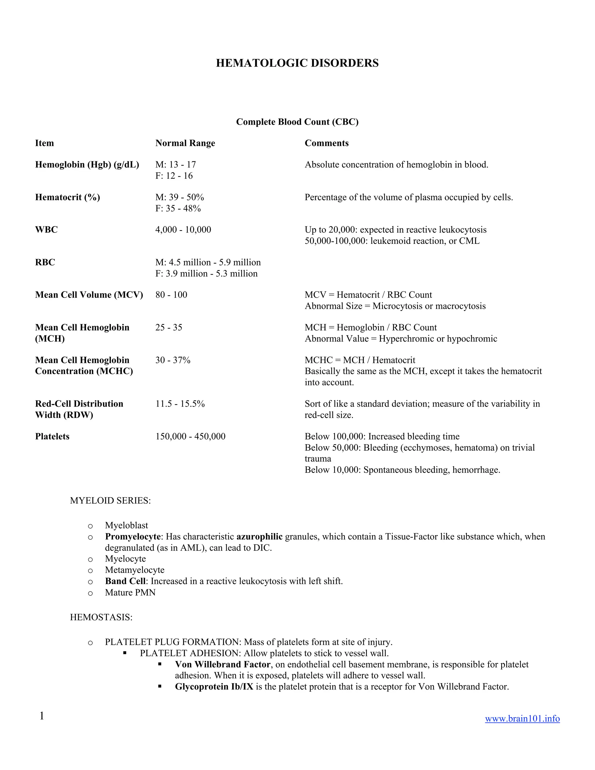
SlideShare a Scribd company logo
1 of 19
Download to read offline
www.brain101.info1
HEMATOLOGIC DISORDERS
Complete Blood Count (CBC)
Item Normal Range Comments
Hemoglobin (Hgb) (g/dL) M: 13 - 17
F: 12 - 16
Absolute concentration of hemoglobin in blood.
Hematocrit (%) M: 39 - 50%
F: 35 - 48%
Percentage of the volume of plasma occupied by cells.
WBC 4,000 - 10,000 Up to 20,000: expected in reactive leukocytosis
50,000-100,000: leukemoid reaction, or CML
RBC M: 4.5 million - 5.9 million
F: 3.9 million - 5.3 million
Mean Cell Volume (MCV) 80 - 100 MCV = Hematocrit / RBC Count
Abnormal Size = Microcytosis or macrocytosis
Mean Cell Hemoglobin
(MCH)
25 - 35 MCH = Hemoglobin / RBC Count
Abnormal Value = Hyperchromic or hypochromic
Mean Cell Hemoglobin
Concentration (MCHC)
30 - 37% MCHC = MCH / Hematocrit
Basically the same as the MCH, except it takes the hematocrit
into account.
Red-Cell Distribution
Width (RDW)
11.5 - 15.5% Sort of like a standard deviation; measure of the variability in
red-cell size.
Platelets 150,000 - 450,000 Below 100,000: Increased bleeding time
Below 50,000: Bleeding (ecchymoses, hematoma) on trivial
trauma
Below 10,000: Spontaneous bleeding, hemorrhage.
MYELOID SERIES:
o Myeloblast
o Promyelocyte: Has characteristic azurophilic granules, which contain a Tissue-Factor like substance which, when
degranulated (as in AML), can lead to DIC.
o Myelocyte
o Metamyelocyte
o Band Cell: Increased in a reactive leukocytosis with left shift.
o Mature PMN
HEMOSTASIS:
o PLATELET PLUG FORMATION: Mass of platelets form at site of injury.
 PLATELET ADHESION: Allow platelets to stick to vessel wall.
 Von Willebrand Factor, on endothelial cell basement membrane, is responsible for platelet
adhesion. When it is exposed, platelets will adhere to vessel wall.
 Glycoprotein Ib/IX is the platelet protein that is a receptor for Von Willebrand Factor.
www.brain101.info2
 PLATELET AGGREGATION: Activate platelets so that they stick to each other.
 Several factors cause the platelets to clump together at the site of injury.
 Collagen: Exposed collagen indicates injury.
 ADP
 Thrombin: Final product of the coagulation pathway causes platelets to stick together.
 Glycoprotein IIb/IIIa: Platelet receptor which binds to Collagen, ADP, thrombin, causing cross-
linking between platelets and thus allowing for aggregation. It also binds to fibrinogen to facilitate
cross-linking.
 Thromboxane-A2 (TXA2) activates these receptors, enabling them to cross-link.
 Fibrin Degradation Products can block this glycoprotein, preventing platelet aggregation.
o COAGULATION
 INTRINSIC PATHWAY: XII, XI, IX, VIII, Kallikrein, Kininogen. Partial Thromboplastin Time
measures its integrity.
 Anionic Surfaces can activate one of three substances in order to start the Intrinsic Pathway: Pre-
kallikrein, Kininogen, and Factor XII
 Factor XII ------> Factor XI ------> Factor IX
 Factor VIII, anionic phospholipid, and Ca+2
are then required to activate Factor X
 EXTRINSIC PATHWAY: Tissue Factor, VII. Prothrombin Time measures its integrity.
 Tissue Factor ------> Factor VII ------> Factor X
 COMMON PATHWAY: X, V, II, I. One way or another, common pathway starts when Factor X is
activated.
 Factor X converts Prothrombin (II) ------> Thrombin
 Factor V, anionic phospholipid, and Ca+2
are required as cofactors.
 Thrombin converts Fibrinogen (I) ------> Fibrin
 Fibrin cross-links platelets and forms cohesive plug.
o FIBRINOLYSIS: Occurs concurrent with the healing process.
 Plasminogen is converted to Plasmin by two factors:
 Tissue Plasminogen Activator (tPA) is released by damaged endothelial cells.
 Urokinase
BLEEDING TESTS:
o BLEEDING TIME:
 PROCEDURE: Simply measure the amount of time it takes the patient to stop bleeding, from have two
1mm incisions on forearm. Use filter paper to dop off the blood every 30 seconds.
 NORMAL: 5-10 minutes.
 ABNORMAL: Long bleeding times are seen with any disorder in forming a platelet plug:
 Diseases affecting the blood vessel wall: vasculitis, amyloidosis, scurvy, hereditary hemorrhagic
telangiectasia, Ehlers-Danlos syndrome
 Diseases causing thrombocytopenia: chemotherapy, ITP, DIC, TTP
 Drugs or diseases affecting platelet function: aspirin, ibuprofen, renal failure
 Von Willebrand Disease.
o PLATELET COUNT:
 NORMAL: 150,000 - 450,000
 Von Willebrand Disease, Aspirin, and Renal Failure all show normal platelet counts, but still have
long bleeding times.
 ABNORMAL:
 Below 100,000: Bleeding time is affected.
 Below 50,000: Bleeding with minor trauma will occur.
 Below 20,000: Spontaneous bleeding will occur; transfusions required.
o ACTIVATED PARTIAL THROMBOPLASTIN TIME (aPTT): Measures the integrity of the Intrinsic and
Common Pathways.
 PROCEDURE: Partial Thromboplastin is added to citrate-anticoagulated plasma. Partial Thromboplastin
consists of:
 Anionic surface (koalin, silica)
 Phospholipid
 Calcium
www.brain101.info3
 NORMAL: 30 second to form a clot in vitro
 ABNORMAL: Hemophilia = defect in intrinsic pathway. 50-100 second to form clot.
 Also abnormal in DIC, some cases of Von Willebrand Disease, and in Heparin therapy.
o PROTHROMBIN TIME (PT): Measures the integrity of the Extrinsic and Common Pathways.
 PROCEDURE: Complete thromboplastin is used, which is all of the reagents above, plus Tissue Factor.
 NORMAL: 12-14 second to form a clot in vitro.
 ABNORMAL: Vitamin-K Deficiency leads to deficiency of factors in the Extrinsic Pathway (Factor VII).
Will show times of 18-20 seconds.
o FIBRIN DEGRADATION PRODUCTS: They are elevated in Disseminated Intravascular Coagulation (DIC),
which shows hyperactive fibrinolysis.
BLEEDING DISORDERS: Congenital and acquired diseases that lead to excessive bleeding or imbalances in hemostasis.
o CONGENITAL DISORDERS:
 VON-WILLEBRAND DISEASE:
 PATHOGENESIS: Autosomal dominant (generally) deficiency of Von Willebrand Factor.
 VWF is normally a carrier protein for Factor VIII, so its deficiency results in shorter half-
life for Factor VIII. This is only symptomatic in severe cases.
 VWF is required for initial platelet-plus formation.
 SUBTYPES:
 Type I: Partial quantitative deficiency
 Type II: Qualitative deficiency, dysfunctional protein.
 Type III: Complete absence, with recessive inheritance. Worst form.
 SYMPTOMS: petechiae, ecchymosis, mucosal hemorrhage, and in some cases hemarthrosis and
hematomas.
 Periorbital Ecchymoses: Racoon Eyes are characteristic finding.
 LABS: These patients will have abnormal bleeding time, but normal platelet count. May also see
prolonged partial thromboplastin time.
 BERNARD SOULIER SYNDROME (GIANT PLATELET SYNDROME): Deficiency of
Glycoprotein Ib/IX, which is the Von Willebrand receptor on platelets.
 GLANZMANN THROMBOASTHENIA: Abnormality in Glycoprotein Complex IIb/IIIa (Platelet
receptor), which impairs clotting. You will see excessive bleeding but will have a normal platelet count.
 HEMOPHILIA-A:
 PATHOGENESIS: X-Linked. Deficiency of Factor-VIII, the cofactor required to convert Factor
X ------> Factor Xa, in the Intrinsic Pathway.
 SYMPTOMS:
 Hemarthrosis: Joint and muscle bleeding with minor trauma, which chronically leads to
osteoarthritis. You also see atrophy of muscle distal to the joint.
 GI bleeds
 Ecchymoses
 LABS: Prolonged Partial Thromboplastin Time (PTT), indicating a defect in the Intrinsic
Pathway.
 TREATMENT: Concentrated Factor-VIII transfusions, or more recently, recombinant Factor-VIII.
 HEMOPHILIA-B: X-Linked. Deficiency of Factor IX, the final intermediate in the Intrinsic Pathway.
 EPIDEMIOLOGY: Rarer than Hemophilia-A
 SYMPTOMS: Identical to Hemophilia-A
 LABS: Prolonged Partial Thromboplastin Time (PTT), indicating a defect in the Intrinsic
Pathway.
 RENDU-OSLER-WEBER SYNDROME (HEREDITARY HEMORRHAGIC TELANGIECTASIA):
Autosomal dominant.
 SYMPTOMS: Recurrent spontaneous hemorrhages from trivial trauma. Epistaxis.
o ACQUIRED DISORDERS:
 VITAMIN-K DEFICIENCY: It will lead to a defect in many coagulation factors of the Extrinsic and
Common Pathways.
 PATHOGENESIS: Vit-K Deficiency is caused by broad-spectrum antibiotics (due to lost
intestinal flora), malnutrition, and fat-malabsorption syndromes.
www.brain101.info4
 Vitamin K is normally involved in forming gamma-carboxyglutamate residues, which are
required to chelate calcium in many calcium-dependent clotting factors.
 SOURCES: 50% from diet, and 50% from intestinal bacterial flora.
 Vitamin-K dependent factors:
 Factor VII: EXTRINSIC pathway factor.
 Factors V, X, II (Prothrombin), and I (Fibrinogen): COMMON pathway factors.
 DISSEMINATED INTRAVASCULAR COAGULATION (DIC):
 PATHOGENESIS: Caused by the pathologic release of pro-coagulant factors into the
bloodstream, which sets off the clotting cascade in the bloodstream.
 Over-activation of the clotting cascade ------> generation of excessive thrombin and
plasmin ------> excessive fibrin degradation products.
 Lots of things can set off the DIC:
 AML, Type M3: The Promyelocytic azurophilic granules contain a tissue-factor
like substance that can cause DIC when released.
 Endotoxic infections, shock, and sepsis. Macrophages can release tissue factor
in circumstances of shock.
 Complications of pregnancy: Retained dead fetus, placental abruption.
 Hemolytic Uremic Syndrome
 LABS: Prolonged PTT, prolonged Prothrombin time, prolonged bleeding time, thrombocytopenia,
and elevated fibrin degradation products.
 TREATMENT: Try to treat the underlying cause. Beyond that, give platelets and plasma (to
replace clotting factors), and heparin (to prevent further intravascular coagulation)
 IDIOPATHIC THROMBOCYTOPENIA PURPURA (ITP): Autoimmune disease against platelets.
 PATHOGENESIS: Anti-platelet auto-antibodies cause the widespread destruction of platelets ---
---> low platelet count.
 LABS: Platelet count is low, but bleeding times are still relatively okay, because the remaining
platelets are hyperfunctional.
 SYMPTOMS: Petechiae, ecchymoses
 SUBTYPES:
 ACUTE ITP: Generally found in children.
 PATHOGENESIS: Usually follows a viral infection.
 SYMPTOMS: Sudden, fulminant onset of symptoms. The disease usually
resolves after 6 months without treatment.
 LABS: Level of thrombocytopenia is severe -- 20,000 platelets or less -- but the
remaining platelets are hyperfunctional.
 CHRONIC ITP: ITP lasting more than 6 months. Occurs in adults, usually females.
 PATHOGENESIS: No preceding antigenic exposure. Associated with HIV
infections, or other diseases, such as SLE, CLL, CML.
 SYMPTOMS: Insidious onset showing mild to moderate thrombocytopenia. No
spontaneous recovery; treatment is usually required.
 TREATMENT: Usually responds to steroids; if that fails, do splenectomy.
 THROMBOTIC THROMBOCYTOPENIC PURPURA (TTP): Simultaneous thrombosis, blood
clotting, and bleeding ------> end-organ infarcts.
 PATHOGENESIS: Patients are thought to release an abnormally large amount of Von
Willebrand Factor (large multimers of it), leading to intravascular activation of clotting cascade.
The VWF molecule has molecular weight of around 10 million rather than 1 million. It can be
idiopathic, or caused by several factors:
 Pregnancy, post-partum state
 Drugs: Cyclosporin, mitomycin
 Infection: HIV, rickettsial
 Collagen Vascular Diseases: Lupus, Sjögren
 Certain Malignancies
 PATHOLOGY:
 Thrombocytopenia, anemia
 Elevated creatinine and BUN, from renal failure.
 Prolonged bleeding times.
 Schistocytes (from microangiopathic hemolysis) found on blood smear.
www.brain101.info5
 SYMPTOMS: The disease is deadly if not treated, and treatment must be undertaken quickly.
Classical pentad of findings:
 Thrombocytopenia: Spontaneous platelet aggregation ------> low platelet count
 Microangiopathic Hemolytic Anemia: RBC's are lysed in small vessels by the presence
of intravascular fibrin clots.
 Fever: It isn't known why, and not all patients will show it.
 Neurologic Symptoms: Arteriolar thromboses in brain; similar to TIA's.
 Renal Failure: Due to thrombosis in renal vasculature. Often a prominent symptom.
 TREATMENT: Plasmapheresis. Machine filters out the clots and the extra large Von Willebrand
Factor. Extra transfusions are required, too.
 HENOCH-SCHONLEIN PURPURA (ALLERGIC PURPURA): Auto-immune attack against vessel-
wall, leading to purpura. Often follows a drug-reaction.
ANEMIAS:
o GENERAL PROPERTIES:
 SYMPTOMS:
 Fatigue
 Pallor resulting form decreased perfusion of skin.
 Light-headedness
 Decreased renal perfusion ------> increased erythropoietin
 Increased Cardiac Output and tachycardia, in compensation. This combined with myocardial
hypoxia can lead to an MI or CHF.
 CLASSIFICATIONS:
 Hypoproliferative: Decreased production of red-cells.
 Reticulocyte Index < 2%
 Hyperproliferative: Increased destruction of red-cells.
 Reticulocyte Index > 2%: Reticulocytes are signs of effective erythropoiesis and red-
blood cell proliferation.
 POLYCHROMASIA: The histological appearance of reticulocytes, which still have
some residual RNA elements left in them. They appear in the standard Wright Stain
blood smear.
o SECONDARY ANEMIAS: Anemias occurring secondary to other, unrelated body systems.
 Renal Failure: Due to lack of erythropoietin, renal failure almost always leads to anemia.
 Anemia of Chronic Disease: Normocytic, normochromic anemia. Very common cause of anemia.
 PATHOGENESIS: Abnormal or excessive utilization of iron, due to chronic infections (Histo,
TB), chronic inflammation (SLE, Rheumatoid Arthritis), or neoplasia.
 SYMPTOMS: Usually only a moderate anemia.
 LABS: Patients will have low serum iron, but they will have adequate iron stores in bone marrow,
which distinguishes it from iron-deficiency anemia.
 Infiltrative Anemia: Anemia resulting when other things infiltrate the bone-marrow, pushing out normal
cellular elements.
 Metastatic Cancer to bone: Lung, breast, and prostate cancers often metastasize to bone.
 Gaucher's Disease
o APLASTIC ANEMIA: Deficiency or complete failure to produce all blood cells.
 PATHOGENESIS: Several things can cause it. They are all external (environmental) causes.
 Ionizing Radiation
 Drugs: Chemotherapy, benzene, chloramphenicol, gold, anti-convulsants, insecticides.
 Viruses: Parvovirus B-19, HCV, HIV-1
 PATHOLOGY: Pancytopenia. The blood-cells that are there retain normal morphology.
o PURE RED CELL APLASIA: Anemia due to isolated depletion of erythroid precursors in the marrow, and may
be acute or chronic.
 PATHOLOGY: Normochromic anemia, normocytic or macrocytic.
 Reticulocytes are decreased or absent because it is hypoproliferative.
www.brain101.info6
 SUBTYPES:
 ACUTE PRCA: Acute pure red cell aplasia often follows a viral illness, notably Parvovirus B19
infection.
 CHRONIC PRCA:
 CHRONIC INHERITED PRCA (DIAMOND-BLACKFAN ANEMIA): Quite
responsive to steroids.
 CHRONIC ACQUIRED PRCA: Immunologic etiology, and may be seen in association
with thymoma.
 TREATMENT: Removal of thymus may result in clinical remission.
o IRON-DEFICIENCY ANEMIA:
 PATHOGENESIS: Several causes.
 Most common cause = chronic or acute blood loss ------> intracellular fluid goes into vascular
space to replace lost fluid ------> relative anemia results.
 Growth, pregnancy, lactation.
 Inadequate dietary intake.
 Increased metabolic requirements, neoplasia.
 PATHOLOGY: Microcytic, hypochromic anemia. Low serum iron.
 Bone-Marrow will show absence of iron.
 Ferritin: Low serum ferritin indicates low body stores of iron. Ferritin is a storage-protein found
in liver, in Kupffer cells.
 However, ferritin is an acute-phase protein, so there are some acute conditions in which
ferritin may be high (inflammation, hepatitis) anyway. Therefore, don't rely on ferritin to
make the diagnosis of iron-deficiency anemia.
 Transferrin: These carrier proteins will be unsaturated and available to bind iron, hence the Total
Iron Binding Capacity (TIBC) is increased with anemia.
 SYMPTOMS: General anemia symptoms. Can see pica, a craving to eat clay.
 Koilonychia: Spoon-shaped nails can be seen.
 Thrombocytosis: May see increased platelets in chronic anemia, due to general Over-activation
(via erythropoietin) of the bone marrow.
o MEGALOBLASTIC ANEMIA: Macrocytic, hyperchromic anemia.
 PATHOGENESIS: The nuclear development of the RBC can't keep up with the cytoplasmic growth,
because of faulty DNA synthesis.
 Folate Deficiency:
 Usually dietary, as in alcoholism
 Increased metabolic demand, as in pregnancy
 Malabsorption (as in Sprue)
 B-12 Deficiency:
 Autoimmune Gastritis ------> Pernicious Anemia
 Fish Tapeworm, Diphyllobothrium Latum
 Malabsorption: Sprue, ileitis, ileal resection.
 PATHOLOGY:
 Morphologic Abnormalities:
 Large RBC's with nuclear-cytoplasmic dyssynchrony
 Tear-Drop Cells
 Hypersegmented Neutrophils: One of the earliest signs of disease. 5 or 6 lobes.
 Ovalocytes: The large RBC's tend to have an oval-shape.
 Howell-Jolly Bodies: Nuclear fragments seen in Megaloblastic anemia.
 LABS: Reticulocyte count is low, because this is a hypoproliferative anemia. Erythropoiesis is
ineffective.
o THALASSEMIAS: Deficient production of hemoglobin.
 EPIDEMIOLOGY: Mediterranean, middle east.
 SYMPTOMS: Hypochromic, microcytic anemia, owing to deficient production of hemoglobin.
 PATHOLOGY:
 Heinz Bodies, precipitated red blood cells, may be found.
 Target Cells, due to increased membrane : cytoplasm ratio, can also be seen.
 alpha-THALASSEMIA: Deficient production of the alpha-globin chain.
www.brain101.info7
 SYMPTOMS: There are four alpha-genes, and severity of disease depends on how many of those
genes are deleted. If all four are deleted, death in utero is inevitable.
 beta-THALASSEMIA: Deficient production of the beta-globin chain.
 SYMPTOMS: Both excess hemolysis and ineffective erythropoiesis occurs, with homozygous
trait.
 Excess alpha-chains precipitate in RBC's, leading to hemolysis.
 HETEROZYGOUS TRAIT: Very mild, with an increased amount of Hemoglobin A2
(alpha2,delta2), because it doesn't contain the beta-chain.
 TREATMENT: Transfusion therapy, plus iron chelators to prevent secondary iron overload from
the therapy.
o MEMBRANE-DEFECTS: Defects in RBC shape
 HEREDITARY SPHEROCYTOSIS:
 PATHOGENESIS: Deficiency in spectrin, plus a variety of other possible problems, leads to
spherical shape of RBC's.
 PATHOLOGY: RBC's can be sequestered in spleen and prematurely removed from circulation.
 Normocytic, hyperchromic (no central pallor -- for unknown reasons) anemia.
 SYMPTOMS: Hemolytic jaundice, hyperproliferative anemia, splenomegaly.
 Pigment gallstones may result from the hemolytic jaundice.
 TREATMENT: Splenectomy is usually curative, since the spleen is responsible for removing the
spherocytes.
 HEREDITARY ELLIPTOCYTOSIS
 ACANTHOCYTOSIS
o GLUCOSE-6-PHOSPHATE DEHYDROGENASE (G6PD) DEFICIENCY:
 PATHOGENESIS: X-Linked Recessive.
 G6PD is an enzyme of the Hexose Monophosphate Shunt pathway. It maintains Glutathione in its
reduced (active) form. Deficiency of this enzyme makes the RBC susceptible to oxidative
damage.
 PATHOLOGY:
 Heinz Bodies are bodies of denatured or precipitated hemoglobin, found in this disease as well as
other diseases.
 SYMPTOMS: Hemolytic anemia brought on by infection or drugs.
 FAVISM: Susceptibility to eating Fava beans ------> potentially lethal hemolytic anemia.
o HEMOGLOBINOPATHIES: Normal levels of hemoglobin, but defects in Hgb structure.
 HEMOGLOBIN-C DISEASE: Second most common hemoglobinopathy.
 PATHOGENESIS: Substitution of Glu ------> Lys at 6th
position of beta-chain.
 SYMPTOMS: Chronic, mild hemolytic anemia.
 PATHOLOGY: See lots of target cells -- formed by excess hemoglobin-accumulation in center of
cell, giving bulls-eye appearance.
 SICKLE CELL DISEASE:
 EPIDEMIOLOGY: Endemic to Sub-saharan Africa, due to heterozygous advantage conferred
against Falciparum Malaria. The advantage is thought to be that infected RBC's preferentially
sickle and are thus taken to the spleen and sequestered, limiting the spread of infection.
 PATHOGENESIS: Point-mutation of Glu ------> Val at 6th
position of beta-globin chain.
 PATHOLOGY: Deoxygenation (low oxygen tension) causes the HbS to polymerize ------> RBC
becomes rigid and non-deformable ------> RBC gets stuck in the microvasculature causing micro-
infarcts.
 The cells can be fine for quite some time, as they go through cycles of deoxygenation and
reoxygenation. After several cycles they may become deformed, and wind up in the
spleen or in an end-organ.
 Howell-Jolly Bodies: Nuclear fragments seen in RBC's.
 SYMPTOMS:
 SICKLE CRISIS: Accelerated sickling of cells due to low O2-tension.
 Infarctive Crisis: Most common type of crisis. End-organ infarcts.
 Aplastic Crisis: Some bacterial infection can depress erythropoiesis, which,
when combined with normal rate of hemolysis, can lead to Aplastic crisis.
www.brain101.info8
 Sequestration Crisis: Reactive hyperplasia of spleen, with sudden pooling of
RBC's and rapid fall in hematocrit. Most common cause of death in early life
from Sickle Cell.
 SPLEEN: Small and fibrotic in chronic disease, due to repeated infarcts. Poor splenic
function leads to propensity for infections.
 Osteomyelitis from infections and microinfarcts in bone marrow.
 Hemolytic Jaundice is frequently seen, which can lead to Pigment (Bilirubin)
Gallstones.
 CNS: Strokes, ischemic attacks, neurologic complications.
 CV: MI, CHF, Cardiomegaly.
 PATHOLOGY: Heinz Bodies, precipitated hemoglobin inside RBC's, are commonly found with
hemoglobinopathies.
o HEMOLYTIC ANEMIAS: Hyperproliferative anemias that result from destruction (either mechanical or immune-
mediated) of RBC's.
 PATHOLOGY:
 Howell-Jolly Bodies: Nuclear fragments seen in RBC's, often found in hemolytic anemias.
 TRAUMATIC HEMOLYTIC ANEMIAS:
 MICROANGIOPATHIC HEMOLYSIS: RBC's being damaged by intravascular fibrin-clots, in
small vessels. DIC, TTP, HUS.
 MACROANGIOPATHIC HEMOLYSIS: Damage by artifical heart valves.
 PATHOLOGY: SCHISTOCYTES are broken-up red blood cells that result from mechanical
hemolysis, classically found in microangiopathic and macroangiopathic anemias.
 ERYTHROBLASTOSIS FETALIS: Hemolytic disease in the fetus or neonate due to maternal antibodies
against fetal RBC's. Hemolytic disease may occur when the maternal antibody is against a fetal antigen of
the RH or ABO blood group system. Alloimmunization of RH negative mother by fetal Rh positive cells
may occur at delivery, and therefore, it is future pregnancies which are at risk of developing hemolytic
disease. The use of RhoGam has greatly reduced the incidence of RH hemolytic disease.
 PAROXYSMAL NOCTURNAL HEMOGLOBINURIA (PNH):
 PATHOGENESIS: Deficiency of functional Decay Accelerating Factor (DAF) on RBC
membranes ------> complement is unregulated and disinhibited ------> complement-mediated lysis
of RBC's ensues.
 Actual genetic defect is not in DAF itself, but in some downstream function needed to get
DAF into the membrane
 LABS: The hemolysis can affect all cell-types. Can see thrombocytopenia and granulocytopenia,
as well as anemia.
 SYMPTOMS: Classic symptom is periodic morning red urine.
 AUTOIMMUNE HEMOLYTIC ANEMIA:
 COOMBS TEST: Add Anti-Rabbit IgG to RBC's and see if you get agglutination. You can then
later determine if it is Warm or Cold autoantibodies.
 PATHOGENESIS: Can be idiopathic, or secondary to some cause.
 SECONDARY AUTOIMMUNE HEMOLYTIC ANEMIA: Various causes
 Drug-induced Hemolytic Anemia: alpha-Methyldopa is the classic drug
which leads to autoantibodies and can result in hemolytic anemia (1%
incidence). Also quinidine.
 SLE
 Cancers, especially CLL.
 PATHOLOGY:
 Polychromasia (high reticulocyte count) indicates effective erythropoiesis.
 Spherocytes are found in autoimmune hemolytic anemia.
 LDH is elevated.
 SUBTYPES: Come in the warm and cold flavors:
 WARM-REACTING HEMOLYTIC ANEMIA: Hemolysis occurs at 37C.
 PATHOGENESIS: 50% idiopathic, and most others associated with CLL or
Lymphocytic Lymphoma.
 PATHOLOGY: Usually IgG antibodies directed against the Rh antigen on
RBC's. Cells will be destroyed by macrophages in the spleen, which recognize
the Fc portion of IgG. It is not complement-fixing.
www.brain101.info9
 COLD-REACTING HEMOLYTIC ANEMIA: Hemolysis only occurs at cool temps
less than 37C
 PATHOLOGY: Usually IgM antibodies, directed against the I (blood-type)
antigens on RBC's. It is complement-fixing, and is sequestered by Kupffer cells
in the liver.
HYPERSPLENISM:
o PATHOGENESIS: Often caused by portal hypertension or liver disease.
o SYMPTOMS: Triad of findings
 Splenomegaly
 Peripheral cytopenia of varying degrees, due to inappropriate sequestration of cells in the big spleen. Can
see thrombocytopenia, anemia, granulocytopenia.
 Compensatory bone-marrow hyperplasia (polychromasia, possible left shift).
HEMOGLOBINS:
Hemoglobin-A alpha2, beta2 Predominant hemoglobin in adults
Hemoglobin-A2 alpha2, delta2 Found in normal adults
Hemoglobin-F alpha2, gamma2 Fetal (cord) hemoglobin, with higher O2-binding affinity
Hemoglobin-S alpha2, beta2
(beta: Glu-6 ---> Val)
Sickle-Cell Hemoglobin. Sickle crisis can result from
low O2-tension.
Hemoglobin-C alpha2, beta2
(beta: Glu-6 ---> Lys)
Hemoglobin-C Disease. Second most common
hemoglobinopathy.
Selected Cluster Designation (CD) Markers:
CD3 T-Cell Receptor (TCR), common to all T-Cells.
CD4 T-Cell Helper
CD8 T-Cell Suppressor, Cytotoxic T-Cells
CD10 CALLA: Common Acute Lymphoblastic Leukemia (ALL) Antigen.
CD20 B-Cell marker
CD34 Stem-Cell Marker
CD45 LCA: Leukocyte Common Antigen, common to all leukocytes.
MYELODYSPLASIA: A group of disorders that are considered to be pre-Leukemic -- precursors to acute leukemia.
o Refractory Anemia: Unexplained, normocytic, normochromic anemia with less than 5% blasts.
o Refractory Anemia with Excess Blasts (RAEB): Refractory Anemia with 5-20% blasts. Greater than 30% blasts is
diagnostic of AML.
www.brain101.info10
o PATHOLOGY: Tend to have some immature red-cells in bone-marrow.
 Ring Sideroblasts are red-cells with iron-laden mitochondria forming rings around the rim of the nucleus.
The rings stain with Prussian Blue.
NON-NEOPLASTIC WBC ABNORMALITIES:
o AGRANULOCYTOSIS: Bad reaction to certain drugs. Severe pancytopenia.
o REACTIVE LEUKOCYTOSIS: Physiologic response to infections, showing high WBC count with a left shift
(i.e. excess bands, some metamyelocytes).
 LEUKEMOID REACTION: A reactive leukocytosis showing a huge increase in the WBC-count, to
50,000 - 100,000. It must be distinguished from Chronic Myelogenous Leukemia, which will show a
similar WBC count with similar differential.
 Toxic Granulation: The presence of this characteristic finding in the cells can be used to
distinguish the Leukemoid Reaction from CML. This is seen especially in burn patients, sepsis,
and severe infection.
 Dohle Bodies: Can also use this finding to rule out CML.
 Leukocyte Alkaline Phosphatase activity is high in the leukemoid reaction, but not in CML.
o NEUTROPHILIC DISORDERS:
 CHRONIC GRANULOMATOUS DISEASE
 MYELOPEROXIDASE DEFICIENCY
 CHEDIAK-HIGASHI SYNDROME
BENIGN LYMPHADENOPATHIES:
o ACUTE LYMPHADENITIS:
 CAT-SCRATCH DISEASE: Bartonella Henselae infection.
 PATHOLOGY: Follicular Hyperplasia, with suppurative, necrotizing granulomatous
inflammation. It looks histologically identical lymphogranuloma venereum (Chlamydia
Trachomatis L1-L3).
o CHRONIC LYMPHADENITIS:
 PATHOGENESIS: You can't tell what is causing it by the histology. Non-specific findings. Some
etiologies:
 INFECTIOUS MONONUCLEOSIS: Shows follicular hyperplasia, indicating a T-Cell
proliferation.
 Also see Atypical Lymphocytes, characteristic of Infectious Mono.
 SYMPTOMS: Pharyngitis, maybe hepatosplenomegaly, lymphadenopathy, particularly
of posterior cervical nodes.
 Rheumatoid Arthritis
 AIDS Lymphadenopathy: Generalized lymphadenopathy maybe the presenting symptom in HIV
infection.
o DERMATOPATHIC LYMPHADENOPATHY: Reactive changes in lymph nodes secondary to a chronic
dermatosis.
LANGERHANS CELL HISTIOCYTOSIS (HISTIOCYTOSIS X):
o Langerhans Cells: Tissue macrophages, derived from bone marrow, found in the skin. Analogous cells are found
all over body, such as Kupffer Cells of liver and Mesangial Cells of kidney.
o PATHOGENESIS: Some people think it is a clonal proliferation, but pathogenesis remains uncertain.
o SUBTYPES:
 EOSINOPHILIC GRANULOMA (Unifocal): 75% of cases. Mildest form, affecting young males. Often
affects lungs, but can affect bones.
 HAND-SCHULLER-CHRISTIAN DISEASE (Multifocal): Same histopathology, but multifocal. Often
affects pituitary gland, but can also affect bones.
 SYMPTOMS: Characteristic triad of Diabetes Insipidus, Exophthalmos, Bony Lesions. But all
three only occur in 15% of cases.
www.brain101.info11
 LETTERER-SIWE DISEASE (Disseminated): Presents with skin rash. Rapidly fatal disease identified
in infants.
o PATHOLOGY:
 Birbeck Granules: Look like tennis-rackets. Characteristic granules that help to identify Langerhans
Histiocytes.
 GRANULOMA: Eosinophils and Langerhans Cells make up the characteristic granulomas, found in all
forms of the disease.
HEMATOLOGIC NEOPLASMS
MYELOPROLIFERATIVE DISORDERS:
o GENERAL PROPERTIES:
 PATHOLOGY:
 Basophilia is characteristically found in all of the myeloproliferative disorders.
 Tear-Drop Cells are characteristically found.
 SYMPTOMS:
 Varying degrees of splenomegaly (extreme in Myeloid Metaplasia)
 Varying degrees of cytopenia: anemia, thrombocytopenia, leukopenia.
o CHRONIC MYELOGENOUS LEUKEMIA (CML):
 PATHOGENESIS: Philadelphia Chromosome is found in all cases of CML. It may, however, result from
a few different translocations.
 t(9:22), abl:bcr is the most common and classic translocation. abl is fused to bcr on Chromosome
22, such that bcr drives the over expression of the abl gene.
 abl codes for Tyrosine Kinase Activity. Hyper activation leads to uncontrolled growth
and cancer.
 The initial error occurs in a multipotential stem-cell, and the cancer can manifest in all
subsequent cell-lineages.
 SYMPTOMS: Splenomegaly, lymphadenopathy. Median 5-yr survival is 3-4 years.
 Chronic Phase: Insidious. Fatigue, fever, sweating.
 Blast Crisis: Leukemic conversion occurs in 70% of cases ------> rapidly progressive anemia,
neutropenia, thrombocytopenia.
 It usually converts to AML (rapid death in matter of months), or it can convert to ALL.
 DIAGNOSIS / PATHOLOGY:
 Leukocytosis: Sustained leukocytosis > 20,000 shows left-shift with myelocyte bulge (too many
myelocytes in peripheral blood). Normally peripheral blood should have no myelocytes.
 Leukocyte Alkaline Phosphatase (LAP): It is absent (low score), which distinguishes CML
from reactive leukocytosis and from the other myeloproliferative disorders.
 CML shows fewer than 30% blasts, or else the diagnosis is changed to AML.
 All cell types can be seen in the peripheral blood.
 Basophilia
 TREATMENT:
 Interferon-alfa has been used recently, to suppress proliferation of progenitor cells.
 Bone-marrow transplant is improving the outlook of CML.
o POLYCYTHEMIA VERA: Idiopathic, erythropoietin-independent growth of red-cells.
 PATHOLOGY:
 Pancellular hyperplasia -- increase in all cell-types: erythrocytosis, thrombocytosis, leukocytosis.
 DIAGNOSIS:
 High hematocrit: Male > 54%, Female > 51%
 Pancytosis with normal differential. Most or all cell-counts are mildly high, and there is no left
shift. This distinguishes it from CML, where you see a marked left shift.
 Decreased or normal erythropoietin.
 LAP is variable, but not absent as in CML.
www.brain101.info12
 SYMPTOMS: Median survival is 13 years.
 Proliferative Phase: Chronic hyperviscosity, major thrombotic complications. Gastric ulcers,
intermittent claudication.
 Spent Phase: Red cells settle down, and you can see a post-proliferative reactive myeloid
metaplasia (myelofibrosis).
 Progression:
 AML progression can occur in 1-3% of cases.
 Can also progress to AMM (myelofibrosis).
 TREATMENT: Repeated phlebotomy to remove excess red cells, plus iron supplements to replace iron lost
from treatments.
o IDIOPATHIC THROMBOCYTHEMIA:
 PATHOGENESIS: Idiopathic increase in Megakaryocytes, leading to increased platelets.
 PATHOLOGY:
 Megakaryocytes show bizarre (malignant) morphologies.
 SYMPTOMS: Median survival 5-8 years (Rubin says 10 years)
 Patients may have a thrombotic or bleeding tendency.
 Leukemic conversion occurs in 2-5% of patients.
 Microcytic hypochromic anemia is common.
 DIAGNOSIS: Diagnose by exclusion. Exclude all the other reasons for having high platelets, and you are
left with essential thrombocythemia.
 Platelet count of 1 - 3 million or more.
o AGNOGENIC MYELOID METAPLASIA (AMM) (IDIOPATHIC MYELOFIBROSIS):
 PATHOGENESIS: Reactive fibrosis in the bone-marrow.
 Clonal hemopoietic cells proliferate initially, and they can go into peripheral blood. In response to
this, polyclonal activation of fibroblasts occurs. They secrete collagen in the bone marrow, causing
fibrosis.
 Fibroblasts are thought to be responding to growth signals, (PDGF, TGF-beta) sent out
by platelets and megakaryocytes.
 AMM can also occur as a response to the other myeloproliferative disorders (CML, Polycythemia
Vera). It can be a common endpoint to any of the myeloproliferative disorders.
 PATHOLOGY:
 Extramedullary Hematopoiesis occurs in liver and spleen, because the bone marrow is fibrotic
and non-functional.
 Leukoerythroblastic Reaction: The space-occupying myelofibrosis causes immature blood cells
to be kicked out into the blood in disproportionate numbers. Specifically you see:
 Promyelocytes (immature granulocyte)
 Nucleated RBC (immature RBC)
 SYMPTOMS:
 Leukocytosis: Especially early on, you will see high WBC count. Later on as the fibrosis becomes
really bad, the WBC may actually be low.
 This distinguishes Myelofibrosis from Hair Cell Leukemia, which shows splenomegaly,
dry tap, and leukopenia.
 Massive Splenomegaly: The biggest spleens you'll ever see.
 Dry Tap: Bone-marrow tap is difficult to do and is almost always dry, due to fibrosis in marrow.
 TREATMENT: No specific treatment.
 Splenic radiation and splenectomy are often done.
 Bone marrow transplantation shows mixed results.
ACUTE MYELOGENOUS LEUKEMIA (AML):
o EPIDEMIOLOGY: Most common leukemia found in adulthood. 4% of newly diagnosed adult cancers.
o PATHOGENESIS: Clonal disorder arising from an aberrant myeloid precursor cell, which includes myeloblasts,
monoblasts, erythroblasts, and megakaryoblasts. Possible causes:
 Myelotoxic agents: Benzene, and chemotherapeutic alkylating agents are most important ones.
 Radiation
 Down Syndrome has propensity to lead to AML. Other chromosomal abnormalities too.
www.brain101.info13
 Myelodysplastic Disorders (Refractory Anemias) are considered to be pre-leukemic. It basically is finding
the same anemias, but with the greater than 30% blasts needed to make a diagnosis.
o PATHOLOGY:
 AUER RODS: Structures characteristic of myeloblasts. Found in AML but not ALL.
o SUBTYPES: FAB divides it into 8 Subtypes: M0, M1 - M7
 M0 AML: Minimal differentiation, with no cytochemical markers.
 M2 AML: AML Without Maturation. Most common subtype.
 M3 AML: ACUTE PROMYELOCYTIC LEUKEMIA (APL)
 SYMPTOMS: DIC. Promyelocytes will show azurophilic granules and Auer rods, which
contain a Tissue-Factor-like substance. When degranulated, this leads to DIC, a very common
complication of APL.
 PATHOGENESIS: t(15:17) is the characteristic translocation. It is treated with retinoic acid,
different than other subtypes.
 M5 AML: Monocytic Leukemia. Clinically most severe type.
 Non-Specific Esterase is a stain that is specific for monocytes, diagnostic of AML Subtype M5.
o DIAGNOSIS: Leukocytosis, with greater than 30% blasts are present in the bone marrow or blood.
 TDT-Negative: Terminal Deoxynucleotidyl Transferase is not present in myeloid cells, distinguishing it
from the lymphoid cells (ALL).
o SYMPTOMS: Granulocytopenia, anemia, thrombocytopenia. All the myeloblasts encroach on normal bone marrow
function.
 GRANULOCYTIC SARCOMA (CHLOROMA): Discrete tumor masses infiltrated into soft tissues.
Occurs especially in bones around face and lymph nodes.
 Stains positive (red) with Chloroacetate Esterase, which is the diagnostic stain.
ACUTE LYMPHOBLASTIC LEUKEMIA (ALL):
o EPIDEMIOLOGY: Childhood. Most common childhood malignancy. 3500 new cases diagnosed in 1995 in USA.
o PATHOGENESIS: The c-myc gene is involved, as in Burkitt Lymphoma.
o IMMUNOTYPES: Several different cell-types can yield ALL. Flow Cytometry can be used to identify the specific
type.
 B-Cell ALL: More common, better prognosis. Subtypes are Pre-Pre-B-ALL, Pre-B-ALL, and Mature B-
ALL (worst prognosis)
 T-Cell ALL: More common in adolescents, worst prognosis.
 Null Cell ALL
o SUBTYPES: 3 Subtypes proposed by the French-American-British (FAB) Group:
 L1 LYMPHOBLASTS: Small, plain cells.
 CLINICAL: Best prognosis, common in the children 3-7 age group.
 L2 LYMPHOBLASTS: Contain prominent nucleoli.
 CLINICAL: Found in infants younger than 1, or common in adolescents (T-Cell immunotype) or
in adults.
 L3 LYMPHOBLASTS: Burkitt's Leukemia. Identical histology to Burkitt's Lymphoma. Larger cells
with vacuoles in cytoplasm, and showing characteristic starry sky appearance.
 CLINICAL: Poor prognosis, found in children 6-11 years of age.
o SYMPTOMS:
 Hepatosplenomegaly
 Generalized Lymphadenopathy, particularly cervical nodes.
 Normocytic normochromic anemia, thrombocytopenia, neutropenia.
 May have CNS involvement.
 Metastasis to testes is common in kids, which changes how the disease would be treated.
o PATHOLOGY:
 60% of cases have cytogenetic abnormalities. Hyperdiploidy is a common abnormality and is a favorable
prognostic indicator.
o DIAGNOSTIC CRITERIA: Greater than 30% blasts must be present in the bone marrow or peripheral blood.
 TDT-Positive: Terminal Deoxynucleotidyl Transferase is present in the lymphoid cells, distinguishing it
from the myeloid cells (AML).
 Myeloperoxidase-negative: Only granulocytes have myeloperoxidase.
 PAS-Positive: Lymphoblasts in general stain positive for PAS.
www.brain101.info14
o PROGNOSTIC FACTORS:
Clinical Features Favorable Prognosis Unfavorable Prognosis
FAB Subtype L1 L2, and especially L3
Immunotype B-Cell (Pre-Pre-B-All, Pre-B-All) T-Cell ALL
Mature B-Cell ALL
WBC Count at Diagnosis Less than 10,000 Greater than 50,000
Age Children 3-7 (will probably be L1 subtype) Children less than 1 (L2
subtype), or older than 10 (L3
subtype)
Race White Black
Sex Female Male
Organ Involvement Minimal Prominent
Cytogenetic Abnormalities Hyperdiploidy
CHRONIC LYMPHOCYTIC LEUKEMIA (CLL):
o EPIDEMIOLOGY: The most common hematologic cancer in the United States. Found in old folks.
o PATHOGENESIS: CLL is a clonal proliferation of immunologically incompetent small lymphocytes, which are
almost always of B-cell phenotype. It is the most common leukemia in Western countries, occurs in the older
population and has an indolent course, with a mean survival of 6 years. CLL involves the bone marrow and
peripheral blood, with an absolute lymphocyte count above 5000 /l, and may infiltrate the liver, spleen and lymph
nodes as well as other organs.
o SUBTYPES: B-Cell CLL is by far (~95%) the most type of CLL in the USA.
o PATHOLOGY:
 Lymphocytosis: High lymphocyte-count diagnostic of CLL
 Lymphocytes above 15,000 is diagnostic.
 Lymphocytes between 5,000 - 15,000 is diagnostic, if monoclonality is present.
 Pan-T-Cell Marker CD5 is expressed, which indicates that it is in immature B-Cell (but still not a blast).
 Smudge-Cells: Cell appearance characteristic of CLL.
o SYMPTOMS: Indolent course, mean survival of 6 years.
 Immunodeficiency: Results from:
 Hypogammaglobulinemia and granulocytopenia ------> pyogenic infections.
 Impaired cellular immunity, due to too many CD8 Suppressor cells, and not enough CD4 cells.
 Thrombocytopenia
 Coombs-positive hemolytic anemia
o PROGNOSIS: Bad prognostic indicators include:
 Diffuse, as opposed to interstitial or nodular histological patterns in the bone marrow.
 The presence of chromosomal abnormalities.
HAIRY-CELL LEUKEMIA: A rare chronic lymphocytic leukemia of B-Cells.
o EPIDEMIOLOGY: The mean age is 50-60 years old, with a male predominance.
o PATHOLOGY: Low-grade B-Cell leukemia. Diffuse infiltrate of hairy cells into bone marrow and spleen.
 Hairy Cells: Cytoplasmic projections on cell-surface of B-Cells.
 Tartrate-Resistant Acid Phosphatase (TRAP): Hairy-Cells are TRAP-Positive.
 This distinguishes the cells from neutrophils which are TRAP-negative.
www.brain101.info15
o SYMPTOMS:
 Presenting Symptoms: Classic symptoms, found in a middle-aged man.
 Massive Splenomegaly: Due to infiltration of hairy cells into spleen
 Pancytopenia
 Dry Tap: Bone-marrow tap is dry, due to reactive fibrosis in the bone marrow.
 No lymphadenopathy
 Prognosis: Chronic and indolent course, but aggressive in 15% of cases. Death usually by infection.
o TREATMENT:
 2-CDA: 2-Deoxycoformycin is a recent treatment that has greatly improved the survival in these patients.
 Interferon alfa also used, to inhibit proliferation of hairy cells.
MULTIPLE MYELOMA: 90% of plasma cell cancers are Multiple Myeloma.
o PATHOGENESIS: Several causative factors
 Genetic predisposition
 Chronic antigenic stimulation: chronic stimulation of B-Cells can cause plasma cell malignancy.
 Chromosomal abnormalities
o OTHER PLASMA CELL CANCERS:
 EXTRAMEDULLARY PLASMACYTOMA: 5% of cancers. Plasma-cell cancer usually occurring in
upper respiratory tract. Up to 20% of cases progress to Multiple Myeloma.
 Solitary Osseus Myeloma: 5% of cancers. Single lytic lesion of bone.
o PATHOLOGY: Monoclonal spikes in the Protein Electrophoresis are found, indicating an abundance of a single
type of antibody.
 ROULEAUX: Paraproteins from the monoclonal antibodies tend to make red-blood cells stick together,
giving the characteristic "roll-of-coins" appearance.
 Bence-Jones Proteins: Immunoglobulin light-chains present in the urine.
 Russell Bodies: Eosinophilic cytoplasmic inclusions of excess immunoglobulin.
 Dutcher Bodies: Eosinophilic nuclear inclusions of excess immunoglobulin.
o SYMPTOMS:
 Lytic Bone Lesions: Characteristic lesions due to cancer cells secreting an osteoclast-activating factor.
 Amyloidosis is characteristic of Multiple Myeloma. Amyloidosis of Multiple Myeloma is considered to be
a primary amyloidosis, since the origin of the amyloid is directly related to the increase in
immunoglobulins.
 Light-Chain Cast Nephropathy: Severe renal involvement, causing mesangioproliferative
glomerulonephritis and tubulointerstitial inflammation.
o DIAGNOSIS:
 Sheets of plasma cells found in the bone marrow.
 Significant Monoclonal M-Component found in serum or urine.
 Monoclonal Gammopathy of Unknown Significance (MGUS) is part of the differential
diagnosis. It is diagnosed when a monoclonal spike is found in the absence of the other diagnostic
features. MGUS later advances to full-blown Multiple Myeloma in many cases.
 Radiologic demonstration of lytic bone lesions, or diffuse demineralization of bone.
NON-HODGKIN LYMPHOMAS:
o STAGING: Progression of tumor is generally: Start in lymph node ------> spleen ------> liver ------> bone marrow --
----> peripheral blood. Only rarely do the tumors disseminate through peripheral blood.
o SYMPTOMS: Painless lymphadenopathy, splenomegaly
 "B" SYMPTOMS: Weight loss, fever, night sweats, fatigue. General symptoms of a malignancy; when
they are prominent they are almost always associated with a worse prognosis.
o PATHOGENESIS: Several disorders are associated with increased risk of Non-Hodgkin Lymphoma
 Sjögren Syndrome
 HIV: Particularly Burkitt Lymphoma
 Congenital immune deficiency syndromes (Wiskott-Aldrich, Ataxia Telangiectasia, SCID, Chediak-
Higashi)
 Hodgkin's Disease (post-treatment, resulting from chemotherapy)
www.brain101.info16
o International Working Formulation: Classifies lymphomas into low, intermediate, high grades, based on the rate
of growth and aggressivity of the tumor
 Low-Grade: Proliferative index < 5%. Indolent course. Tough to treat by chemotherapy because it's slow-
growing.
 Intermediate-Grade: Proliferative Index 5-10%
 High-Grade: Proliferative Index > 10%, often much greater than 10%, as in Burkitt Lymphoma.
Aggressive course, but easier to treat by chemotherapy.
o PATHOLOGY:
 Nodular (Follicular): Mostly low-grade. Histological pattern carrier a better prognosis.
 Follicular Large Cell Lymphoma is the only follicular tumor placed in the intermediate grade
category; all others are low-grade.
 Diffuse: Mostly high grade, carrying a worse prognosis.
LOW-GRADE LYMPHOMAS:
o SMALL LYMPHOCYTIC LYMPHOMA (SLL): Equivalent to Chronic Lymphocytic Leukemia (CLL)
 SYMPTOMS: Low-grade, indolent lymphoma.
 One third will see dissemination to blood ------> clinical picture identical to CLL.
 WALDENSTROM MACROGLOBULINEMIA: SLL with Plasmacytoid Differentiation most often
presents with a monoclonal gammopathy -- secretion of monoclonal IgM. SYMPTOMS:
 Hyperviscosity Syndrome is seen secondary to the monoclonal gammopathy: Peripheral
neuropathy, headache, deafness, paresis, coma.
 It is caused by high molecular weight paraprotein in the blood.
 Tendency to bleed, due to reduced Factor VIII (which is mopped up by all the extra
paraprotein).
 Mean survival = 4-5 years
o MALT LYMPHOMA (MALTOMA):
o FOLLICULAR (CENTER-CELL) LYMPHOMAS: Follicular (as opposed to diffuse) generally indicates high
level of differentiation, slow growth, and a low-grade.
 PATHOGENESIS:
 t(14:18) translocation results in over-expression of bcl-2 oncogene. This results in inhibition of
apoptosis ------> uncontrolled growth. Found in 90% of cases.
 PATHOLOGY: Generally, the larger the cells, the more aggressive is the tumor.
 SUBTYPES:
 FOLLICULAR SMALL-CLEAVED CELL LYMPHOMA:
 FOLLICULAR MIXED SMALL CLEAVED AND LARGE-CELL LYMPHOMA:
INTERMEDIATE-GRADE LYMPHOMAS:
o FOLLICULAR LARGE-CELL LYMPHOMA: This is the only subtype of follicular lymphoma associated with
an aggressive clinical course (hence it is intermediate grade). It may be classified with the other follicular
lymphomas in future.
o DIFFUSE LARGE CELL LYMPHOMA: Relatively common tumor. Sometimes grouped with the High-Grade
Immunoblastic Lymphoma.
 PATHOLOGY: Both CLEAVED and NON-CLEAVED cells are present.
 SYMPTOMS: They are intermediate grade, yet they are still fairly aggressive.
 Extranodal Sites: Tumor usually presents at an extranodal site, such as stomach, terminal ileum,
thyroid, bone marrow.
 Can be widespread at time od diagnosis, hence prognosis ain't so good.
HIGH GRADE LYMPHOMAS:
o LARGE-CELL IMMUNOBLASTIC LYMPHOMA:
 PATHOGENESIS: Can be associated with auto-immune disorders (RA, Sjögren's), or other immune
disorders.
 AIDS patients often get this lymphoma.
www.brain101.info17
 PATHOLOGY:
 Immunoblasts are identified by huge nucleoli in the center of the cell.
o LYMPHOBLASTIC LYMPHOMA: Analogous tumor to Acute Lymphocytic Leukemia.
 EPIDEMIOLOGY: Adolescent and young-adult males.
 PATHOLOGY: Usually T-Cell lineage, which distinguishes this tumor from most others.
 Sea of blasts, immature cells.
 TDT-Positive, as the cells are of lymphocytic lineage.
 SYMPTOMS: Often presents as big mass in mediastinum, suggesting origin the thymus.
 Often spreads to bone marrow and disseminates as Leukemia.
 Late spread shows involvement of CNS and leptomeninges.
 Overall poor prognosis.
o DIFFUSE SMALL NON-CLEAVED CELL LYMPHOMA (BURKITT LYMPHOMA): Extremely fast-
growing B-Cell Lymphoma, analogous to Acute Lymphocytic Leukemia (ALL), Type L3 (Burkitt's Leukemia).
 PATHOGENESIS:
 c-myc: Translocation t(8,14) is present in 80% of cases. c-myc is activated by its proximity to the
heavy-chain gene. This results in a dominant monoclonal colony of B-Cells, which are selected for
based on the mutation.
 EBV: EBV infection occurs in 80% of endemic Burkitt lymphomas, in children in Africa.
 HIV: Burkitt Lymphoma is the most common lymphoma seen in AIDS population.
 PATHOLOGY: Burkitt Lymphoma is the fastest growing tumor known to mankind.
 Starry sky: Characteristic appearance of Burkitt's. This appearance results from tingible-body
macrophages, which swallow up dead tumor cells, throughout the tumor.
 SUBTYPES:
 Endemic Burkitt Lymphoma: Africa; 80% EBV. Lymphadenopathy presents as mass in jaw, in
children in Africa.
 Sporadic Burkitt Lymphoma: USA; Only 15% EBV. Often present with abdominal masses
rather than mass in jaw.
 Burkitt-Like Lymphoma: Rare
MISCELLANEOUS LYMPHOMAS:
o CUTANEOUS T-CELL LYMPHOMA (MYCOSIS FUNGOIDES): Primary lymphoma of the skin.
 PATHOGENESIS:
 PATHOLOGY: T-Cell origin.
 Epidermotropism: T-Cells migrate up into the epidermis.
 Pautrier Microabscesses: Characteristic microabscesses of cancer cells, found in epidermis, in
late-stage disease.
 SYMPTOMS: Has a chronic, indolent course.
 STAGES of Disease:
 Patch Stage: Benign chromic dermatoses. Cannot yet be diagnosed as Mycosis
Fungoides.
 Plaque Stage: Well demarcated plaque; can usually be diagnosed at this stage.
 Tumor Stage: Microabscesses and epidermotropism occur. Most common on face and in
body folds; frequently ulcerate.
 SÉZARY SYNDROME: Generalized erythroderma (intense and widespread reddening of skin),
secondary to leukemic dissemination of the cancer.
 Sézary Cells in blood have a perinuclear ring of PAS-positive vacuoles.
 Pruritus and recurrent cutaneous infections are the most common chronic symptoms.
o ADULT T-CELL LEUKEMIA / LYMPHOMA: Caused by the HTLV retrovirus, endemic to Carribean and West
Africa. Disease is widespread and prognosis is poor.
www.brain101.info18
EQUIVALENT NEOPLASMS:
Leukemia Lymphoma
Chronic Lymphocytic Leukemia (CLL) (Low-Grade)
Small Lymphocytic Lymphoma (SLL)
Acute Lymphoblastic Leukemia (ALL) (High-Grade)
Lymphoblastic Lymphoma
Acute Lymphoblastic Leukemia (ALL), Type L3 = Burkitt
Leukemia
(High-Grade)
Diffuse Small Non-Cleaved Lymphoma =
Burkitt Lymphoma
HODGKIN'S -vs- NON-HODGKIN
Clinical Feature Hodgkin's Disease Non-Hodgkin's Lymphoma
Lymphadenopathy Localized lymphadenopathy Generalized Lymphadenopathy
Spread of Tumor Always an orderly, contiguous spread Spreads everywhere
unpredictably
Involved nodes Mesenteric nodes, Waldeyer's Ring Predominantly Waldeyer's Ring
Extranodal Involvement Uncommon Common (30% of cases)
HODGKIN'S LYMPHOMA:
o EPIDEMIOLOGY: Relatively rare disease.
 Bimodal Age Distribution: 15-35, than older than 50.
 15-35: Generally get the Nodular Sclerosis subtype, with a good prognosis.
 > 50: Generally get the Mixed Cellularity subtype, with a worse prognosis.
o PATHOLOGY: The entire disease is based on the histopathology, the presence of characteristic Reed-Sternberg
cells.
 REED-STERNBERG CELL: Large, binucleated cell with giant eosinophilic inclusions. Resembles owl's
eyes.
 Latest research says it probably is a B-Cell lineage, but origin is still uncertain.
 Reed-Sternberg Cells are the only malignant cells. All other cells express disease only in their
reaction to Reed-Sternberg cells.
o PROGNOSIS:
 Histologic Subtype: Generally, the more background (non-malignant) lymphocytes you have in the tumor,
the better the prognosis.
 B-Symptoms: The presence of fever, night sweats, correlates with a bad prognosis.
 Ann-Arbor Staging System: Stages I-IV, used to stage Hodgkin's Disease. Stage at diagnosis correlates
with prognosis.
 Stage-I: Single node, or single primary site.
 Stage-II: Two or more sites, on the same side of the diaphragm, or localized contiguous
involvement.
 Stage-III: Both sides of diaphragm, involving spleen.
 Stage-IV: Disseminated.
www.brain101.info19
o SUBTYPES:
 LYMPHOCYTE-PREDOMINANT: Least common subtype; good prognosis. This may later by grouped
with Non-Hodgkin's B-Cell Lymphomas.
 CLINICAL: Excellent prognosis, because of lots of non-malignant lymphocytes and few Reed-
Sternberg cells.
 MIXED CELLULARITY: Found in 40's to 50's; intermediate prognosis.
 LYMPHOCYTE DEPLETED: Found in elderly men. Poorest prognosis.
 NODULAR SCLEROSIS: Good prognosis.
 SYMPTOMS: Usually found in young women.
 Often presents with mediastinal lymphadenopathy, which distinguishes it from other
forms of Hodgkins. Can also present with standard cervical lymphadenopathy.
 PATHOLOGY:
 LACUNAR CELL is a unique form of Reed-Sternberg cell, pathognomonic for this
subtype of Hodgkin's Disease.
o SYMPTOMS: Lymphadenopathy.
 Immunodeficiency: Usually presents at time of diagnosis. They're not sure which comes first -- the disease
or the immune deficiency.
 Anergy to skin tests is often noted at diagnosis, or as time progresses.
 B-Symptoms correlate with poor prognosis and are present in 40%.
o TREATMENT: Chemotherapy is usually effective. But, secondary malignancies subsequent to the chemotherapy
are especially high with Hodgkin's Disease.
 Common Secondary Tumors: AML, and Non-Hodgkin's Lymphoma.

More Related Content

What's hot

Chronic pyelonephritis
Chronic pyelonephritisChronic pyelonephritis
Chronic pyelonephritisLaya Pillai
 
Histology and pathology of lymph nodes
Histology and pathology of lymph nodesHistology and pathology of lymph nodes
Histology and pathology of lymph nodesAnjum Baker
 
Lymphoproliferative disorders
Lymphoproliferative disordersLymphoproliferative disorders
Lymphoproliferative disordersTai Alakawy
 
Pathology of Biliary & Pancreatic Disorders - Quiz
Pathology of Biliary & Pancreatic Disorders - QuizPathology of Biliary & Pancreatic Disorders - Quiz
Pathology of Biliary & Pancreatic Disorders - QuizShashidhar Venkatesh Murthy
 
Abdominal wall defects (dr.tanvir)
Abdominal wall defects (dr.tanvir)Abdominal wall defects (dr.tanvir)
Abdominal wall defects (dr.tanvir)Dr.Tanvir Ahmed
 
Hemostasis and Coagulation
Hemostasis and Coagulation Hemostasis and Coagulation
Hemostasis and Coagulation Ahmed Abdel-Aal
 
Bleeding and Thrombotic Disorders
Bleeding and Thrombotic Disorders Bleeding and Thrombotic Disorders
Bleeding and Thrombotic Disorders Rakesh Verma
 
Patterns in histopathology
Patterns in histopathologyPatterns in histopathology
Patterns in histopathologyAnkita Baghel
 
Acute leukemias aml-csbrp
Acute leukemias aml-csbrpAcute leukemias aml-csbrp
Acute leukemias aml-csbrpPrasad CSBR
 
Thyroid surgical anatomy
Thyroid surgical anatomyThyroid surgical anatomy
Thyroid surgical anatomyikramdr01
 
Secondary hemostasis - coagulation cascades and lab tests
Secondary hemostasis - coagulation cascades and lab testsSecondary hemostasis - coagulation cascades and lab tests
Secondary hemostasis - coagulation cascades and lab testsharveenbhusari
 
Bleeding vs coagulation disorders
Bleeding vs coagulation disordersBleeding vs coagulation disorders
Bleeding vs coagulation disordersRavi Kumar Meena
 
Pancytopenia
PancytopeniaPancytopenia
Pancytopeniabasiohack
 

What's hot (20)

PLASMA CELL DISORERS
PLASMA CELL DISORERSPLASMA CELL DISORERS
PLASMA CELL DISORERS
 
Approach to bleeding disorders
Approach to  bleeding disordersApproach to  bleeding disorders
Approach to bleeding disorders
 
Chronic pyelonephritis
Chronic pyelonephritisChronic pyelonephritis
Chronic pyelonephritis
 
Hydatid cyst
Hydatid cystHydatid cyst
Hydatid cyst
 
Histology and pathology of lymph nodes
Histology and pathology of lymph nodesHistology and pathology of lymph nodes
Histology and pathology of lymph nodes
 
Lymphoproliferative disorders
Lymphoproliferative disordersLymphoproliferative disorders
Lymphoproliferative disorders
 
Pathology of Biliary & Pancreatic Disorders - Quiz
Pathology of Biliary & Pancreatic Disorders - QuizPathology of Biliary & Pancreatic Disorders - Quiz
Pathology of Biliary & Pancreatic Disorders - Quiz
 
Abdominal wall defects (dr.tanvir)
Abdominal wall defects (dr.tanvir)Abdominal wall defects (dr.tanvir)
Abdominal wall defects (dr.tanvir)
 
Thrombophilia
ThrombophiliaThrombophilia
Thrombophilia
 
Hemostasis and Coagulation
Hemostasis and Coagulation Hemostasis and Coagulation
Hemostasis and Coagulation
 
Bleeding and Thrombotic Disorders
Bleeding and Thrombotic Disorders Bleeding and Thrombotic Disorders
Bleeding and Thrombotic Disorders
 
Patterns in histopathology
Patterns in histopathologyPatterns in histopathology
Patterns in histopathology
 
Coagulation cascade
Coagulation cascadeCoagulation cascade
Coagulation cascade
 
Acute leukemias aml-csbrp
Acute leukemias aml-csbrpAcute leukemias aml-csbrp
Acute leukemias aml-csbrp
 
Thyroid surgical anatomy
Thyroid surgical anatomyThyroid surgical anatomy
Thyroid surgical anatomy
 
Secondary hemostasis - coagulation cascades and lab tests
Secondary hemostasis - coagulation cascades and lab testsSecondary hemostasis - coagulation cascades and lab tests
Secondary hemostasis - coagulation cascades and lab tests
 
Bleeding vs coagulation disorders
Bleeding vs coagulation disordersBleeding vs coagulation disorders
Bleeding vs coagulation disorders
 
Hereditary Spherocytosis -HS
Hereditary Spherocytosis -HSHereditary Spherocytosis -HS
Hereditary Spherocytosis -HS
 
Groin swellings- Introduction
Groin swellings- IntroductionGroin swellings- Introduction
Groin swellings- Introduction
 
Pancytopenia
PancytopeniaPancytopenia
Pancytopenia
 

Viewers also liked

Comprehensive survey of human genetic diseases
Comprehensive survey of human genetic diseasesComprehensive survey of human genetic diseases
Comprehensive survey of human genetic diseasesEnharmonic Melodies
 
Guia del tratamiento_integral_de_la_espasticidad
Guia del tratamiento_integral_de_la_espasticidadGuia del tratamiento_integral_de_la_espasticidad
Guia del tratamiento_integral_de_la_espasticidadNorma Obaid
 
USMLE Step 1 Guide 2014
USMLE Step 1 Guide 2014USMLE Step 1 Guide 2014
USMLE Step 1 Guide 2014USMLEstep1
 
Top 100 Diseases USMLE
Top 100 Diseases USMLETop 100 Diseases USMLE
Top 100 Diseases USMLENorma Obaid
 
Usmle presentation final
Usmle presentation   finalUsmle presentation   final
Usmle presentation finaljaidev86
 
Step 1 Clues 1135 Slides
Step 1 Clues 1135 SlidesStep 1 Clues 1135 Slides
Step 1 Clues 1135 Slidesstepitupmsm
 
Scored 99 percentile on usmle step 1
Scored 99 percentile on usmle step 1Scored 99 percentile on usmle step 1
Scored 99 percentile on usmle step 1Rahul Singh
 
Clinical chemistry review sheet for mlt certification and ascp
Clinical chemistry review sheet for mlt certification and ascpClinical chemistry review sheet for mlt certification and ascp
Clinical chemistry review sheet for mlt certification and ascpDonna Kim
 
USMLE Step 1 clues 2- 800 slides
USMLE Step 1 clues 2- 800 slidesUSMLE Step 1 clues 2- 800 slides
USMLE Step 1 clues 2- 800 slidesstepitupmsm
 
PowerPoint Tutorial Presentation - 100 Pictures
PowerPoint Tutorial Presentation - 100 PicturesPowerPoint Tutorial Presentation - 100 Pictures
PowerPoint Tutorial Presentation - 100 PicturesNiezette -
 
100 growth hacks 100 days | 1 to 10
100 growth hacks 100 days | 1 to 10100 growth hacks 100 days | 1 to 10
100 growth hacks 100 days | 1 to 10Robin Yjord
 

Viewers also liked (14)

Comprehensive survey of human genetic diseases
Comprehensive survey of human genetic diseasesComprehensive survey of human genetic diseases
Comprehensive survey of human genetic diseases
 
Guia del tratamiento_integral_de_la_espasticidad
Guia del tratamiento_integral_de_la_espasticidadGuia del tratamiento_integral_de_la_espasticidad
Guia del tratamiento_integral_de_la_espasticidad
 
USMLE Step 1 Guide 2014
USMLE Step 1 Guide 2014USMLE Step 1 Guide 2014
USMLE Step 1 Guide 2014
 
Top 100 Diseases USMLE
Top 100 Diseases USMLETop 100 Diseases USMLE
Top 100 Diseases USMLE
 
Usmle presentation final
Usmle presentation   finalUsmle presentation   final
Usmle presentation final
 
Step 1 Clues 1135 Slides
Step 1 Clues 1135 SlidesStep 1 Clues 1135 Slides
Step 1 Clues 1135 Slides
 
Goljan pathology audio transcripts
Goljan pathology audio transcriptsGoljan pathology audio transcripts
Goljan pathology audio transcripts
 
Scored 99 percentile on usmle step 1
Scored 99 percentile on usmle step 1Scored 99 percentile on usmle step 1
Scored 99 percentile on usmle step 1
 
Usmle Step 1 Bible
Usmle Step 1 BibleUsmle Step 1 Bible
Usmle Step 1 Bible
 
Clinical chemistry review sheet for mlt certification and ascp
Clinical chemistry review sheet for mlt certification and ascpClinical chemistry review sheet for mlt certification and ascp
Clinical chemistry review sheet for mlt certification and ascp
 
Usmle step by step 1
Usmle step by step 1Usmle step by step 1
Usmle step by step 1
 
USMLE Step 1 clues 2- 800 slides
USMLE Step 1 clues 2- 800 slidesUSMLE Step 1 clues 2- 800 slides
USMLE Step 1 clues 2- 800 slides
 
PowerPoint Tutorial Presentation - 100 Pictures
PowerPoint Tutorial Presentation - 100 PicturesPowerPoint Tutorial Presentation - 100 Pictures
PowerPoint Tutorial Presentation - 100 Pictures
 
100 growth hacks 100 days | 1 to 10
100 growth hacks 100 days | 1 to 10100 growth hacks 100 days | 1 to 10
100 growth hacks 100 days | 1 to 10
 

Similar to Pathophysiology hematology usmle review notes

ix of hemorrhagic disease.pptx
ix of hemorrhagic disease.pptxix of hemorrhagic disease.pptx
ix of hemorrhagic disease.pptxRavi Kothari
 
Coagulation disorders
Coagulation disordersCoagulation disorders
Coagulation disordersVijay Shankar
 
02a Surgical hemostasis
02a Surgical hemostasis02a Surgical hemostasis
02a Surgical hemostasisTantasurgery
 
4. hemostasis, bleeding & BT.pptx
4. hemostasis, bleeding & BT.pptx4. hemostasis, bleeding & BT.pptx
4. hemostasis, bleeding & BT.pptxMohammadKhan656704
 
Hemostatsis & blood transfusion
Hemostatsis & blood transfusionHemostatsis & blood transfusion
Hemostatsis & blood transfusionABDUL QADEER MEMON
 
Hemostasis and coagulation of blood For M.Sc & Basic Medical Students by Pand...
Hemostasis and coagulation of blood For M.Sc & Basic Medical Students by Pand...Hemostasis and coagulation of blood For M.Sc & Basic Medical Students by Pand...
Hemostasis and coagulation of blood For M.Sc & Basic Medical Students by Pand...Pandian M
 
Coagulation Mechanism and blood disorders
Coagulation Mechanism and blood disordersCoagulation Mechanism and blood disorders
Coagulation Mechanism and blood disordersSreenivasa Reddy Thalla
 
Hematology Board Review 2 .pdf
Hematology Board Review 2 .pdfHematology Board Review 2 .pdf
Hematology Board Review 2 .pdfEdwinOkon2
 
Seminar on hemostatsis
Seminar on hemostatsisSeminar on hemostatsis
Seminar on hemostatsisaasthakadiyan
 
Coagulation disorders Pathology. Dr. Umme HAbiba
Coagulation disorders Pathology. Dr. Umme HAbibaCoagulation disorders Pathology. Dr. Umme HAbiba
Coagulation disorders Pathology. Dr. Umme HAbibaHabibah Chaudhary
 
NORMAL HEMOSTASIS, PLATELET AND VASCULAR DISORDERS
NORMAL HEMOSTASIS, PLATELET AND VASCULAR DISORDERSNORMAL HEMOSTASIS, PLATELET AND VASCULAR DISORDERS
NORMAL HEMOSTASIS, PLATELET AND VASCULAR DISORDERSAmosiRichard
 
GIẢM TIỂU CẦU - HỘI CHỨNG HELLP
GIẢM TIỂU CẦU - HỘI CHỨNG HELLPGIẢM TIỂU CẦU - HỘI CHỨNG HELLP
GIẢM TIỂU CẦU - HỘI CHỨNG HELLPSoM
 
Platelet and coagulation disorder
Platelet and coagulation disorderPlatelet and coagulation disorder
Platelet and coagulation disordersartika Amran
 
Blood transfusion (2)
Blood transfusion  (2)Blood transfusion  (2)
Blood transfusion (2)SADAFQAYYUM5
 
HEMOSTASIS /stages of hemostasis / Formation of platelet plug/ Mechanism of b...
HEMOSTASIS /stages of hemostasis / Formation of platelet plug/ Mechanism of b...HEMOSTASIS /stages of hemostasis / Formation of platelet plug/ Mechanism of b...
HEMOSTASIS /stages of hemostasis / Formation of platelet plug/ Mechanism of b...Bharath S R
 

Similar to Pathophysiology hematology usmle review notes (20)

ix of hemorrhagic disease.pptx
ix of hemorrhagic disease.pptxix of hemorrhagic disease.pptx
ix of hemorrhagic disease.pptx
 
Coagulation disorders
Coagulation disordersCoagulation disorders
Coagulation disorders
 
02a Surgical hemostasis
02a Surgical hemostasis02a Surgical hemostasis
02a Surgical hemostasis
 
4. hemostasis, bleeding & BT.pptx
4. hemostasis, bleeding & BT.pptx4. hemostasis, bleeding & BT.pptx
4. hemostasis, bleeding & BT.pptx
 
Hemostatsis & blood transfusion
Hemostatsis & blood transfusionHemostatsis & blood transfusion
Hemostatsis & blood transfusion
 
Hemostasis and coagulation of blood For M.Sc & Basic Medical Students by Pand...
Hemostasis and coagulation of blood For M.Sc & Basic Medical Students by Pand...Hemostasis and coagulation of blood For M.Sc & Basic Medical Students by Pand...
Hemostasis and coagulation of blood For M.Sc & Basic Medical Students by Pand...
 
Coagulation Mechanism and blood disorders
Coagulation Mechanism and blood disordersCoagulation Mechanism and blood disorders
Coagulation Mechanism and blood disorders
 
Hematology Board Review 2 .pdf
Hematology Board Review 2 .pdfHematology Board Review 2 .pdf
Hematology Board Review 2 .pdf
 
Seminar on hemostatsis
Seminar on hemostatsisSeminar on hemostatsis
Seminar on hemostatsis
 
Coagulation factors
Coagulation factorsCoagulation factors
Coagulation factors
 
Coagulation disorders Pathology. Dr. Umme HAbiba
Coagulation disorders Pathology. Dr. Umme HAbibaCoagulation disorders Pathology. Dr. Umme HAbiba
Coagulation disorders Pathology. Dr. Umme HAbiba
 
NORMAL HEMOSTASIS, PLATELET AND VASCULAR DISORDERS
NORMAL HEMOSTASIS, PLATELET AND VASCULAR DISORDERSNORMAL HEMOSTASIS, PLATELET AND VASCULAR DISORDERS
NORMAL HEMOSTASIS, PLATELET AND VASCULAR DISORDERS
 
GIẢM TIỂU CẦU - HỘI CHỨNG HELLP
GIẢM TIỂU CẦU - HỘI CHỨNG HELLPGIẢM TIỂU CẦU - HỘI CHỨNG HELLP
GIẢM TIỂU CẦU - HỘI CHỨNG HELLP
 
Hemostasis Seminar .pptx
Hemostasis Seminar .pptxHemostasis Seminar .pptx
Hemostasis Seminar .pptx
 
Platelet and coagulation disorder
Platelet and coagulation disorderPlatelet and coagulation disorder
Platelet and coagulation disorder
 
Blood Coagulation Cascade
Blood Coagulation CascadeBlood Coagulation Cascade
Blood Coagulation Cascade
 
Hemorrhagic Syndromes
Hemorrhagic SyndromesHemorrhagic Syndromes
Hemorrhagic Syndromes
 
Thrombocytopenia
ThrombocytopeniaThrombocytopenia
Thrombocytopenia
 
Blood transfusion (2)
Blood transfusion  (2)Blood transfusion  (2)
Blood transfusion (2)
 
HEMOSTASIS /stages of hemostasis / Formation of platelet plug/ Mechanism of b...
HEMOSTASIS /stages of hemostasis / Formation of platelet plug/ Mechanism of b...HEMOSTASIS /stages of hemostasis / Formation of platelet plug/ Mechanism of b...
HEMOSTASIS /stages of hemostasis / Formation of platelet plug/ Mechanism of b...
 

More from sarosem

pathophysiology- terminologies bsc nursing slides notes
pathophysiology-  terminologies bsc nursing slides notespathophysiology-  terminologies bsc nursing slides notes
pathophysiology- terminologies bsc nursing slides notessarosem
 
General medicine health assistant syllabus 2010
General medicine health assistant syllabus 2010General medicine health assistant syllabus 2010
General medicine health assistant syllabus 2010sarosem
 
Yoga anatomy
Yoga anatomyYoga anatomy
Yoga anatomysarosem
 
drugs for respiratory for PCL Nursing
drugs for respiratory for PCL Nursingdrugs for respiratory for PCL Nursing
drugs for respiratory for PCL Nursingsarosem
 
drugs on cns for PCL Nursing
drugs on cns for PCL Nursingdrugs on cns for PCL Nursing
drugs on cns for PCL Nursingsarosem
 
drugs acting on skin For PCL Nursing
drugs acting on skin For PCL Nursingdrugs acting on skin For PCL Nursing
drugs acting on skin For PCL Nursingsarosem
 
Orthopaedic infection management dr.saroj
Orthopaedic infection management dr.sarojOrthopaedic infection management dr.saroj
Orthopaedic infection management dr.sarojsarosem
 
Gout and RA Drugs
Gout and RA DrugsGout and RA Drugs
Gout and RA Drugssarosem
 
Drugs used in intestinal infestation
Drugs used in intestinal infestationDrugs used in intestinal infestation
Drugs used in intestinal infestationsarosem
 
Drugs in muculoskelatal
Drugs in muculoskelatalDrugs in muculoskelatal
Drugs in muculoskelatalsarosem
 
Anti tubercular drugs
Anti tubercular drugsAnti tubercular drugs
Anti tubercular drugssarosem
 
Drugsused in Cardiovascular System
Drugsused in Cardiovascular SystemDrugsused in Cardiovascular System
Drugsused in Cardiovascular Systemsarosem
 
Drugs for treating shock
Drugs for treating shockDrugs for treating shock
Drugs for treating shocksarosem
 
Drugs used in gastrointestinal system for PCL
Drugs used in gastrointestinal system for PCLDrugs used in gastrointestinal system for PCL
Drugs used in gastrointestinal system for PCLsarosem
 
Introduction to pharmacology for PCL Nursing
Introduction to pharmacology for PCL NursingIntroduction to pharmacology for PCL Nursing
Introduction to pharmacology for PCL Nursingsarosem
 
Drugs used in gastrointestinal system
Drugs used in gastrointestinal systemDrugs used in gastrointestinal system
Drugs used in gastrointestinal systemsarosem
 
Antimicrobial agents Nursing ppt
Antimicrobial agents Nursing pptAntimicrobial agents Nursing ppt
Antimicrobial agents Nursing pptsarosem
 
Palliative care nepal
Palliative care nepalPalliative care nepal
Palliative care nepalsarosem
 
Inflamation-Pathology lecture notes
Inflamation-Pathology lecture notesInflamation-Pathology lecture notes
Inflamation-Pathology lecture notessarosem
 
Duty of doctors medico legal aspects
Duty of doctors medico legal aspectsDuty of doctors medico legal aspects
Duty of doctors medico legal aspectssarosem
 

More from sarosem (20)

pathophysiology- terminologies bsc nursing slides notes
pathophysiology-  terminologies bsc nursing slides notespathophysiology-  terminologies bsc nursing slides notes
pathophysiology- terminologies bsc nursing slides notes
 
General medicine health assistant syllabus 2010
General medicine health assistant syllabus 2010General medicine health assistant syllabus 2010
General medicine health assistant syllabus 2010
 
Yoga anatomy
Yoga anatomyYoga anatomy
Yoga anatomy
 
drugs for respiratory for PCL Nursing
drugs for respiratory for PCL Nursingdrugs for respiratory for PCL Nursing
drugs for respiratory for PCL Nursing
 
drugs on cns for PCL Nursing
drugs on cns for PCL Nursingdrugs on cns for PCL Nursing
drugs on cns for PCL Nursing
 
drugs acting on skin For PCL Nursing
drugs acting on skin For PCL Nursingdrugs acting on skin For PCL Nursing
drugs acting on skin For PCL Nursing
 
Orthopaedic infection management dr.saroj
Orthopaedic infection management dr.sarojOrthopaedic infection management dr.saroj
Orthopaedic infection management dr.saroj
 
Gout and RA Drugs
Gout and RA DrugsGout and RA Drugs
Gout and RA Drugs
 
Drugs used in intestinal infestation
Drugs used in intestinal infestationDrugs used in intestinal infestation
Drugs used in intestinal infestation
 
Drugs in muculoskelatal
Drugs in muculoskelatalDrugs in muculoskelatal
Drugs in muculoskelatal
 
Anti tubercular drugs
Anti tubercular drugsAnti tubercular drugs
Anti tubercular drugs
 
Drugsused in Cardiovascular System
Drugsused in Cardiovascular SystemDrugsused in Cardiovascular System
Drugsused in Cardiovascular System
 
Drugs for treating shock
Drugs for treating shockDrugs for treating shock
Drugs for treating shock
 
Drugs used in gastrointestinal system for PCL
Drugs used in gastrointestinal system for PCLDrugs used in gastrointestinal system for PCL
Drugs used in gastrointestinal system for PCL
 
Introduction to pharmacology for PCL Nursing
Introduction to pharmacology for PCL NursingIntroduction to pharmacology for PCL Nursing
Introduction to pharmacology for PCL Nursing
 
Drugs used in gastrointestinal system
Drugs used in gastrointestinal systemDrugs used in gastrointestinal system
Drugs used in gastrointestinal system
 
Antimicrobial agents Nursing ppt
Antimicrobial agents Nursing pptAntimicrobial agents Nursing ppt
Antimicrobial agents Nursing ppt
 
Palliative care nepal
Palliative care nepalPalliative care nepal
Palliative care nepal
 
Inflamation-Pathology lecture notes
Inflamation-Pathology lecture notesInflamation-Pathology lecture notes
Inflamation-Pathology lecture notes
 
Duty of doctors medico legal aspects
Duty of doctors medico legal aspectsDuty of doctors medico legal aspects
Duty of doctors medico legal aspects
 

Recently uploaded

Blooming Together_ Growing a Community Garden Worksheet.docx
Blooming Together_ Growing a Community Garden Worksheet.docxBlooming Together_ Growing a Community Garden Worksheet.docx
Blooming Together_ Growing a Community Garden Worksheet.docxUnboundStockton
 
Pharmacognosy Flower 3. Compositae 2023.pdf
Pharmacognosy Flower 3. Compositae 2023.pdfPharmacognosy Flower 3. Compositae 2023.pdf
Pharmacognosy Flower 3. Compositae 2023.pdfMahmoud M. Sallam
 
Earth Day Presentation wow hello nice great
Earth Day Presentation wow hello nice greatEarth Day Presentation wow hello nice great
Earth Day Presentation wow hello nice greatYousafMalik24
 
Final demo Grade 9 for demo Plan dessert.pptx
Final demo Grade 9 for demo Plan dessert.pptxFinal demo Grade 9 for demo Plan dessert.pptx
Final demo Grade 9 for demo Plan dessert.pptxAvyJaneVismanos
 
Computed Fields and api Depends in the Odoo 17
Computed Fields and api Depends in the Odoo 17Computed Fields and api Depends in the Odoo 17
Computed Fields and api Depends in the Odoo 17Celine George
 
Like-prefer-love -hate+verb+ing & silent letters & citizenship text.pdf
Like-prefer-love -hate+verb+ing & silent letters & citizenship text.pdfLike-prefer-love -hate+verb+ing & silent letters & citizenship text.pdf
Like-prefer-love -hate+verb+ing & silent letters & citizenship text.pdfMr Bounab Samir
 
How to Make a Pirate ship Primary Education.pptx
How to Make a Pirate ship Primary Education.pptxHow to Make a Pirate ship Primary Education.pptx
How to Make a Pirate ship Primary Education.pptxmanuelaromero2013
 
Difference Between Search & Browse Methods in Odoo 17
Difference Between Search & Browse Methods in Odoo 17Difference Between Search & Browse Methods in Odoo 17
Difference Between Search & Browse Methods in Odoo 17Celine George
 
AmericanHighSchoolsprezentacijaoskolama.
AmericanHighSchoolsprezentacijaoskolama.AmericanHighSchoolsprezentacijaoskolama.
AmericanHighSchoolsprezentacijaoskolama.arsicmarija21
 
CELL CYCLE Division Science 8 quarter IV.pptx
CELL CYCLE Division Science 8 quarter IV.pptxCELL CYCLE Division Science 8 quarter IV.pptx
CELL CYCLE Division Science 8 quarter IV.pptxJiesonDelaCerna
 
POINT- BIOCHEMISTRY SEM 2 ENZYMES UNIT 5.pptx
POINT- BIOCHEMISTRY SEM 2 ENZYMES UNIT 5.pptxPOINT- BIOCHEMISTRY SEM 2 ENZYMES UNIT 5.pptx
POINT- BIOCHEMISTRY SEM 2 ENZYMES UNIT 5.pptxSayali Powar
 
MARGINALIZATION (Different learners in Marginalized Group
MARGINALIZATION (Different learners in Marginalized GroupMARGINALIZATION (Different learners in Marginalized Group
MARGINALIZATION (Different learners in Marginalized GroupJonathanParaisoCruz
 
Crayon Activity Handout For the Crayon A
Crayon Activity Handout For the Crayon ACrayon Activity Handout For the Crayon A
Crayon Activity Handout For the Crayon AUnboundStockton
 
Gas measurement O2,Co2,& ph) 04/2024.pptx
Gas measurement O2,Co2,& ph) 04/2024.pptxGas measurement O2,Co2,& ph) 04/2024.pptx
Gas measurement O2,Co2,& ph) 04/2024.pptxDr.Ibrahim Hassaan
 
ENGLISH 7_Q4_LESSON 2_ Employing a Variety of Strategies for Effective Interp...
ENGLISH 7_Q4_LESSON 2_ Employing a Variety of Strategies for Effective Interp...ENGLISH 7_Q4_LESSON 2_ Employing a Variety of Strategies for Effective Interp...
ENGLISH 7_Q4_LESSON 2_ Employing a Variety of Strategies for Effective Interp...JhezDiaz1
 
ECONOMIC CONTEXT - LONG FORM TV DRAMA - PPT
ECONOMIC CONTEXT - LONG FORM TV DRAMA - PPTECONOMIC CONTEXT - LONG FORM TV DRAMA - PPT
ECONOMIC CONTEXT - LONG FORM TV DRAMA - PPTiammrhaywood
 
Presiding Officer Training module 2024 lok sabha elections
Presiding Officer Training module 2024 lok sabha electionsPresiding Officer Training module 2024 lok sabha elections
Presiding Officer Training module 2024 lok sabha electionsanshu789521
 
What is Model Inheritance in Odoo 17 ERP
What is Model Inheritance in Odoo 17 ERPWhat is Model Inheritance in Odoo 17 ERP
What is Model Inheritance in Odoo 17 ERPCeline George
 

Recently uploaded (20)

Blooming Together_ Growing a Community Garden Worksheet.docx
Blooming Together_ Growing a Community Garden Worksheet.docxBlooming Together_ Growing a Community Garden Worksheet.docx
Blooming Together_ Growing a Community Garden Worksheet.docx
 
Pharmacognosy Flower 3. Compositae 2023.pdf
Pharmacognosy Flower 3. Compositae 2023.pdfPharmacognosy Flower 3. Compositae 2023.pdf
Pharmacognosy Flower 3. Compositae 2023.pdf
 
Earth Day Presentation wow hello nice great
Earth Day Presentation wow hello nice greatEarth Day Presentation wow hello nice great
Earth Day Presentation wow hello nice great
 
Final demo Grade 9 for demo Plan dessert.pptx
Final demo Grade 9 for demo Plan dessert.pptxFinal demo Grade 9 for demo Plan dessert.pptx
Final demo Grade 9 for demo Plan dessert.pptx
 
Computed Fields and api Depends in the Odoo 17
Computed Fields and api Depends in the Odoo 17Computed Fields and api Depends in the Odoo 17
Computed Fields and api Depends in the Odoo 17
 
Like-prefer-love -hate+verb+ing & silent letters & citizenship text.pdf
Like-prefer-love -hate+verb+ing & silent letters & citizenship text.pdfLike-prefer-love -hate+verb+ing & silent letters & citizenship text.pdf
Like-prefer-love -hate+verb+ing & silent letters & citizenship text.pdf
 
How to Make a Pirate ship Primary Education.pptx
How to Make a Pirate ship Primary Education.pptxHow to Make a Pirate ship Primary Education.pptx
How to Make a Pirate ship Primary Education.pptx
 
Difference Between Search & Browse Methods in Odoo 17
Difference Between Search & Browse Methods in Odoo 17Difference Between Search & Browse Methods in Odoo 17
Difference Between Search & Browse Methods in Odoo 17
 
Model Call Girl in Tilak Nagar Delhi reach out to us at 🔝9953056974🔝
Model Call Girl in Tilak Nagar Delhi reach out to us at 🔝9953056974🔝Model Call Girl in Tilak Nagar Delhi reach out to us at 🔝9953056974🔝
Model Call Girl in Tilak Nagar Delhi reach out to us at 🔝9953056974🔝
 
AmericanHighSchoolsprezentacijaoskolama.
AmericanHighSchoolsprezentacijaoskolama.AmericanHighSchoolsprezentacijaoskolama.
AmericanHighSchoolsprezentacijaoskolama.
 
CELL CYCLE Division Science 8 quarter IV.pptx
CELL CYCLE Division Science 8 quarter IV.pptxCELL CYCLE Division Science 8 quarter IV.pptx
CELL CYCLE Division Science 8 quarter IV.pptx
 
POINT- BIOCHEMISTRY SEM 2 ENZYMES UNIT 5.pptx
POINT- BIOCHEMISTRY SEM 2 ENZYMES UNIT 5.pptxPOINT- BIOCHEMISTRY SEM 2 ENZYMES UNIT 5.pptx
POINT- BIOCHEMISTRY SEM 2 ENZYMES UNIT 5.pptx
 
MARGINALIZATION (Different learners in Marginalized Group
MARGINALIZATION (Different learners in Marginalized GroupMARGINALIZATION (Different learners in Marginalized Group
MARGINALIZATION (Different learners in Marginalized Group
 
9953330565 Low Rate Call Girls In Rohini Delhi NCR
9953330565 Low Rate Call Girls In Rohini  Delhi NCR9953330565 Low Rate Call Girls In Rohini  Delhi NCR
9953330565 Low Rate Call Girls In Rohini Delhi NCR
 
Crayon Activity Handout For the Crayon A
Crayon Activity Handout For the Crayon ACrayon Activity Handout For the Crayon A
Crayon Activity Handout For the Crayon A
 
Gas measurement O2,Co2,& ph) 04/2024.pptx
Gas measurement O2,Co2,& ph) 04/2024.pptxGas measurement O2,Co2,& ph) 04/2024.pptx
Gas measurement O2,Co2,& ph) 04/2024.pptx
 
ENGLISH 7_Q4_LESSON 2_ Employing a Variety of Strategies for Effective Interp...
ENGLISH 7_Q4_LESSON 2_ Employing a Variety of Strategies for Effective Interp...ENGLISH 7_Q4_LESSON 2_ Employing a Variety of Strategies for Effective Interp...
ENGLISH 7_Q4_LESSON 2_ Employing a Variety of Strategies for Effective Interp...
 
ECONOMIC CONTEXT - LONG FORM TV DRAMA - PPT
ECONOMIC CONTEXT - LONG FORM TV DRAMA - PPTECONOMIC CONTEXT - LONG FORM TV DRAMA - PPT
ECONOMIC CONTEXT - LONG FORM TV DRAMA - PPT
 
Presiding Officer Training module 2024 lok sabha elections
Presiding Officer Training module 2024 lok sabha electionsPresiding Officer Training module 2024 lok sabha elections
Presiding Officer Training module 2024 lok sabha elections
 
What is Model Inheritance in Odoo 17 ERP
What is Model Inheritance in Odoo 17 ERPWhat is Model Inheritance in Odoo 17 ERP
What is Model Inheritance in Odoo 17 ERP
 

Pathophysiology hematology usmle review notes

  • 1. www.brain101.info1 HEMATOLOGIC DISORDERS Complete Blood Count (CBC) Item Normal Range Comments Hemoglobin (Hgb) (g/dL) M: 13 - 17 F: 12 - 16 Absolute concentration of hemoglobin in blood. Hematocrit (%) M: 39 - 50% F: 35 - 48% Percentage of the volume of plasma occupied by cells. WBC 4,000 - 10,000 Up to 20,000: expected in reactive leukocytosis 50,000-100,000: leukemoid reaction, or CML RBC M: 4.5 million - 5.9 million F: 3.9 million - 5.3 million Mean Cell Volume (MCV) 80 - 100 MCV = Hematocrit / RBC Count Abnormal Size = Microcytosis or macrocytosis Mean Cell Hemoglobin (MCH) 25 - 35 MCH = Hemoglobin / RBC Count Abnormal Value = Hyperchromic or hypochromic Mean Cell Hemoglobin Concentration (MCHC) 30 - 37% MCHC = MCH / Hematocrit Basically the same as the MCH, except it takes the hematocrit into account. Red-Cell Distribution Width (RDW) 11.5 - 15.5% Sort of like a standard deviation; measure of the variability in red-cell size. Platelets 150,000 - 450,000 Below 100,000: Increased bleeding time Below 50,000: Bleeding (ecchymoses, hematoma) on trivial trauma Below 10,000: Spontaneous bleeding, hemorrhage. MYELOID SERIES: o Myeloblast o Promyelocyte: Has characteristic azurophilic granules, which contain a Tissue-Factor like substance which, when degranulated (as in AML), can lead to DIC. o Myelocyte o Metamyelocyte o Band Cell: Increased in a reactive leukocytosis with left shift. o Mature PMN HEMOSTASIS: o PLATELET PLUG FORMATION: Mass of platelets form at site of injury.  PLATELET ADHESION: Allow platelets to stick to vessel wall.  Von Willebrand Factor, on endothelial cell basement membrane, is responsible for platelet adhesion. When it is exposed, platelets will adhere to vessel wall.  Glycoprotein Ib/IX is the platelet protein that is a receptor for Von Willebrand Factor.
  • 2. www.brain101.info2  PLATELET AGGREGATION: Activate platelets so that they stick to each other.  Several factors cause the platelets to clump together at the site of injury.  Collagen: Exposed collagen indicates injury.  ADP  Thrombin: Final product of the coagulation pathway causes platelets to stick together.  Glycoprotein IIb/IIIa: Platelet receptor which binds to Collagen, ADP, thrombin, causing cross- linking between platelets and thus allowing for aggregation. It also binds to fibrinogen to facilitate cross-linking.  Thromboxane-A2 (TXA2) activates these receptors, enabling them to cross-link.  Fibrin Degradation Products can block this glycoprotein, preventing platelet aggregation. o COAGULATION  INTRINSIC PATHWAY: XII, XI, IX, VIII, Kallikrein, Kininogen. Partial Thromboplastin Time measures its integrity.  Anionic Surfaces can activate one of three substances in order to start the Intrinsic Pathway: Pre- kallikrein, Kininogen, and Factor XII  Factor XII ------> Factor XI ------> Factor IX  Factor VIII, anionic phospholipid, and Ca+2 are then required to activate Factor X  EXTRINSIC PATHWAY: Tissue Factor, VII. Prothrombin Time measures its integrity.  Tissue Factor ------> Factor VII ------> Factor X  COMMON PATHWAY: X, V, II, I. One way or another, common pathway starts when Factor X is activated.  Factor X converts Prothrombin (II) ------> Thrombin  Factor V, anionic phospholipid, and Ca+2 are required as cofactors.  Thrombin converts Fibrinogen (I) ------> Fibrin  Fibrin cross-links platelets and forms cohesive plug. o FIBRINOLYSIS: Occurs concurrent with the healing process.  Plasminogen is converted to Plasmin by two factors:  Tissue Plasminogen Activator (tPA) is released by damaged endothelial cells.  Urokinase BLEEDING TESTS: o BLEEDING TIME:  PROCEDURE: Simply measure the amount of time it takes the patient to stop bleeding, from have two 1mm incisions on forearm. Use filter paper to dop off the blood every 30 seconds.  NORMAL: 5-10 minutes.  ABNORMAL: Long bleeding times are seen with any disorder in forming a platelet plug:  Diseases affecting the blood vessel wall: vasculitis, amyloidosis, scurvy, hereditary hemorrhagic telangiectasia, Ehlers-Danlos syndrome  Diseases causing thrombocytopenia: chemotherapy, ITP, DIC, TTP  Drugs or diseases affecting platelet function: aspirin, ibuprofen, renal failure  Von Willebrand Disease. o PLATELET COUNT:  NORMAL: 150,000 - 450,000  Von Willebrand Disease, Aspirin, and Renal Failure all show normal platelet counts, but still have long bleeding times.  ABNORMAL:  Below 100,000: Bleeding time is affected.  Below 50,000: Bleeding with minor trauma will occur.  Below 20,000: Spontaneous bleeding will occur; transfusions required. o ACTIVATED PARTIAL THROMBOPLASTIN TIME (aPTT): Measures the integrity of the Intrinsic and Common Pathways.  PROCEDURE: Partial Thromboplastin is added to citrate-anticoagulated plasma. Partial Thromboplastin consists of:  Anionic surface (koalin, silica)  Phospholipid  Calcium
  • 3. www.brain101.info3  NORMAL: 30 second to form a clot in vitro  ABNORMAL: Hemophilia = defect in intrinsic pathway. 50-100 second to form clot.  Also abnormal in DIC, some cases of Von Willebrand Disease, and in Heparin therapy. o PROTHROMBIN TIME (PT): Measures the integrity of the Extrinsic and Common Pathways.  PROCEDURE: Complete thromboplastin is used, which is all of the reagents above, plus Tissue Factor.  NORMAL: 12-14 second to form a clot in vitro.  ABNORMAL: Vitamin-K Deficiency leads to deficiency of factors in the Extrinsic Pathway (Factor VII). Will show times of 18-20 seconds. o FIBRIN DEGRADATION PRODUCTS: They are elevated in Disseminated Intravascular Coagulation (DIC), which shows hyperactive fibrinolysis. BLEEDING DISORDERS: Congenital and acquired diseases that lead to excessive bleeding or imbalances in hemostasis. o CONGENITAL DISORDERS:  VON-WILLEBRAND DISEASE:  PATHOGENESIS: Autosomal dominant (generally) deficiency of Von Willebrand Factor.  VWF is normally a carrier protein for Factor VIII, so its deficiency results in shorter half- life for Factor VIII. This is only symptomatic in severe cases.  VWF is required for initial platelet-plus formation.  SUBTYPES:  Type I: Partial quantitative deficiency  Type II: Qualitative deficiency, dysfunctional protein.  Type III: Complete absence, with recessive inheritance. Worst form.  SYMPTOMS: petechiae, ecchymosis, mucosal hemorrhage, and in some cases hemarthrosis and hematomas.  Periorbital Ecchymoses: Racoon Eyes are characteristic finding.  LABS: These patients will have abnormal bleeding time, but normal platelet count. May also see prolonged partial thromboplastin time.  BERNARD SOULIER SYNDROME (GIANT PLATELET SYNDROME): Deficiency of Glycoprotein Ib/IX, which is the Von Willebrand receptor on platelets.  GLANZMANN THROMBOASTHENIA: Abnormality in Glycoprotein Complex IIb/IIIa (Platelet receptor), which impairs clotting. You will see excessive bleeding but will have a normal platelet count.  HEMOPHILIA-A:  PATHOGENESIS: X-Linked. Deficiency of Factor-VIII, the cofactor required to convert Factor X ------> Factor Xa, in the Intrinsic Pathway.  SYMPTOMS:  Hemarthrosis: Joint and muscle bleeding with minor trauma, which chronically leads to osteoarthritis. You also see atrophy of muscle distal to the joint.  GI bleeds  Ecchymoses  LABS: Prolonged Partial Thromboplastin Time (PTT), indicating a defect in the Intrinsic Pathway.  TREATMENT: Concentrated Factor-VIII transfusions, or more recently, recombinant Factor-VIII.  HEMOPHILIA-B: X-Linked. Deficiency of Factor IX, the final intermediate in the Intrinsic Pathway.  EPIDEMIOLOGY: Rarer than Hemophilia-A  SYMPTOMS: Identical to Hemophilia-A  LABS: Prolonged Partial Thromboplastin Time (PTT), indicating a defect in the Intrinsic Pathway.  RENDU-OSLER-WEBER SYNDROME (HEREDITARY HEMORRHAGIC TELANGIECTASIA): Autosomal dominant.  SYMPTOMS: Recurrent spontaneous hemorrhages from trivial trauma. Epistaxis. o ACQUIRED DISORDERS:  VITAMIN-K DEFICIENCY: It will lead to a defect in many coagulation factors of the Extrinsic and Common Pathways.  PATHOGENESIS: Vit-K Deficiency is caused by broad-spectrum antibiotics (due to lost intestinal flora), malnutrition, and fat-malabsorption syndromes.
  • 4. www.brain101.info4  Vitamin K is normally involved in forming gamma-carboxyglutamate residues, which are required to chelate calcium in many calcium-dependent clotting factors.  SOURCES: 50% from diet, and 50% from intestinal bacterial flora.  Vitamin-K dependent factors:  Factor VII: EXTRINSIC pathway factor.  Factors V, X, II (Prothrombin), and I (Fibrinogen): COMMON pathway factors.  DISSEMINATED INTRAVASCULAR COAGULATION (DIC):  PATHOGENESIS: Caused by the pathologic release of pro-coagulant factors into the bloodstream, which sets off the clotting cascade in the bloodstream.  Over-activation of the clotting cascade ------> generation of excessive thrombin and plasmin ------> excessive fibrin degradation products.  Lots of things can set off the DIC:  AML, Type M3: The Promyelocytic azurophilic granules contain a tissue-factor like substance that can cause DIC when released.  Endotoxic infections, shock, and sepsis. Macrophages can release tissue factor in circumstances of shock.  Complications of pregnancy: Retained dead fetus, placental abruption.  Hemolytic Uremic Syndrome  LABS: Prolonged PTT, prolonged Prothrombin time, prolonged bleeding time, thrombocytopenia, and elevated fibrin degradation products.  TREATMENT: Try to treat the underlying cause. Beyond that, give platelets and plasma (to replace clotting factors), and heparin (to prevent further intravascular coagulation)  IDIOPATHIC THROMBOCYTOPENIA PURPURA (ITP): Autoimmune disease against platelets.  PATHOGENESIS: Anti-platelet auto-antibodies cause the widespread destruction of platelets --- ---> low platelet count.  LABS: Platelet count is low, but bleeding times are still relatively okay, because the remaining platelets are hyperfunctional.  SYMPTOMS: Petechiae, ecchymoses  SUBTYPES:  ACUTE ITP: Generally found in children.  PATHOGENESIS: Usually follows a viral infection.  SYMPTOMS: Sudden, fulminant onset of symptoms. The disease usually resolves after 6 months without treatment.  LABS: Level of thrombocytopenia is severe -- 20,000 platelets or less -- but the remaining platelets are hyperfunctional.  CHRONIC ITP: ITP lasting more than 6 months. Occurs in adults, usually females.  PATHOGENESIS: No preceding antigenic exposure. Associated with HIV infections, or other diseases, such as SLE, CLL, CML.  SYMPTOMS: Insidious onset showing mild to moderate thrombocytopenia. No spontaneous recovery; treatment is usually required.  TREATMENT: Usually responds to steroids; if that fails, do splenectomy.  THROMBOTIC THROMBOCYTOPENIC PURPURA (TTP): Simultaneous thrombosis, blood clotting, and bleeding ------> end-organ infarcts.  PATHOGENESIS: Patients are thought to release an abnormally large amount of Von Willebrand Factor (large multimers of it), leading to intravascular activation of clotting cascade. The VWF molecule has molecular weight of around 10 million rather than 1 million. It can be idiopathic, or caused by several factors:  Pregnancy, post-partum state  Drugs: Cyclosporin, mitomycin  Infection: HIV, rickettsial  Collagen Vascular Diseases: Lupus, Sjögren  Certain Malignancies  PATHOLOGY:  Thrombocytopenia, anemia  Elevated creatinine and BUN, from renal failure.  Prolonged bleeding times.  Schistocytes (from microangiopathic hemolysis) found on blood smear.
  • 5. www.brain101.info5  SYMPTOMS: The disease is deadly if not treated, and treatment must be undertaken quickly. Classical pentad of findings:  Thrombocytopenia: Spontaneous platelet aggregation ------> low platelet count  Microangiopathic Hemolytic Anemia: RBC's are lysed in small vessels by the presence of intravascular fibrin clots.  Fever: It isn't known why, and not all patients will show it.  Neurologic Symptoms: Arteriolar thromboses in brain; similar to TIA's.  Renal Failure: Due to thrombosis in renal vasculature. Often a prominent symptom.  TREATMENT: Plasmapheresis. Machine filters out the clots and the extra large Von Willebrand Factor. Extra transfusions are required, too.  HENOCH-SCHONLEIN PURPURA (ALLERGIC PURPURA): Auto-immune attack against vessel- wall, leading to purpura. Often follows a drug-reaction. ANEMIAS: o GENERAL PROPERTIES:  SYMPTOMS:  Fatigue  Pallor resulting form decreased perfusion of skin.  Light-headedness  Decreased renal perfusion ------> increased erythropoietin  Increased Cardiac Output and tachycardia, in compensation. This combined with myocardial hypoxia can lead to an MI or CHF.  CLASSIFICATIONS:  Hypoproliferative: Decreased production of red-cells.  Reticulocyte Index < 2%  Hyperproliferative: Increased destruction of red-cells.  Reticulocyte Index > 2%: Reticulocytes are signs of effective erythropoiesis and red- blood cell proliferation.  POLYCHROMASIA: The histological appearance of reticulocytes, which still have some residual RNA elements left in them. They appear in the standard Wright Stain blood smear. o SECONDARY ANEMIAS: Anemias occurring secondary to other, unrelated body systems.  Renal Failure: Due to lack of erythropoietin, renal failure almost always leads to anemia.  Anemia of Chronic Disease: Normocytic, normochromic anemia. Very common cause of anemia.  PATHOGENESIS: Abnormal or excessive utilization of iron, due to chronic infections (Histo, TB), chronic inflammation (SLE, Rheumatoid Arthritis), or neoplasia.  SYMPTOMS: Usually only a moderate anemia.  LABS: Patients will have low serum iron, but they will have adequate iron stores in bone marrow, which distinguishes it from iron-deficiency anemia.  Infiltrative Anemia: Anemia resulting when other things infiltrate the bone-marrow, pushing out normal cellular elements.  Metastatic Cancer to bone: Lung, breast, and prostate cancers often metastasize to bone.  Gaucher's Disease o APLASTIC ANEMIA: Deficiency or complete failure to produce all blood cells.  PATHOGENESIS: Several things can cause it. They are all external (environmental) causes.  Ionizing Radiation  Drugs: Chemotherapy, benzene, chloramphenicol, gold, anti-convulsants, insecticides.  Viruses: Parvovirus B-19, HCV, HIV-1  PATHOLOGY: Pancytopenia. The blood-cells that are there retain normal morphology. o PURE RED CELL APLASIA: Anemia due to isolated depletion of erythroid precursors in the marrow, and may be acute or chronic.  PATHOLOGY: Normochromic anemia, normocytic or macrocytic.  Reticulocytes are decreased or absent because it is hypoproliferative.
  • 6. www.brain101.info6  SUBTYPES:  ACUTE PRCA: Acute pure red cell aplasia often follows a viral illness, notably Parvovirus B19 infection.  CHRONIC PRCA:  CHRONIC INHERITED PRCA (DIAMOND-BLACKFAN ANEMIA): Quite responsive to steroids.  CHRONIC ACQUIRED PRCA: Immunologic etiology, and may be seen in association with thymoma.  TREATMENT: Removal of thymus may result in clinical remission. o IRON-DEFICIENCY ANEMIA:  PATHOGENESIS: Several causes.  Most common cause = chronic or acute blood loss ------> intracellular fluid goes into vascular space to replace lost fluid ------> relative anemia results.  Growth, pregnancy, lactation.  Inadequate dietary intake.  Increased metabolic requirements, neoplasia.  PATHOLOGY: Microcytic, hypochromic anemia. Low serum iron.  Bone-Marrow will show absence of iron.  Ferritin: Low serum ferritin indicates low body stores of iron. Ferritin is a storage-protein found in liver, in Kupffer cells.  However, ferritin is an acute-phase protein, so there are some acute conditions in which ferritin may be high (inflammation, hepatitis) anyway. Therefore, don't rely on ferritin to make the diagnosis of iron-deficiency anemia.  Transferrin: These carrier proteins will be unsaturated and available to bind iron, hence the Total Iron Binding Capacity (TIBC) is increased with anemia.  SYMPTOMS: General anemia symptoms. Can see pica, a craving to eat clay.  Koilonychia: Spoon-shaped nails can be seen.  Thrombocytosis: May see increased platelets in chronic anemia, due to general Over-activation (via erythropoietin) of the bone marrow. o MEGALOBLASTIC ANEMIA: Macrocytic, hyperchromic anemia.  PATHOGENESIS: The nuclear development of the RBC can't keep up with the cytoplasmic growth, because of faulty DNA synthesis.  Folate Deficiency:  Usually dietary, as in alcoholism  Increased metabolic demand, as in pregnancy  Malabsorption (as in Sprue)  B-12 Deficiency:  Autoimmune Gastritis ------> Pernicious Anemia  Fish Tapeworm, Diphyllobothrium Latum  Malabsorption: Sprue, ileitis, ileal resection.  PATHOLOGY:  Morphologic Abnormalities:  Large RBC's with nuclear-cytoplasmic dyssynchrony  Tear-Drop Cells  Hypersegmented Neutrophils: One of the earliest signs of disease. 5 or 6 lobes.  Ovalocytes: The large RBC's tend to have an oval-shape.  Howell-Jolly Bodies: Nuclear fragments seen in Megaloblastic anemia.  LABS: Reticulocyte count is low, because this is a hypoproliferative anemia. Erythropoiesis is ineffective. o THALASSEMIAS: Deficient production of hemoglobin.  EPIDEMIOLOGY: Mediterranean, middle east.  SYMPTOMS: Hypochromic, microcytic anemia, owing to deficient production of hemoglobin.  PATHOLOGY:  Heinz Bodies, precipitated red blood cells, may be found.  Target Cells, due to increased membrane : cytoplasm ratio, can also be seen.  alpha-THALASSEMIA: Deficient production of the alpha-globin chain.
  • 7. www.brain101.info7  SYMPTOMS: There are four alpha-genes, and severity of disease depends on how many of those genes are deleted. If all four are deleted, death in utero is inevitable.  beta-THALASSEMIA: Deficient production of the beta-globin chain.  SYMPTOMS: Both excess hemolysis and ineffective erythropoiesis occurs, with homozygous trait.  Excess alpha-chains precipitate in RBC's, leading to hemolysis.  HETEROZYGOUS TRAIT: Very mild, with an increased amount of Hemoglobin A2 (alpha2,delta2), because it doesn't contain the beta-chain.  TREATMENT: Transfusion therapy, plus iron chelators to prevent secondary iron overload from the therapy. o MEMBRANE-DEFECTS: Defects in RBC shape  HEREDITARY SPHEROCYTOSIS:  PATHOGENESIS: Deficiency in spectrin, plus a variety of other possible problems, leads to spherical shape of RBC's.  PATHOLOGY: RBC's can be sequestered in spleen and prematurely removed from circulation.  Normocytic, hyperchromic (no central pallor -- for unknown reasons) anemia.  SYMPTOMS: Hemolytic jaundice, hyperproliferative anemia, splenomegaly.  Pigment gallstones may result from the hemolytic jaundice.  TREATMENT: Splenectomy is usually curative, since the spleen is responsible for removing the spherocytes.  HEREDITARY ELLIPTOCYTOSIS  ACANTHOCYTOSIS o GLUCOSE-6-PHOSPHATE DEHYDROGENASE (G6PD) DEFICIENCY:  PATHOGENESIS: X-Linked Recessive.  G6PD is an enzyme of the Hexose Monophosphate Shunt pathway. It maintains Glutathione in its reduced (active) form. Deficiency of this enzyme makes the RBC susceptible to oxidative damage.  PATHOLOGY:  Heinz Bodies are bodies of denatured or precipitated hemoglobin, found in this disease as well as other diseases.  SYMPTOMS: Hemolytic anemia brought on by infection or drugs.  FAVISM: Susceptibility to eating Fava beans ------> potentially lethal hemolytic anemia. o HEMOGLOBINOPATHIES: Normal levels of hemoglobin, but defects in Hgb structure.  HEMOGLOBIN-C DISEASE: Second most common hemoglobinopathy.  PATHOGENESIS: Substitution of Glu ------> Lys at 6th position of beta-chain.  SYMPTOMS: Chronic, mild hemolytic anemia.  PATHOLOGY: See lots of target cells -- formed by excess hemoglobin-accumulation in center of cell, giving bulls-eye appearance.  SICKLE CELL DISEASE:  EPIDEMIOLOGY: Endemic to Sub-saharan Africa, due to heterozygous advantage conferred against Falciparum Malaria. The advantage is thought to be that infected RBC's preferentially sickle and are thus taken to the spleen and sequestered, limiting the spread of infection.  PATHOGENESIS: Point-mutation of Glu ------> Val at 6th position of beta-globin chain.  PATHOLOGY: Deoxygenation (low oxygen tension) causes the HbS to polymerize ------> RBC becomes rigid and non-deformable ------> RBC gets stuck in the microvasculature causing micro- infarcts.  The cells can be fine for quite some time, as they go through cycles of deoxygenation and reoxygenation. After several cycles they may become deformed, and wind up in the spleen or in an end-organ.  Howell-Jolly Bodies: Nuclear fragments seen in RBC's.  SYMPTOMS:  SICKLE CRISIS: Accelerated sickling of cells due to low O2-tension.  Infarctive Crisis: Most common type of crisis. End-organ infarcts.  Aplastic Crisis: Some bacterial infection can depress erythropoiesis, which, when combined with normal rate of hemolysis, can lead to Aplastic crisis.
  • 8. www.brain101.info8  Sequestration Crisis: Reactive hyperplasia of spleen, with sudden pooling of RBC's and rapid fall in hematocrit. Most common cause of death in early life from Sickle Cell.  SPLEEN: Small and fibrotic in chronic disease, due to repeated infarcts. Poor splenic function leads to propensity for infections.  Osteomyelitis from infections and microinfarcts in bone marrow.  Hemolytic Jaundice is frequently seen, which can lead to Pigment (Bilirubin) Gallstones.  CNS: Strokes, ischemic attacks, neurologic complications.  CV: MI, CHF, Cardiomegaly.  PATHOLOGY: Heinz Bodies, precipitated hemoglobin inside RBC's, are commonly found with hemoglobinopathies. o HEMOLYTIC ANEMIAS: Hyperproliferative anemias that result from destruction (either mechanical or immune- mediated) of RBC's.  PATHOLOGY:  Howell-Jolly Bodies: Nuclear fragments seen in RBC's, often found in hemolytic anemias.  TRAUMATIC HEMOLYTIC ANEMIAS:  MICROANGIOPATHIC HEMOLYSIS: RBC's being damaged by intravascular fibrin-clots, in small vessels. DIC, TTP, HUS.  MACROANGIOPATHIC HEMOLYSIS: Damage by artifical heart valves.  PATHOLOGY: SCHISTOCYTES are broken-up red blood cells that result from mechanical hemolysis, classically found in microangiopathic and macroangiopathic anemias.  ERYTHROBLASTOSIS FETALIS: Hemolytic disease in the fetus or neonate due to maternal antibodies against fetal RBC's. Hemolytic disease may occur when the maternal antibody is against a fetal antigen of the RH or ABO blood group system. Alloimmunization of RH negative mother by fetal Rh positive cells may occur at delivery, and therefore, it is future pregnancies which are at risk of developing hemolytic disease. The use of RhoGam has greatly reduced the incidence of RH hemolytic disease.  PAROXYSMAL NOCTURNAL HEMOGLOBINURIA (PNH):  PATHOGENESIS: Deficiency of functional Decay Accelerating Factor (DAF) on RBC membranes ------> complement is unregulated and disinhibited ------> complement-mediated lysis of RBC's ensues.  Actual genetic defect is not in DAF itself, but in some downstream function needed to get DAF into the membrane  LABS: The hemolysis can affect all cell-types. Can see thrombocytopenia and granulocytopenia, as well as anemia.  SYMPTOMS: Classic symptom is periodic morning red urine.  AUTOIMMUNE HEMOLYTIC ANEMIA:  COOMBS TEST: Add Anti-Rabbit IgG to RBC's and see if you get agglutination. You can then later determine if it is Warm or Cold autoantibodies.  PATHOGENESIS: Can be idiopathic, or secondary to some cause.  SECONDARY AUTOIMMUNE HEMOLYTIC ANEMIA: Various causes  Drug-induced Hemolytic Anemia: alpha-Methyldopa is the classic drug which leads to autoantibodies and can result in hemolytic anemia (1% incidence). Also quinidine.  SLE  Cancers, especially CLL.  PATHOLOGY:  Polychromasia (high reticulocyte count) indicates effective erythropoiesis.  Spherocytes are found in autoimmune hemolytic anemia.  LDH is elevated.  SUBTYPES: Come in the warm and cold flavors:  WARM-REACTING HEMOLYTIC ANEMIA: Hemolysis occurs at 37C.  PATHOGENESIS: 50% idiopathic, and most others associated with CLL or Lymphocytic Lymphoma.  PATHOLOGY: Usually IgG antibodies directed against the Rh antigen on RBC's. Cells will be destroyed by macrophages in the spleen, which recognize the Fc portion of IgG. It is not complement-fixing.
  • 9. www.brain101.info9  COLD-REACTING HEMOLYTIC ANEMIA: Hemolysis only occurs at cool temps less than 37C  PATHOLOGY: Usually IgM antibodies, directed against the I (blood-type) antigens on RBC's. It is complement-fixing, and is sequestered by Kupffer cells in the liver. HYPERSPLENISM: o PATHOGENESIS: Often caused by portal hypertension or liver disease. o SYMPTOMS: Triad of findings  Splenomegaly  Peripheral cytopenia of varying degrees, due to inappropriate sequestration of cells in the big spleen. Can see thrombocytopenia, anemia, granulocytopenia.  Compensatory bone-marrow hyperplasia (polychromasia, possible left shift). HEMOGLOBINS: Hemoglobin-A alpha2, beta2 Predominant hemoglobin in adults Hemoglobin-A2 alpha2, delta2 Found in normal adults Hemoglobin-F alpha2, gamma2 Fetal (cord) hemoglobin, with higher O2-binding affinity Hemoglobin-S alpha2, beta2 (beta: Glu-6 ---> Val) Sickle-Cell Hemoglobin. Sickle crisis can result from low O2-tension. Hemoglobin-C alpha2, beta2 (beta: Glu-6 ---> Lys) Hemoglobin-C Disease. Second most common hemoglobinopathy. Selected Cluster Designation (CD) Markers: CD3 T-Cell Receptor (TCR), common to all T-Cells. CD4 T-Cell Helper CD8 T-Cell Suppressor, Cytotoxic T-Cells CD10 CALLA: Common Acute Lymphoblastic Leukemia (ALL) Antigen. CD20 B-Cell marker CD34 Stem-Cell Marker CD45 LCA: Leukocyte Common Antigen, common to all leukocytes. MYELODYSPLASIA: A group of disorders that are considered to be pre-Leukemic -- precursors to acute leukemia. o Refractory Anemia: Unexplained, normocytic, normochromic anemia with less than 5% blasts. o Refractory Anemia with Excess Blasts (RAEB): Refractory Anemia with 5-20% blasts. Greater than 30% blasts is diagnostic of AML.
  • 10. www.brain101.info10 o PATHOLOGY: Tend to have some immature red-cells in bone-marrow.  Ring Sideroblasts are red-cells with iron-laden mitochondria forming rings around the rim of the nucleus. The rings stain with Prussian Blue. NON-NEOPLASTIC WBC ABNORMALITIES: o AGRANULOCYTOSIS: Bad reaction to certain drugs. Severe pancytopenia. o REACTIVE LEUKOCYTOSIS: Physiologic response to infections, showing high WBC count with a left shift (i.e. excess bands, some metamyelocytes).  LEUKEMOID REACTION: A reactive leukocytosis showing a huge increase in the WBC-count, to 50,000 - 100,000. It must be distinguished from Chronic Myelogenous Leukemia, which will show a similar WBC count with similar differential.  Toxic Granulation: The presence of this characteristic finding in the cells can be used to distinguish the Leukemoid Reaction from CML. This is seen especially in burn patients, sepsis, and severe infection.  Dohle Bodies: Can also use this finding to rule out CML.  Leukocyte Alkaline Phosphatase activity is high in the leukemoid reaction, but not in CML. o NEUTROPHILIC DISORDERS:  CHRONIC GRANULOMATOUS DISEASE  MYELOPEROXIDASE DEFICIENCY  CHEDIAK-HIGASHI SYNDROME BENIGN LYMPHADENOPATHIES: o ACUTE LYMPHADENITIS:  CAT-SCRATCH DISEASE: Bartonella Henselae infection.  PATHOLOGY: Follicular Hyperplasia, with suppurative, necrotizing granulomatous inflammation. It looks histologically identical lymphogranuloma venereum (Chlamydia Trachomatis L1-L3). o CHRONIC LYMPHADENITIS:  PATHOGENESIS: You can't tell what is causing it by the histology. Non-specific findings. Some etiologies:  INFECTIOUS MONONUCLEOSIS: Shows follicular hyperplasia, indicating a T-Cell proliferation.  Also see Atypical Lymphocytes, characteristic of Infectious Mono.  SYMPTOMS: Pharyngitis, maybe hepatosplenomegaly, lymphadenopathy, particularly of posterior cervical nodes.  Rheumatoid Arthritis  AIDS Lymphadenopathy: Generalized lymphadenopathy maybe the presenting symptom in HIV infection. o DERMATOPATHIC LYMPHADENOPATHY: Reactive changes in lymph nodes secondary to a chronic dermatosis. LANGERHANS CELL HISTIOCYTOSIS (HISTIOCYTOSIS X): o Langerhans Cells: Tissue macrophages, derived from bone marrow, found in the skin. Analogous cells are found all over body, such as Kupffer Cells of liver and Mesangial Cells of kidney. o PATHOGENESIS: Some people think it is a clonal proliferation, but pathogenesis remains uncertain. o SUBTYPES:  EOSINOPHILIC GRANULOMA (Unifocal): 75% of cases. Mildest form, affecting young males. Often affects lungs, but can affect bones.  HAND-SCHULLER-CHRISTIAN DISEASE (Multifocal): Same histopathology, but multifocal. Often affects pituitary gland, but can also affect bones.  SYMPTOMS: Characteristic triad of Diabetes Insipidus, Exophthalmos, Bony Lesions. But all three only occur in 15% of cases.
  • 11. www.brain101.info11  LETTERER-SIWE DISEASE (Disseminated): Presents with skin rash. Rapidly fatal disease identified in infants. o PATHOLOGY:  Birbeck Granules: Look like tennis-rackets. Characteristic granules that help to identify Langerhans Histiocytes.  GRANULOMA: Eosinophils and Langerhans Cells make up the characteristic granulomas, found in all forms of the disease. HEMATOLOGIC NEOPLASMS MYELOPROLIFERATIVE DISORDERS: o GENERAL PROPERTIES:  PATHOLOGY:  Basophilia is characteristically found in all of the myeloproliferative disorders.  Tear-Drop Cells are characteristically found.  SYMPTOMS:  Varying degrees of splenomegaly (extreme in Myeloid Metaplasia)  Varying degrees of cytopenia: anemia, thrombocytopenia, leukopenia. o CHRONIC MYELOGENOUS LEUKEMIA (CML):  PATHOGENESIS: Philadelphia Chromosome is found in all cases of CML. It may, however, result from a few different translocations.  t(9:22), abl:bcr is the most common and classic translocation. abl is fused to bcr on Chromosome 22, such that bcr drives the over expression of the abl gene.  abl codes for Tyrosine Kinase Activity. Hyper activation leads to uncontrolled growth and cancer.  The initial error occurs in a multipotential stem-cell, and the cancer can manifest in all subsequent cell-lineages.  SYMPTOMS: Splenomegaly, lymphadenopathy. Median 5-yr survival is 3-4 years.  Chronic Phase: Insidious. Fatigue, fever, sweating.  Blast Crisis: Leukemic conversion occurs in 70% of cases ------> rapidly progressive anemia, neutropenia, thrombocytopenia.  It usually converts to AML (rapid death in matter of months), or it can convert to ALL.  DIAGNOSIS / PATHOLOGY:  Leukocytosis: Sustained leukocytosis > 20,000 shows left-shift with myelocyte bulge (too many myelocytes in peripheral blood). Normally peripheral blood should have no myelocytes.  Leukocyte Alkaline Phosphatase (LAP): It is absent (low score), which distinguishes CML from reactive leukocytosis and from the other myeloproliferative disorders.  CML shows fewer than 30% blasts, or else the diagnosis is changed to AML.  All cell types can be seen in the peripheral blood.  Basophilia  TREATMENT:  Interferon-alfa has been used recently, to suppress proliferation of progenitor cells.  Bone-marrow transplant is improving the outlook of CML. o POLYCYTHEMIA VERA: Idiopathic, erythropoietin-independent growth of red-cells.  PATHOLOGY:  Pancellular hyperplasia -- increase in all cell-types: erythrocytosis, thrombocytosis, leukocytosis.  DIAGNOSIS:  High hematocrit: Male > 54%, Female > 51%  Pancytosis with normal differential. Most or all cell-counts are mildly high, and there is no left shift. This distinguishes it from CML, where you see a marked left shift.  Decreased or normal erythropoietin.  LAP is variable, but not absent as in CML.
  • 12. www.brain101.info12  SYMPTOMS: Median survival is 13 years.  Proliferative Phase: Chronic hyperviscosity, major thrombotic complications. Gastric ulcers, intermittent claudication.  Spent Phase: Red cells settle down, and you can see a post-proliferative reactive myeloid metaplasia (myelofibrosis).  Progression:  AML progression can occur in 1-3% of cases.  Can also progress to AMM (myelofibrosis).  TREATMENT: Repeated phlebotomy to remove excess red cells, plus iron supplements to replace iron lost from treatments. o IDIOPATHIC THROMBOCYTHEMIA:  PATHOGENESIS: Idiopathic increase in Megakaryocytes, leading to increased platelets.  PATHOLOGY:  Megakaryocytes show bizarre (malignant) morphologies.  SYMPTOMS: Median survival 5-8 years (Rubin says 10 years)  Patients may have a thrombotic or bleeding tendency.  Leukemic conversion occurs in 2-5% of patients.  Microcytic hypochromic anemia is common.  DIAGNOSIS: Diagnose by exclusion. Exclude all the other reasons for having high platelets, and you are left with essential thrombocythemia.  Platelet count of 1 - 3 million or more. o AGNOGENIC MYELOID METAPLASIA (AMM) (IDIOPATHIC MYELOFIBROSIS):  PATHOGENESIS: Reactive fibrosis in the bone-marrow.  Clonal hemopoietic cells proliferate initially, and they can go into peripheral blood. In response to this, polyclonal activation of fibroblasts occurs. They secrete collagen in the bone marrow, causing fibrosis.  Fibroblasts are thought to be responding to growth signals, (PDGF, TGF-beta) sent out by platelets and megakaryocytes.  AMM can also occur as a response to the other myeloproliferative disorders (CML, Polycythemia Vera). It can be a common endpoint to any of the myeloproliferative disorders.  PATHOLOGY:  Extramedullary Hematopoiesis occurs in liver and spleen, because the bone marrow is fibrotic and non-functional.  Leukoerythroblastic Reaction: The space-occupying myelofibrosis causes immature blood cells to be kicked out into the blood in disproportionate numbers. Specifically you see:  Promyelocytes (immature granulocyte)  Nucleated RBC (immature RBC)  SYMPTOMS:  Leukocytosis: Especially early on, you will see high WBC count. Later on as the fibrosis becomes really bad, the WBC may actually be low.  This distinguishes Myelofibrosis from Hair Cell Leukemia, which shows splenomegaly, dry tap, and leukopenia.  Massive Splenomegaly: The biggest spleens you'll ever see.  Dry Tap: Bone-marrow tap is difficult to do and is almost always dry, due to fibrosis in marrow.  TREATMENT: No specific treatment.  Splenic radiation and splenectomy are often done.  Bone marrow transplantation shows mixed results. ACUTE MYELOGENOUS LEUKEMIA (AML): o EPIDEMIOLOGY: Most common leukemia found in adulthood. 4% of newly diagnosed adult cancers. o PATHOGENESIS: Clonal disorder arising from an aberrant myeloid precursor cell, which includes myeloblasts, monoblasts, erythroblasts, and megakaryoblasts. Possible causes:  Myelotoxic agents: Benzene, and chemotherapeutic alkylating agents are most important ones.  Radiation  Down Syndrome has propensity to lead to AML. Other chromosomal abnormalities too.
  • 13. www.brain101.info13  Myelodysplastic Disorders (Refractory Anemias) are considered to be pre-leukemic. It basically is finding the same anemias, but with the greater than 30% blasts needed to make a diagnosis. o PATHOLOGY:  AUER RODS: Structures characteristic of myeloblasts. Found in AML but not ALL. o SUBTYPES: FAB divides it into 8 Subtypes: M0, M1 - M7  M0 AML: Minimal differentiation, with no cytochemical markers.  M2 AML: AML Without Maturation. Most common subtype.  M3 AML: ACUTE PROMYELOCYTIC LEUKEMIA (APL)  SYMPTOMS: DIC. Promyelocytes will show azurophilic granules and Auer rods, which contain a Tissue-Factor-like substance. When degranulated, this leads to DIC, a very common complication of APL.  PATHOGENESIS: t(15:17) is the characteristic translocation. It is treated with retinoic acid, different than other subtypes.  M5 AML: Monocytic Leukemia. Clinically most severe type.  Non-Specific Esterase is a stain that is specific for monocytes, diagnostic of AML Subtype M5. o DIAGNOSIS: Leukocytosis, with greater than 30% blasts are present in the bone marrow or blood.  TDT-Negative: Terminal Deoxynucleotidyl Transferase is not present in myeloid cells, distinguishing it from the lymphoid cells (ALL). o SYMPTOMS: Granulocytopenia, anemia, thrombocytopenia. All the myeloblasts encroach on normal bone marrow function.  GRANULOCYTIC SARCOMA (CHLOROMA): Discrete tumor masses infiltrated into soft tissues. Occurs especially in bones around face and lymph nodes.  Stains positive (red) with Chloroacetate Esterase, which is the diagnostic stain. ACUTE LYMPHOBLASTIC LEUKEMIA (ALL): o EPIDEMIOLOGY: Childhood. Most common childhood malignancy. 3500 new cases diagnosed in 1995 in USA. o PATHOGENESIS: The c-myc gene is involved, as in Burkitt Lymphoma. o IMMUNOTYPES: Several different cell-types can yield ALL. Flow Cytometry can be used to identify the specific type.  B-Cell ALL: More common, better prognosis. Subtypes are Pre-Pre-B-ALL, Pre-B-ALL, and Mature B- ALL (worst prognosis)  T-Cell ALL: More common in adolescents, worst prognosis.  Null Cell ALL o SUBTYPES: 3 Subtypes proposed by the French-American-British (FAB) Group:  L1 LYMPHOBLASTS: Small, plain cells.  CLINICAL: Best prognosis, common in the children 3-7 age group.  L2 LYMPHOBLASTS: Contain prominent nucleoli.  CLINICAL: Found in infants younger than 1, or common in adolescents (T-Cell immunotype) or in adults.  L3 LYMPHOBLASTS: Burkitt's Leukemia. Identical histology to Burkitt's Lymphoma. Larger cells with vacuoles in cytoplasm, and showing characteristic starry sky appearance.  CLINICAL: Poor prognosis, found in children 6-11 years of age. o SYMPTOMS:  Hepatosplenomegaly  Generalized Lymphadenopathy, particularly cervical nodes.  Normocytic normochromic anemia, thrombocytopenia, neutropenia.  May have CNS involvement.  Metastasis to testes is common in kids, which changes how the disease would be treated. o PATHOLOGY:  60% of cases have cytogenetic abnormalities. Hyperdiploidy is a common abnormality and is a favorable prognostic indicator. o DIAGNOSTIC CRITERIA: Greater than 30% blasts must be present in the bone marrow or peripheral blood.  TDT-Positive: Terminal Deoxynucleotidyl Transferase is present in the lymphoid cells, distinguishing it from the myeloid cells (AML).  Myeloperoxidase-negative: Only granulocytes have myeloperoxidase.  PAS-Positive: Lymphoblasts in general stain positive for PAS.
  • 14. www.brain101.info14 o PROGNOSTIC FACTORS: Clinical Features Favorable Prognosis Unfavorable Prognosis FAB Subtype L1 L2, and especially L3 Immunotype B-Cell (Pre-Pre-B-All, Pre-B-All) T-Cell ALL Mature B-Cell ALL WBC Count at Diagnosis Less than 10,000 Greater than 50,000 Age Children 3-7 (will probably be L1 subtype) Children less than 1 (L2 subtype), or older than 10 (L3 subtype) Race White Black Sex Female Male Organ Involvement Minimal Prominent Cytogenetic Abnormalities Hyperdiploidy CHRONIC LYMPHOCYTIC LEUKEMIA (CLL): o EPIDEMIOLOGY: The most common hematologic cancer in the United States. Found in old folks. o PATHOGENESIS: CLL is a clonal proliferation of immunologically incompetent small lymphocytes, which are almost always of B-cell phenotype. It is the most common leukemia in Western countries, occurs in the older population and has an indolent course, with a mean survival of 6 years. CLL involves the bone marrow and peripheral blood, with an absolute lymphocyte count above 5000 /l, and may infiltrate the liver, spleen and lymph nodes as well as other organs. o SUBTYPES: B-Cell CLL is by far (~95%) the most type of CLL in the USA. o PATHOLOGY:  Lymphocytosis: High lymphocyte-count diagnostic of CLL  Lymphocytes above 15,000 is diagnostic.  Lymphocytes between 5,000 - 15,000 is diagnostic, if monoclonality is present.  Pan-T-Cell Marker CD5 is expressed, which indicates that it is in immature B-Cell (but still not a blast).  Smudge-Cells: Cell appearance characteristic of CLL. o SYMPTOMS: Indolent course, mean survival of 6 years.  Immunodeficiency: Results from:  Hypogammaglobulinemia and granulocytopenia ------> pyogenic infections.  Impaired cellular immunity, due to too many CD8 Suppressor cells, and not enough CD4 cells.  Thrombocytopenia  Coombs-positive hemolytic anemia o PROGNOSIS: Bad prognostic indicators include:  Diffuse, as opposed to interstitial or nodular histological patterns in the bone marrow.  The presence of chromosomal abnormalities. HAIRY-CELL LEUKEMIA: A rare chronic lymphocytic leukemia of B-Cells. o EPIDEMIOLOGY: The mean age is 50-60 years old, with a male predominance. o PATHOLOGY: Low-grade B-Cell leukemia. Diffuse infiltrate of hairy cells into bone marrow and spleen.  Hairy Cells: Cytoplasmic projections on cell-surface of B-Cells.  Tartrate-Resistant Acid Phosphatase (TRAP): Hairy-Cells are TRAP-Positive.  This distinguishes the cells from neutrophils which are TRAP-negative.
  • 15. www.brain101.info15 o SYMPTOMS:  Presenting Symptoms: Classic symptoms, found in a middle-aged man.  Massive Splenomegaly: Due to infiltration of hairy cells into spleen  Pancytopenia  Dry Tap: Bone-marrow tap is dry, due to reactive fibrosis in the bone marrow.  No lymphadenopathy  Prognosis: Chronic and indolent course, but aggressive in 15% of cases. Death usually by infection. o TREATMENT:  2-CDA: 2-Deoxycoformycin is a recent treatment that has greatly improved the survival in these patients.  Interferon alfa also used, to inhibit proliferation of hairy cells. MULTIPLE MYELOMA: 90% of plasma cell cancers are Multiple Myeloma. o PATHOGENESIS: Several causative factors  Genetic predisposition  Chronic antigenic stimulation: chronic stimulation of B-Cells can cause plasma cell malignancy.  Chromosomal abnormalities o OTHER PLASMA CELL CANCERS:  EXTRAMEDULLARY PLASMACYTOMA: 5% of cancers. Plasma-cell cancer usually occurring in upper respiratory tract. Up to 20% of cases progress to Multiple Myeloma.  Solitary Osseus Myeloma: 5% of cancers. Single lytic lesion of bone. o PATHOLOGY: Monoclonal spikes in the Protein Electrophoresis are found, indicating an abundance of a single type of antibody.  ROULEAUX: Paraproteins from the monoclonal antibodies tend to make red-blood cells stick together, giving the characteristic "roll-of-coins" appearance.  Bence-Jones Proteins: Immunoglobulin light-chains present in the urine.  Russell Bodies: Eosinophilic cytoplasmic inclusions of excess immunoglobulin.  Dutcher Bodies: Eosinophilic nuclear inclusions of excess immunoglobulin. o SYMPTOMS:  Lytic Bone Lesions: Characteristic lesions due to cancer cells secreting an osteoclast-activating factor.  Amyloidosis is characteristic of Multiple Myeloma. Amyloidosis of Multiple Myeloma is considered to be a primary amyloidosis, since the origin of the amyloid is directly related to the increase in immunoglobulins.  Light-Chain Cast Nephropathy: Severe renal involvement, causing mesangioproliferative glomerulonephritis and tubulointerstitial inflammation. o DIAGNOSIS:  Sheets of plasma cells found in the bone marrow.  Significant Monoclonal M-Component found in serum or urine.  Monoclonal Gammopathy of Unknown Significance (MGUS) is part of the differential diagnosis. It is diagnosed when a monoclonal spike is found in the absence of the other diagnostic features. MGUS later advances to full-blown Multiple Myeloma in many cases.  Radiologic demonstration of lytic bone lesions, or diffuse demineralization of bone. NON-HODGKIN LYMPHOMAS: o STAGING: Progression of tumor is generally: Start in lymph node ------> spleen ------> liver ------> bone marrow -- ----> peripheral blood. Only rarely do the tumors disseminate through peripheral blood. o SYMPTOMS: Painless lymphadenopathy, splenomegaly  "B" SYMPTOMS: Weight loss, fever, night sweats, fatigue. General symptoms of a malignancy; when they are prominent they are almost always associated with a worse prognosis. o PATHOGENESIS: Several disorders are associated with increased risk of Non-Hodgkin Lymphoma  Sjögren Syndrome  HIV: Particularly Burkitt Lymphoma  Congenital immune deficiency syndromes (Wiskott-Aldrich, Ataxia Telangiectasia, SCID, Chediak- Higashi)  Hodgkin's Disease (post-treatment, resulting from chemotherapy)
  • 16. www.brain101.info16 o International Working Formulation: Classifies lymphomas into low, intermediate, high grades, based on the rate of growth and aggressivity of the tumor  Low-Grade: Proliferative index < 5%. Indolent course. Tough to treat by chemotherapy because it's slow- growing.  Intermediate-Grade: Proliferative Index 5-10%  High-Grade: Proliferative Index > 10%, often much greater than 10%, as in Burkitt Lymphoma. Aggressive course, but easier to treat by chemotherapy. o PATHOLOGY:  Nodular (Follicular): Mostly low-grade. Histological pattern carrier a better prognosis.  Follicular Large Cell Lymphoma is the only follicular tumor placed in the intermediate grade category; all others are low-grade.  Diffuse: Mostly high grade, carrying a worse prognosis. LOW-GRADE LYMPHOMAS: o SMALL LYMPHOCYTIC LYMPHOMA (SLL): Equivalent to Chronic Lymphocytic Leukemia (CLL)  SYMPTOMS: Low-grade, indolent lymphoma.  One third will see dissemination to blood ------> clinical picture identical to CLL.  WALDENSTROM MACROGLOBULINEMIA: SLL with Plasmacytoid Differentiation most often presents with a monoclonal gammopathy -- secretion of monoclonal IgM. SYMPTOMS:  Hyperviscosity Syndrome is seen secondary to the monoclonal gammopathy: Peripheral neuropathy, headache, deafness, paresis, coma.  It is caused by high molecular weight paraprotein in the blood.  Tendency to bleed, due to reduced Factor VIII (which is mopped up by all the extra paraprotein).  Mean survival = 4-5 years o MALT LYMPHOMA (MALTOMA): o FOLLICULAR (CENTER-CELL) LYMPHOMAS: Follicular (as opposed to diffuse) generally indicates high level of differentiation, slow growth, and a low-grade.  PATHOGENESIS:  t(14:18) translocation results in over-expression of bcl-2 oncogene. This results in inhibition of apoptosis ------> uncontrolled growth. Found in 90% of cases.  PATHOLOGY: Generally, the larger the cells, the more aggressive is the tumor.  SUBTYPES:  FOLLICULAR SMALL-CLEAVED CELL LYMPHOMA:  FOLLICULAR MIXED SMALL CLEAVED AND LARGE-CELL LYMPHOMA: INTERMEDIATE-GRADE LYMPHOMAS: o FOLLICULAR LARGE-CELL LYMPHOMA: This is the only subtype of follicular lymphoma associated with an aggressive clinical course (hence it is intermediate grade). It may be classified with the other follicular lymphomas in future. o DIFFUSE LARGE CELL LYMPHOMA: Relatively common tumor. Sometimes grouped with the High-Grade Immunoblastic Lymphoma.  PATHOLOGY: Both CLEAVED and NON-CLEAVED cells are present.  SYMPTOMS: They are intermediate grade, yet they are still fairly aggressive.  Extranodal Sites: Tumor usually presents at an extranodal site, such as stomach, terminal ileum, thyroid, bone marrow.  Can be widespread at time od diagnosis, hence prognosis ain't so good. HIGH GRADE LYMPHOMAS: o LARGE-CELL IMMUNOBLASTIC LYMPHOMA:  PATHOGENESIS: Can be associated with auto-immune disorders (RA, Sjögren's), or other immune disorders.  AIDS patients often get this lymphoma.
  • 17. www.brain101.info17  PATHOLOGY:  Immunoblasts are identified by huge nucleoli in the center of the cell. o LYMPHOBLASTIC LYMPHOMA: Analogous tumor to Acute Lymphocytic Leukemia.  EPIDEMIOLOGY: Adolescent and young-adult males.  PATHOLOGY: Usually T-Cell lineage, which distinguishes this tumor from most others.  Sea of blasts, immature cells.  TDT-Positive, as the cells are of lymphocytic lineage.  SYMPTOMS: Often presents as big mass in mediastinum, suggesting origin the thymus.  Often spreads to bone marrow and disseminates as Leukemia.  Late spread shows involvement of CNS and leptomeninges.  Overall poor prognosis. o DIFFUSE SMALL NON-CLEAVED CELL LYMPHOMA (BURKITT LYMPHOMA): Extremely fast- growing B-Cell Lymphoma, analogous to Acute Lymphocytic Leukemia (ALL), Type L3 (Burkitt's Leukemia).  PATHOGENESIS:  c-myc: Translocation t(8,14) is present in 80% of cases. c-myc is activated by its proximity to the heavy-chain gene. This results in a dominant monoclonal colony of B-Cells, which are selected for based on the mutation.  EBV: EBV infection occurs in 80% of endemic Burkitt lymphomas, in children in Africa.  HIV: Burkitt Lymphoma is the most common lymphoma seen in AIDS population.  PATHOLOGY: Burkitt Lymphoma is the fastest growing tumor known to mankind.  Starry sky: Characteristic appearance of Burkitt's. This appearance results from tingible-body macrophages, which swallow up dead tumor cells, throughout the tumor.  SUBTYPES:  Endemic Burkitt Lymphoma: Africa; 80% EBV. Lymphadenopathy presents as mass in jaw, in children in Africa.  Sporadic Burkitt Lymphoma: USA; Only 15% EBV. Often present with abdominal masses rather than mass in jaw.  Burkitt-Like Lymphoma: Rare MISCELLANEOUS LYMPHOMAS: o CUTANEOUS T-CELL LYMPHOMA (MYCOSIS FUNGOIDES): Primary lymphoma of the skin.  PATHOGENESIS:  PATHOLOGY: T-Cell origin.  Epidermotropism: T-Cells migrate up into the epidermis.  Pautrier Microabscesses: Characteristic microabscesses of cancer cells, found in epidermis, in late-stage disease.  SYMPTOMS: Has a chronic, indolent course.  STAGES of Disease:  Patch Stage: Benign chromic dermatoses. Cannot yet be diagnosed as Mycosis Fungoides.  Plaque Stage: Well demarcated plaque; can usually be diagnosed at this stage.  Tumor Stage: Microabscesses and epidermotropism occur. Most common on face and in body folds; frequently ulcerate.  SÉZARY SYNDROME: Generalized erythroderma (intense and widespread reddening of skin), secondary to leukemic dissemination of the cancer.  Sézary Cells in blood have a perinuclear ring of PAS-positive vacuoles.  Pruritus and recurrent cutaneous infections are the most common chronic symptoms. o ADULT T-CELL LEUKEMIA / LYMPHOMA: Caused by the HTLV retrovirus, endemic to Carribean and West Africa. Disease is widespread and prognosis is poor.
  • 18. www.brain101.info18 EQUIVALENT NEOPLASMS: Leukemia Lymphoma Chronic Lymphocytic Leukemia (CLL) (Low-Grade) Small Lymphocytic Lymphoma (SLL) Acute Lymphoblastic Leukemia (ALL) (High-Grade) Lymphoblastic Lymphoma Acute Lymphoblastic Leukemia (ALL), Type L3 = Burkitt Leukemia (High-Grade) Diffuse Small Non-Cleaved Lymphoma = Burkitt Lymphoma HODGKIN'S -vs- NON-HODGKIN Clinical Feature Hodgkin's Disease Non-Hodgkin's Lymphoma Lymphadenopathy Localized lymphadenopathy Generalized Lymphadenopathy Spread of Tumor Always an orderly, contiguous spread Spreads everywhere unpredictably Involved nodes Mesenteric nodes, Waldeyer's Ring Predominantly Waldeyer's Ring Extranodal Involvement Uncommon Common (30% of cases) HODGKIN'S LYMPHOMA: o EPIDEMIOLOGY: Relatively rare disease.  Bimodal Age Distribution: 15-35, than older than 50.  15-35: Generally get the Nodular Sclerosis subtype, with a good prognosis.  > 50: Generally get the Mixed Cellularity subtype, with a worse prognosis. o PATHOLOGY: The entire disease is based on the histopathology, the presence of characteristic Reed-Sternberg cells.  REED-STERNBERG CELL: Large, binucleated cell with giant eosinophilic inclusions. Resembles owl's eyes.  Latest research says it probably is a B-Cell lineage, but origin is still uncertain.  Reed-Sternberg Cells are the only malignant cells. All other cells express disease only in their reaction to Reed-Sternberg cells. o PROGNOSIS:  Histologic Subtype: Generally, the more background (non-malignant) lymphocytes you have in the tumor, the better the prognosis.  B-Symptoms: The presence of fever, night sweats, correlates with a bad prognosis.  Ann-Arbor Staging System: Stages I-IV, used to stage Hodgkin's Disease. Stage at diagnosis correlates with prognosis.  Stage-I: Single node, or single primary site.  Stage-II: Two or more sites, on the same side of the diaphragm, or localized contiguous involvement.  Stage-III: Both sides of diaphragm, involving spleen.  Stage-IV: Disseminated.
  • 19. www.brain101.info19 o SUBTYPES:  LYMPHOCYTE-PREDOMINANT: Least common subtype; good prognosis. This may later by grouped with Non-Hodgkin's B-Cell Lymphomas.  CLINICAL: Excellent prognosis, because of lots of non-malignant lymphocytes and few Reed- Sternberg cells.  MIXED CELLULARITY: Found in 40's to 50's; intermediate prognosis.  LYMPHOCYTE DEPLETED: Found in elderly men. Poorest prognosis.  NODULAR SCLEROSIS: Good prognosis.  SYMPTOMS: Usually found in young women.  Often presents with mediastinal lymphadenopathy, which distinguishes it from other forms of Hodgkins. Can also present with standard cervical lymphadenopathy.  PATHOLOGY:  LACUNAR CELL is a unique form of Reed-Sternberg cell, pathognomonic for this subtype of Hodgkin's Disease. o SYMPTOMS: Lymphadenopathy.  Immunodeficiency: Usually presents at time of diagnosis. They're not sure which comes first -- the disease or the immune deficiency.  Anergy to skin tests is often noted at diagnosis, or as time progresses.  B-Symptoms correlate with poor prognosis and are present in 40%. o TREATMENT: Chemotherapy is usually effective. But, secondary malignancies subsequent to the chemotherapy are especially high with Hodgkin's Disease.  Common Secondary Tumors: AML, and Non-Hodgkin's Lymphoma.