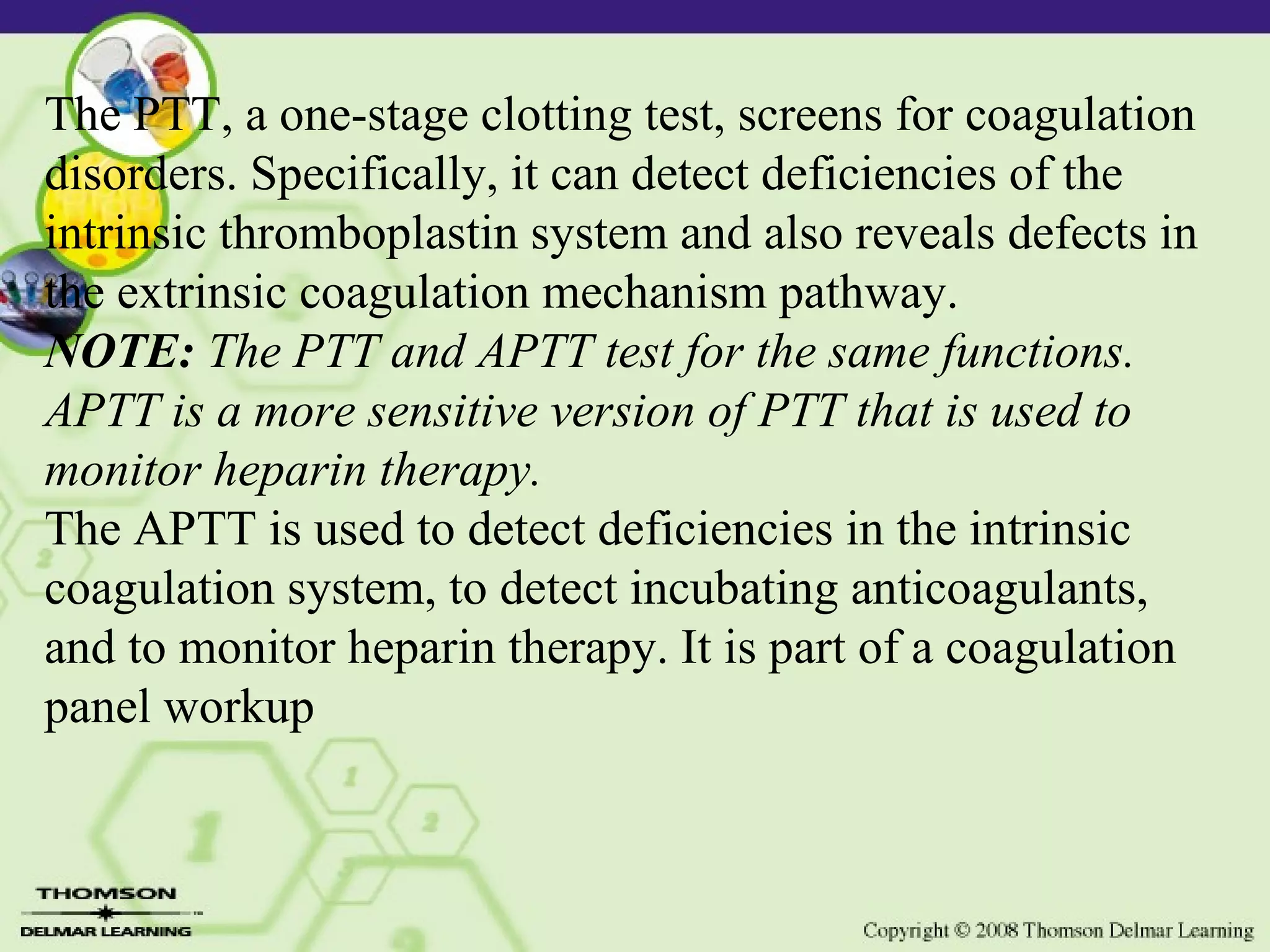
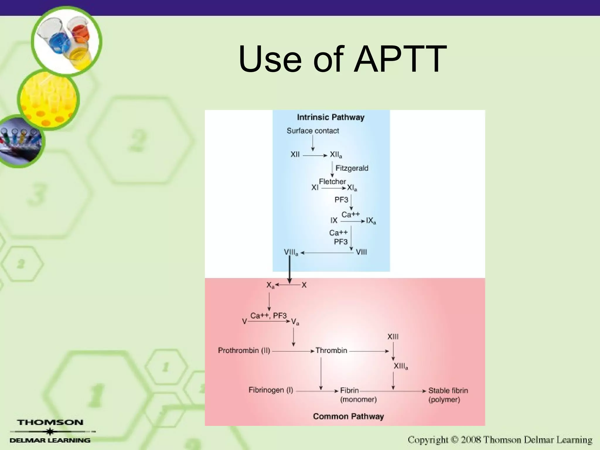
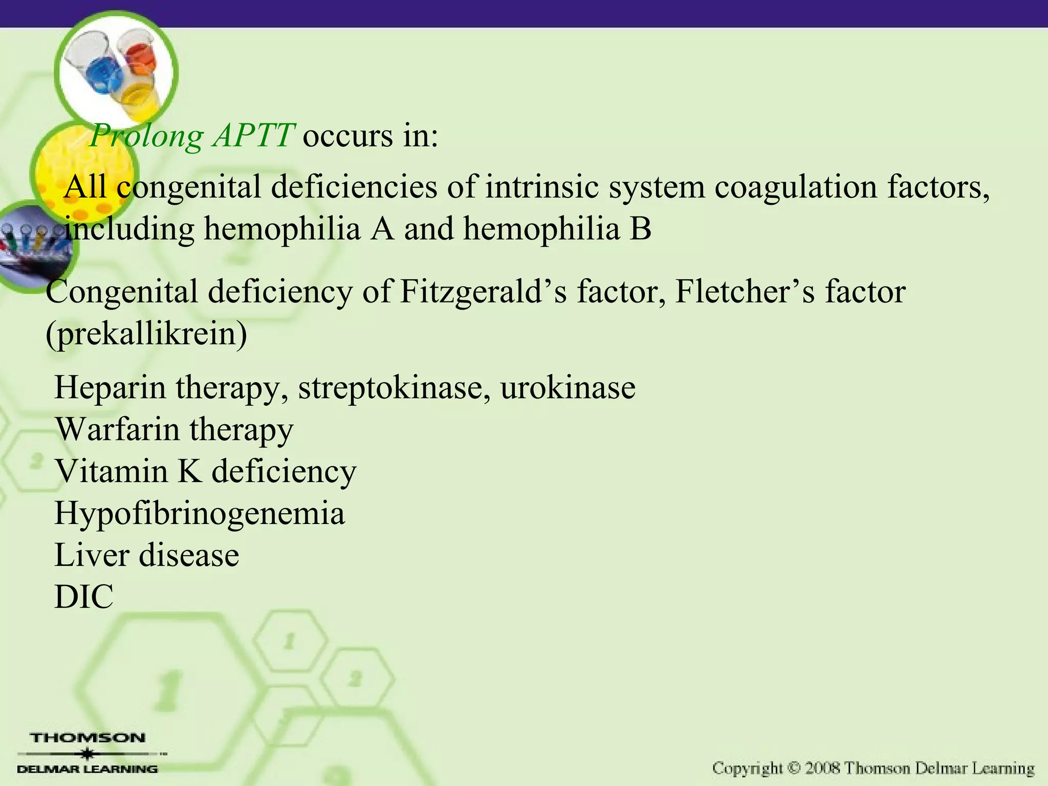
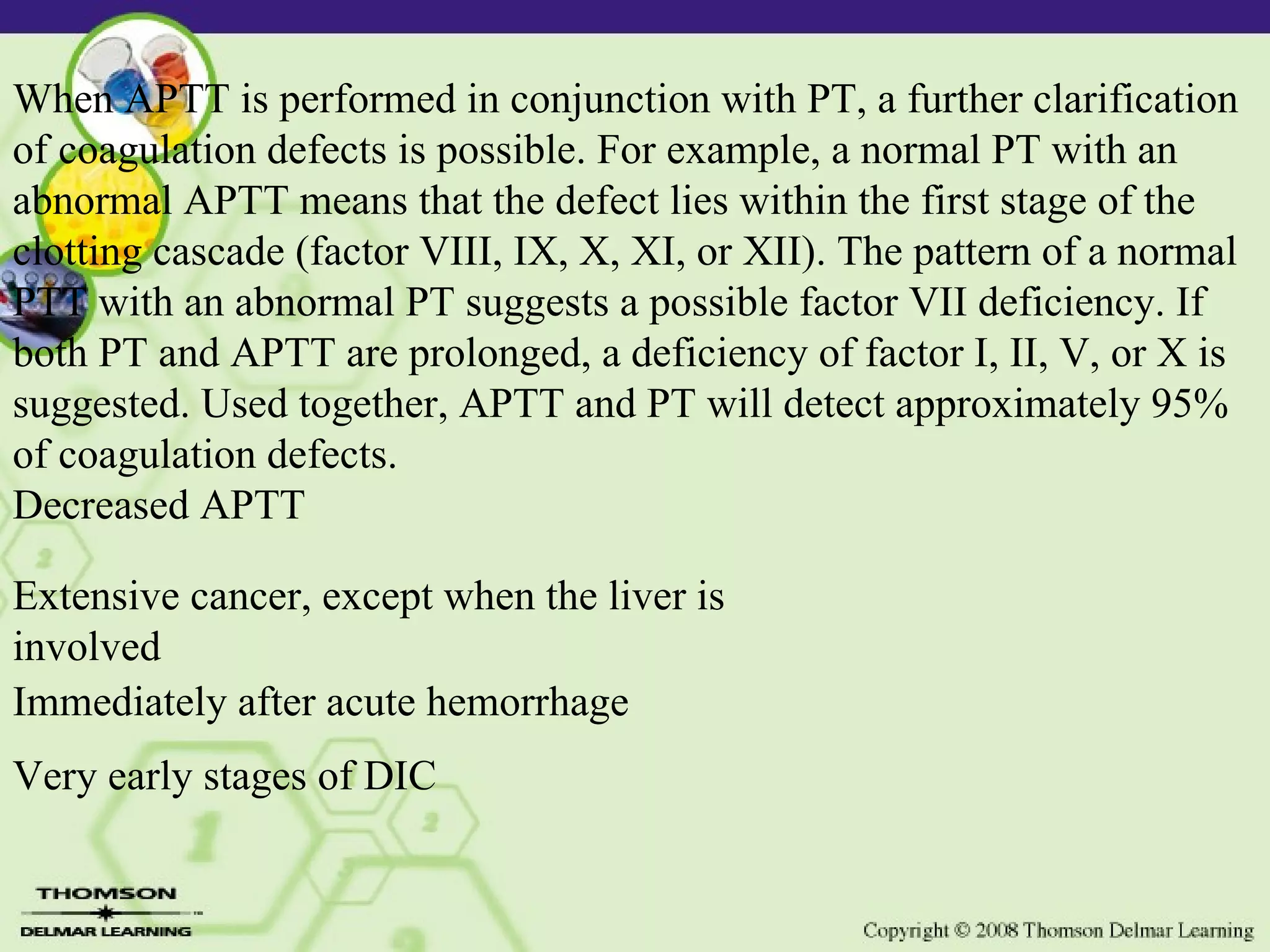
Lesson 3-5 Activated Partial Thromboplastin Time
The PTT, a one-stage clotting test, screens for coagulation disorders. Specifically, it can detect deficiencies of the intrinsic thromboplastin system and also reveals defects in the extrinsic coagulation mechanism pathway. NOTE:  The PTT and APTT test for the same functions. APTT is a more sensitive version of PTT that is used to monitor heparin therapy.  The APTT is used to detect deficiencies in the intrinsic coagulation system, to detect incubating anticoagulants, and to monitor heparin therapy. It is part of a coagulation panel workup
Normal  Critical Value APTT > 70 seconds signifies spontaneous bleeding Obtain a 5-mL venous blood sample and anticoagulate with sodium citrate. Do not draw blood samples from a heparin lock or heparinized catheter. •   APTT: 21.0–35.0 seconds •   Check with your laboratory for therapeutic range values during heparin therapy (2–2.5 times normal).
Use of APTT Screen for intrinsic and common pathways Monitor heparin therapy Uses “partial thromboplastin” reagent
Use of APTT
Prolong APTT  occurs in:  All congenital deficiencies of intrinsic system coagulation factors, including hemophilia A and hemophilia B Congenital deficiency of Fitzgerald’s factor, Fletcher’s factor (prekallikrein) Heparin therapy, streptokinase, urokinase Warfarin therapy Vitamin K deficiency Hypofibrinogenemia Liver disease DIC
When APTT is performed in conjunction with PT, a further clarification of coagulation defects is possible. For example, a normal PT with an abnormal APTT means that the defect lies within the first stage of the clotting cascade (factor VIII, IX, X, XI, or XII). The pattern of a normal PTT with an abnormal PT suggests a possible factor VII deficiency. If both PT and APTT are prolonged, a deficiency of factor I, II, V, or X is suggested. Used together, APTT and PT will detect approximately 95% of coagulation defects. Decreased APTT Extensive cancer, except when the liver is involved Immediately after acute hemorrhage Very early stages of DIC

Lesson 3 5 Laboratory Aptt

  • 1.
    Lesson 3-5 ActivatedPartial Thromboplastin Time
  • 2.
    The PTT, aone-stage clotting test, screens for coagulation disorders. Specifically, it can detect deficiencies of the intrinsic thromboplastin system and also reveals defects in the extrinsic coagulation mechanism pathway. NOTE: The PTT and APTT test for the same functions. APTT is a more sensitive version of PTT that is used to monitor heparin therapy. The APTT is used to detect deficiencies in the intrinsic coagulation system, to detect incubating anticoagulants, and to monitor heparin therapy. It is part of a coagulation panel workup
  • 3.
    Normal CriticalValue APTT > 70 seconds signifies spontaneous bleeding Obtain a 5-mL venous blood sample and anticoagulate with sodium citrate. Do not draw blood samples from a heparin lock or heparinized catheter. •   APTT: 21.0–35.0 seconds •   Check with your laboratory for therapeutic range values during heparin therapy (2–2.5 times normal).
  • 4.
    Use of APTTScreen for intrinsic and common pathways Monitor heparin therapy Uses “partial thromboplastin” reagent
  • 5.
  • 6.
    Prolong APTT occurs in: All congenital deficiencies of intrinsic system coagulation factors, including hemophilia A and hemophilia B Congenital deficiency of Fitzgerald’s factor, Fletcher’s factor (prekallikrein) Heparin therapy, streptokinase, urokinase Warfarin therapy Vitamin K deficiency Hypofibrinogenemia Liver disease DIC
  • 7.
    When APTT isperformed in conjunction with PT, a further clarification of coagulation defects is possible. For example, a normal PT with an abnormal APTT means that the defect lies within the first stage of the clotting cascade (factor VIII, IX, X, XI, or XII). The pattern of a normal PTT with an abnormal PT suggests a possible factor VII deficiency. If both PT and APTT are prolonged, a deficiency of factor I, II, V, or X is suggested. Used together, APTT and PT will detect approximately 95% of coagulation defects. Decreased APTT Extensive cancer, except when the liver is involved Immediately after acute hemorrhage Very early stages of DIC