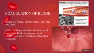
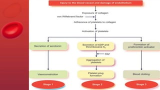
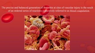
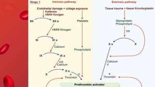
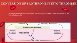
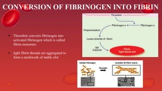
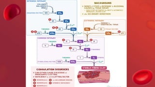
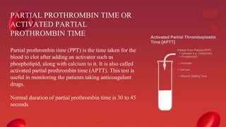
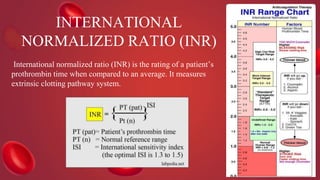
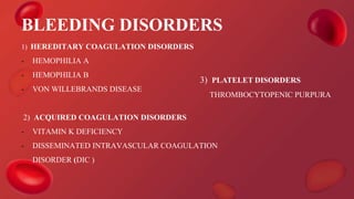
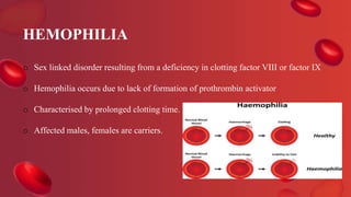
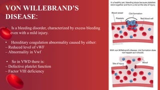
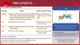
SlideShare a Scribd company logo
BLOOD COAGULATION
Dr. SUSHMITHA SHANKAR
POST GRADUATE STUDENT
DEPARTMENT OF PERIODONTOLOGY
CONTENTS
1. INTRODUCTION
2. HEMOSTASIS
3. FACTORS INVOLVED IN BLOOD COAGULATION
4. THE CLOTTING MECHANISM
5. CLOT RETRACTION
6. FIBRINOLYSIS
7. ANTICOAGULANTS
8. TESTS FOR BLOOD CLOTTING
9. BLEEDING DISORDERS
10. THROMBOSIS
11. MANAGEMENT OF PATIENTS WITH BLEEDING DISORDERS
12. COVID AND BLOOD CLOTTING
INTRODUCTION
Fluidity is essential for circulation of blood and Clotting
is required as a defense during blood loss. Blood when
shed, loses its fluidity in few seconds and becomes jelly
like. This phenomenon is called coagulation.
Hemostasis is the process of forming clots in the walls
of damaged blood vessels and preventing blood loss
while maintaining blood in a fluid state within the
vascular system.
Morawitz in 1904 described the basic mechanism of
blood clotting.
It occurs in three stages:
1. Vasoconstriction
2. Platelet plug formation
3. Coagulation of blood.
HEMOSTASIS
VASOCONSTRICTION
PLATELET PLUG FORMATION
 Platelets get adhered to the collagen of ruptured
blood vessel and secrete adenosine diphosphate
(ADP) and Thromboxane A2.
 These two substances attract more and more platelets
and activate them.
 Platelet aggregation is accelerated by platelet
activating factor
COAGULATION OF BLOOD
 During this process, the fibrinogen is converted
into fibrin.
 Fibrin threads get attached to the loose platelet
plug, which blocks the ruptured part of
blood vessels and prevents further blood loss
completely
The precise and balanced generation of thrombin at sites of vascular injury is the result
of an ordered series of reactions collectively referred to as blood coagulation.
FACTOR I :FIBRINOGEN
Source : Liver
Pathway : Both extrinsic and intrinsic
Activator : Thrombin
FACTOR II :PROTHROMBIN
Source : Liver
Pathway : Both Extrinsic and Intrinsic
Activator : Prothrombin activator
FACTOR III:THROMBOPLASTIN /
TISSUE FACTOR
Source : Platelets(Intrinsic) and Damaged
endothelium lining of the blood vessel
(Extrinsic)
Pathway : Both extrinsic and intrinsic
Activator : Injury to blood vessel
FACTOR IV: CALCIUM
Source : Bone and by absorption of food in GIT
(intrinsic) and the blood vessel ( Extrinsic)
Pathway : Both extrinsic and intrinsic
FACTOR V :PROACCELERIN/
LABILE FACTOR
Source : Liver and platelets
Pathway : Both extrinsic and intrinsic
Activator : Thrombin
FACTOR VII :PROCONVERTIN /
SERUM PROTHROMBIN
CONVERSION ACCELERATOR /
STABLE FACTOR
Source : Liver
Pathway : Extrinsic
Activator : factor III (Tissue factor)
FACTOR VIII :ANTIHEMOPHILIC
FACTOR
Source : Endothelium lining blood vessel and
platelets (plug)
Pathway : Extrinsic
Activator : Thrombin
FACTOR IX :CHRISTMAS FACTOR/
PLASMA THROMBOPLASTIN
COMPONENT
Source : Liver
Activator : Factor XI and Calcium
Deficiency leads to : Hemophilia B
FACTOR X :STUART-PROWER
FACTOR/ANTIHEMOPHILIC
FACTOR B
Source : Liver
Pathway : Intrinsic, Extrinsic
Activator : Factor VII (Extrinsic) / Factor IX +
Factor VIII + Calcium (intrinsic)
FACTOR XI :PLASMA
THROMBOPLASTIN ANTECEDENT /
ANTIHEMOPHILIC FACTOR C
Source : Liver
Pathway : Intrinsic
Activator : Factor XII + Prekallikrein and Kininogen
FACTOR XII :HAGEMAN FACTOR
Source : Liver
Pathway : Intrinsic
Activator : Contact with collagen in the torn wall of
blood vessels
FACTOR XIII:FIBRIN
STABILIZING FACTOR
Source : Liver
Activator : Thrombin and calcium
THE CLOTTING MECHANISM
 The fundamental reaction is conversion of the soluble
plasma protein fibrinogen to insoluble fibrin.
 How is this fibrin formed?
 And, what does this Fibrin do?
In general blood clotting occurs in three stages:
 Formation of prothrombin activator
 Conversion of prothrombin into thrombin
 Conversion of fibrinogen into fibrin
ENZYME CASCADE THEORY
FORMATION OF PROTHROMBIN
ACTIVATOR
• Formation of prothrombin activator occurs through two pathways:
i. Intrinsic pathway
ii. Extrinsic pathway.
CONVERSION OF PROTHROMBIN INTO THROMBIN
 Prothrombin activator converts prothrombin into thrombin in the presence of calcium ions (factor IV).
 Positive feedback effect
CONVERSION OF FIBRINOGEN INTO FIBRIN
 Thrombin converts fibrinogen into
activated fibrinogen which is called
fibrin monomer.
 tight fibrin threads are aggregated to
form a meshwork of stable clot
CLOT RETRACTION
• The process involving the contraction of blood clot and oozing of serum is called clot retraction.
FIBRINOLYSIS
Lysis of blood clot inside the blood
vessel is called fibrinolysis.
Formation of Plasmin :
Plasmin is formed from inactivated
glycoprotein called plasminogen.
Plasminogen is converted into plasmin by
tissue plasminogen activator (t-PA),
lysosomal enzymes and thrombin.
The t-PA and lysosomal enzymes are
released from damaged tissues and damaged
endothelium.
Thrombin is derived from blood.
ANTICOAGULANTS:
The substances which prevent or postpone
coagulation of blood are anticoagulants.
Thrombomodulin, heparin in blood, continuous blood flow, smooth endothelium lining,
clotting factors are in inactive state
* Heparin
* Coumarin derivatives – warfarin, dicoumoral
* EDTA
* Oxalate compounds
* Citrates
* Other substances which prevent blood clotting –
Peptone, C-type lectin, hirudin
Various anticoagulants commonly used are
HEPARIN
Heparin is a naturally produced
anticoagulant in the body.
It is produced by mast cells and
basophils.
OTHER SUBSTANCES WHICH PREVENT
BLOOD CLOTTING
Physical methods:
• cold
• collecting blood in a container with smooth
surface
Procoagulants:
• Thrombin
• Snake venom
• Extract from lungs and thymus
• Calcium or sodium alginate
• Oxidized cellulose
TESTS FOR BLOOD CLOTTING
1. Bleeding time
2. Clotting time
3. Thrombin time.
4. Prothrombin time
5. Activated partial prothrombin time
6. International normalized ratio
BLEEDING TIME
Bleeding time (BT) is the time interval from
oozing of blood after a cut or injury till arrest of
bleeding. Usually, it is determined by Duke
method using blotting paper or filter paper
method. Its normal duration is 3 to 6 minutes.
CLOTTING TIME
Clotting time (CT) is the time interval from oozing of blood after a cut or injury
till the formation of clot. It is usually determined by capillary tube method. Its
normal duration is 3 to 8 minutes.
THROMBIN TIME
 Thrombin time (TT) is the time taken for the blood to clot after adding
thrombin to it.
 Normal duration of thrombin time is 12 to 20 seconds.
 It is prolonged in heparin therapy and during dysfibrinogenimia
PROTHROMBIN TIME
 Prothrombin time (PT) is the time taken by blood to clot
after adding tissue thromboplastin to it.
 Prothrombin time indicates the total quantity of
prothrombin present in the blood.
 Normal duration of prothrombin time is 10 to 12 seconds.
PARTIAL PROTHROMBIN TIME OR
ACTIVATED PARTIAL
PROTHROMBIN TIME
Partial prothrombin time (PPT) is the time taken for the
blood to clot after adding an activator such as
phospholipid, along with calcium to it. It is also called
activated partial prothrombin time (APTT). This test is
useful in monitoring the patients taking anticoagulant
drugs.
Normal duration of partial prothrombin time is 30 to 45
seconds
INTERNATIONAL
NORMALIZED RATIO (INR)
International normalized ratio (INR) is the rating of a patient’s
prothrombin time when compared to an average. It measures
extrinsic clotting pathway system.
>4.0
• No surgical treatment until the INR is reduced
3.5-4.0
• Emergency minor surgical procedures only
• Avoid block anesthesia injections
3.0-3.4
• Minor surgical procedures like extraction, gingivoplasty
2.5-2.9
• Multiple extraction, single bony impaction, periodontal
flap surgery, SRP
1.5-2.4
• All surgical procedures
INR
VALUE
RECOMMENDATIONS CONCERNING INVASIVE
TREATMENT
BLEEDING DISORDERS
1) HEREDITARY COAGULATION DISORDERS
- HEMOPHILIA A
- HEMOPHILIA B
- VON WILLEBRANDS DISEASE
2) ACQUIRED COAGULATION DISORDERS
- VITAMIN K DEFICIENCY
- DISSEMINATED INTRAVASCULAR COAGULATION
DISORDER (DIC )
3) PLATELET DISORDERS
THROMBOCYTOPENIC PURPURA
HEMOPHILIA
o Sex linked disorder resulting from a deficiency in clotting factor VIII or factor IX
o Hemophilia occurs due to lack of formation of prothrombin activator
o Characterised by prolonged clotting time.
o Affected males, females are carriers.
Types of hemophilia: Depending upon the deficiency of the factor involved,
hemophilia is classified into three types:
Hemophilia A or classic hemophilia
Hemophilia B or Christmas disease
Hemophilia C or factor XI deficiency
SYMPTOMS OF HEMOPHILIA
Treatment for hemophilia
• Effective therapy for classical hemophilia involves replacement of
missing clotting factor.
• Prolonged bleeding due to injuries
• Hemorrhage
• Bleeding in joints followed by swelling
and pain
• Appearance of blood in urine.
ORAL
MANIFESTATIONS:
• Petechiae
• Ecchymoses
• Spontaneous gingival bleeding
PERIODONTAL CONSIDERATIONS:
• In all but severe hemophiliacs, scaling can be carried out without LA.
• Periodontal surgery requires LA and factor VIII replacement to a level between 50 to 75%
HEMOPHILIA- A
• TREATMENT:
- Factor replacement
- Bleeding is treated by administration of factor VIII concentrate by intravenous infusion.
- Major surgery: the factor VIII should be raised to 100% preoperatively and maintained
above 50% until healing has occurred.
HEMOPHILIA B
• Hemophilia B is treated with factor IX concentrates
VON WILLEBRAND’S
DISEASE:
• Is a bleeding disorder, characterized by excess bleeding
even with a mild injury.
• Hereditary coagulation abnormality caused by either:
– Reduced level of vWF
– Abnormality in Vwf
• So in VWD there is:
– Defective platelet function
– Factor VIII deficiency
Clinical features:
– Typically there is mucus membrane bleeding
– The severity of symptoms are variable with types
• Type 1, 2 usually mild symptoms
• Type 3 severe symptoms
– Type 1 VWD
– Type 2 VWD
– Type 3 VWD
VWD has been classified into three types:
ORAL MANIFESTATIONS :
• Gingival bleeding
• Uncontrollable bleeding during dental procedures.
PERIODONTAL
CONSIDERATIONS:
 Gingival haemorrhage
 Even minor gingival manipulation can
lead to uncontrollable bleeding.
TREATMENT: –
ACQUIRED COAGULATION DISORDERS
1.VITAMIN K DEFICIENCY
ORAL MANIFESTATIONS :
Spontaneous gingival haemorrhages
Gingival bleeding is seen after brushing
- Important for formation of factors II, VII, IX and X and
proteins C.
- Without it, these factors cannot bind to calcium.
2. DISSEMINATED INTRAVASCULAR COAGULATION
DISORDER (DIC)
 There is widespread deposition of fibrin within blood vessels with consumption of coagulation factors
and platelets occurs as a consequence of many disorders which release procoagulant material into the
circulation.
TREATMENT
-Treat underlying cause
– Supportive therapy with fresh frozen plasma (FFP) and
platelet concentrates
– Cryoprecipitate can be used
CLINICAL FEATURES:
– Bleeding, particularly from venipuncture
– Purpura
– Generalized bleeding in GIT, oropharynx, lungs, urogenital tract, vaginal bleeding
PLATELET DISORDERS :
• Platelet disorders may be the result of alteration in platelet numbers, either
- decreased (thrombocytopenia)
- increased (thrombocythemia)
• Thrombocytopenia has platelet count < 1,50,000
• Platelet disorders may be:
Quantitative- Thrombocytopenic purpura
Qualitative- Disorders of platelet function
THROMBOCYTOPENIC PURPURA
• Thrombocytopenia is a bleeding disorder characterized by a platelet count below
the normal range.
• It may be:
- Auto- immune or Idiopathic
- Drug induced
IDIOPATHIC THROMBOCYTOPENIC PURPURA
• Also known as auto – immune thrombocytopenic purpura.
• most common
• Can lead to purpura and prolonged bleeding.
DRUG INDUCED THROMBOCYTOPENIC PURPURA
• Reactions of drugs or toxins resulting in a low platelet count and bleeding tendency.
• Common cause are:
• Quinidine
• Sulfonamides
• Heavy alcohol consumption
GENERAL FEATURES
• Abrupt appearance of petechial hemorrhages.
• Purpura
• Bruises after trauma
THROMBOSIS
● Thrombosis or intravascular blood
clotting refers to coagulation of blood
inside the blood vessels.
● Causes of Thrombosis:
1. Injury to blood vessels
2. Roughened endothelial lining
3. Sluggishness of blood flow
4. Agglutination of RBCs
5. Toxic thrombosis
6. Congenital absence of protein C
● Complications of Thrombosis:
1. Thrombus
2. Embolism and embolus
3. Ischemia
4. Necrosis
5. Infarction
MANAGEMENT OF PATIENTS WITH BLEEDING DISORDERS
Consultation with a Hematologist
Prophylactic Factor Replacement Therapy
Medication Review
Platelet Count
Thorough Oral Examination
Patient Education
Bleeding Control Measures
pre-operative precautions
Intraoperative precautions
Local Anesthesia
Use of Haemostatic Agents
Avoidance of Trauma
Use of Sutures
Monitoring of Vital Signs
Post-operative care
Use of Hemostatic Agents
Avoidance of Trauma
Pain Management
Follow-up Appointments:
Prophylactic Factor Replacement Therapy
Monitoring of Vital Signs
Patient Education:
Intraoperative Bleeding During Open Flap Debridement and Regenerative
Periodontal Surgery
Hadar Zigdon,*† Liran Levin,*† Margarita Filatov
Background: The objective of this study was to measure the intraoperative bleeding during periodontal flap
surgery.
Methods: Patients scheduled for periodontal surgery were recruited for this study. Data regarding smoking
habits, general health, and medications were collected. The amount of the local anesthetic that was injected
was then recorded, as well as the number of teeth in the operative field and the duration of the procedure.
During surgery, the liquids from the oral cavity were suctioned and collected into a sterile empty vial. To
calculate the net amount of blood volume in the liquids, colorimetric assay using capillary blood fructosamine
as a reference molecule was used.
Results: Twenty-six patients were included in this study. The amount of blood lost during the procedure
ranged from 6.0 to 145.1 mL, with an overall mean of 59.47 – 38.2 mL. Patients taking aspirin (acetylsalicylic
acid) showed mean blood loss of 43.26 – 31.5 mL, whereas the mean blood loss among patients that did not
use this medication was higher (65.4 – 39.4 mL) but not statistically significant. Local anesthetic amount,
surgical field size, and the operation duration did not relate to blood-loss volume. The mean blood loss among
current smokers was significantly higher (96.47 – 44.2 mL) compared to former (12 – 8.4 mL) or never (54.74
– 30.5 mL, P = 0.011) smokers.
Conclusion: The results of the current study support previous papers and confirm that blood loss during
periodontal surgery is minimal.
Blood Loss During Periodontal Flap Surgery
by DAVID A . BAAB, D.D.S., M.S.D.* WILLIAM F . AMMONS, JR., D.D.S., M.S.D.* HERBERT
SELIPSKY, B.D.S., H.D.D., M.S.D.*
Berdon published the first report on hemorrhage during periodontal surgery, he measured blood loss during 50
gingivectomies involving a surgical field of 5 to 14 teeth. He established that approximately 5 ml to 149 ml of blood
was lost. The quantity of hemorrhage from two patients undergoing full-mouth gingivectomies under a general
anesthetic was also measured and found to be 435 ml and 624 ml respectively. Observation and supportive therapy was
recommended for those patients with the "higher volume" of blood loss. No specific guidelines for fluid replacement
were recommended
Mclvor and Wengraf studied blood loss colorimetrically during gingivectomies and isolated periodontal flap procedures
on 14 patients. Three patients had small, one- or two-tooth, mucoperiosteal flaps elevated and osteoplasty performed.
Blood loss for these three patients ranged from 12 ml to 62 ml, while the other patients lost 0.7 ml to 18 ml of blood
during gingivectomies. Based upon this data, they speculated that one could expect a 10-fold increase in blood loss per
tooth during a periodontal flap procedure as compared to a gingivectomy
Periodontal flap surgery in the mandible
resulted in an average blood loss of 151 ml
per segment, while the corresponding
procedure in the maxilla resulted in an
average blood loss of 110 ml per segment.
The greatest average blood loss per segment
occurred from the mandibular right posterior
area, while surgery in the maxillary left
posterior area resulted in the least average
blood loss per segment
Sex*
Females 121.7 (±116 )
Males 156.4 (±113 )
Area of surgery
Upper right 121.5 (±103 )
Upper left 109.9 (±147 )
Lower left 136.5 (±129 )
Lower right 160.1 (±149 )
AVERAGE
BLOOD LOSS
Type of surgery
Flap Curettage 171.8 (±126 )
Osseous surgery 140.4 (±141 )
COVID AND BLOOD CLOTTING
o Patients with coronavirus disease (COVID-19) have elevated D-dimer levels. Early
reports describe high venous thromboembolism (VTE) and disseminated intravascular
coagulation (DIC) rates, but data are limited.
o In support of this hypothesis are recent autopsy studies of COVID-19 patients
demonstrating the presence of fibrin thrombi within distended small vessels and
capillaries and extensive extracellular fibrin deposition
o COVID-19 was associated with similar rates of thrombosis and bleeding as seen in
hospitalized patients with similar degrees of critical illness. Elevated D-dimer levels at
initial presentation predicted bleeding complications, thrombotic complications, critical
illness, and death. Beyond D-dimer, thrombosis was primarily associated with
inflammatory markers rather than coagulation parameters
CONCLUSION
REFERENCES
 Essentials of medical physiology – Sembulingam
 Text book of Medical Physiology , Guyton, 12th edition,
 Dental management of patients with inherited bleeding disorders: a multidisciplinary approach
 Guidelines for dental treatment of patients with inherited bleeding disorders Andrew Brewer, Maria Elvira Correa
 A.K Jain Human physiology for BDS – 5th edition
 Intraoperative Bleeding During Open Flap Debridement and Regenerative Periodontal Surgery Hadar Zigdon,*†
Liran Levin,*† Margarita Filatov
 Periodontology 2000 vol44
 Blood Loss During Periodontal Flap Surgery by DAVID A . BAAB, D.D.S., M.S.D.* WILLIAM F . AMMONS,
JR., D.D.S., M.S.D.* HERBERT SELIPSKY, B.D.S., H.D.D., M.S.D.*
blood coagulation.pptx

More Related Content

What's hot

Hemostasis
HemostasisHemostasis
Blood transfusion
Blood transfusionBlood transfusion
Blood transfusion
Aashissh Shah
 
Normal hemostasis and coagulation
Normal hemostasis and coagulationNormal hemostasis and coagulation
Normal hemostasis and coagulation
wendwesen alemu
 
Hemostasis
HemostasisHemostasis
Hemostasis
katejohnpunag
 
Bleeding timeclotting-time-pt-and-ptt
Bleeding timeclotting-time-pt-and-pttBleeding timeclotting-time-pt-and-ptt
Bleeding timeclotting-time-pt-and-ptt
AKHTAR HUSSAIN
 
Apheresis
ApheresisApheresis
CME: Bleeding Disorders - Case Scenarios
CME: Bleeding Disorders - Case ScenariosCME: Bleeding Disorders - Case Scenarios
CME: Bleeding Disorders - Case Scenarios
Stanley Medical College, Department of Medicine
 
Fibrinolytic system
Fibrinolytic systemFibrinolytic system
Fibrinolytic system
Ibrahim khidir ibrahim osman
 
Bleeding and clotting disorders
Bleeding and clotting disordersBleeding and clotting disorders
Bleeding and clotting disorders
UMAMAHISHAQ
 
Blood coagulation
Blood coagulationBlood coagulation
Blood coagulation
SATYA DAS
 
Blood components preparation and therapeutic uses final
Blood components preparation and therapeutic uses finalBlood components preparation and therapeutic uses final
Blood components preparation and therapeutic uses final
globalsoin
 
Approach to bleeding disorders part II
Approach to bleeding disorders part IIApproach to bleeding disorders part II
Approach to bleeding disorders part II
drbcnayak
 
Platelet transfusion
Platelet transfusionPlatelet transfusion
Platelet transfusion
egyfellow
 
hi
hihi
seminar on Blood transfusion
 seminar on Blood transfusion  seminar on Blood transfusion
seminar on Blood transfusion
Biswajit Deka
 
Hemostasis and blood coagulation general pathology
Hemostasis and blood  coagulation general pathologyHemostasis and blood  coagulation general pathology
Hemostasis and blood coagulation general pathology
Siganga Siganga
 
Bleeding disorders Causes, Types, and Diagnosis
Bleeding disorders Causes, Types, and DiagnosisBleeding disorders Causes, Types, and Diagnosis
Bleeding disorders Causes, Types, and Diagnosis
Dr Medical
 
Fresh frozen plasma
Fresh frozen plasmaFresh frozen plasma
Fresh frozen plasma
Praveen Nagula
 
Coagulation tests
Coagulation testsCoagulation tests
Coagulation tests
temis cola
 
platelet function, disorders and its assesment
platelet function, disorders and its assesmentplatelet function, disorders and its assesment
platelet function, disorders and its assesment
Figo Khan
 

What's hot (20)

Hemostasis
HemostasisHemostasis
Hemostasis
 
Blood transfusion
Blood transfusionBlood transfusion
Blood transfusion
 
Normal hemostasis and coagulation
Normal hemostasis and coagulationNormal hemostasis and coagulation
Normal hemostasis and coagulation
 
Hemostasis
HemostasisHemostasis
Hemostasis
 
Bleeding timeclotting-time-pt-and-ptt
Bleeding timeclotting-time-pt-and-pttBleeding timeclotting-time-pt-and-ptt
Bleeding timeclotting-time-pt-and-ptt
 
Apheresis
ApheresisApheresis
Apheresis
 
CME: Bleeding Disorders - Case Scenarios
CME: Bleeding Disorders - Case ScenariosCME: Bleeding Disorders - Case Scenarios
CME: Bleeding Disorders - Case Scenarios
 
Fibrinolytic system
Fibrinolytic systemFibrinolytic system
Fibrinolytic system
 
Bleeding and clotting disorders
Bleeding and clotting disordersBleeding and clotting disorders
Bleeding and clotting disorders
 
Blood coagulation
Blood coagulationBlood coagulation
Blood coagulation
 
Blood components preparation and therapeutic uses final
Blood components preparation and therapeutic uses finalBlood components preparation and therapeutic uses final
Blood components preparation and therapeutic uses final
 
Approach to bleeding disorders part II
Approach to bleeding disorders part IIApproach to bleeding disorders part II
Approach to bleeding disorders part II
 
Platelet transfusion
Platelet transfusionPlatelet transfusion
Platelet transfusion
 
hi
hihi
hi
 
seminar on Blood transfusion
 seminar on Blood transfusion  seminar on Blood transfusion
seminar on Blood transfusion
 
Hemostasis and blood coagulation general pathology
Hemostasis and blood  coagulation general pathologyHemostasis and blood  coagulation general pathology
Hemostasis and blood coagulation general pathology
 
Bleeding disorders Causes, Types, and Diagnosis
Bleeding disorders Causes, Types, and DiagnosisBleeding disorders Causes, Types, and Diagnosis
Bleeding disorders Causes, Types, and Diagnosis
 
Fresh frozen plasma
Fresh frozen plasmaFresh frozen plasma
Fresh frozen plasma
 
Coagulation tests
Coagulation testsCoagulation tests
Coagulation tests
 
platelet function, disorders and its assesment
platelet function, disorders and its assesmentplatelet function, disorders and its assesment
platelet function, disorders and its assesment
 

Similar to blood coagulation.pptx

BLOOD COAGULATION + BLOOD GROUP.pptx
BLOOD COAGULATION + BLOOD GROUP.pptxBLOOD COAGULATION + BLOOD GROUP.pptx
BLOOD COAGULATION + BLOOD GROUP.pptx
Dr Shyam Chandak
 
Hemostasis and coagulation of blood For M.Sc & Basic Medical Students by Pand...
Hemostasis and coagulation of blood For M.Sc & Basic Medical Students by Pand...Hemostasis and coagulation of blood For M.Sc & Basic Medical Students by Pand...
Hemostasis and coagulation of blood For M.Sc & Basic Medical Students by Pand...
Pandian M
 
PC of Blood and Blood forming agents.pdf
PC of Blood and Blood forming agents.pdfPC of Blood and Blood forming agents.pdf
PC of Blood and Blood forming agents.pdf
RAMDAS BHAT
 
Hemostasis and coagulation of blood by Pandian M, Tutor, Dept of Physiology, ...
Hemostasis and coagulation of blood by Pandian M, Tutor, Dept of Physiology, ...Hemostasis and coagulation of blood by Pandian M, Tutor, Dept of Physiology, ...
Hemostasis and coagulation of blood by Pandian M, Tutor, Dept of Physiology, ...
Pandian M
 
Coagulation Mechanism and blood disorders
Coagulation Mechanism and blood disordersCoagulation Mechanism and blood disorders
Coagulation Mechanism and blood disorders
Sreenivasa Reddy Thalla
 
Mechanism of blood clotting and blood dyscrasias
Mechanism of blood clotting and blood dyscrasiasMechanism of blood clotting and blood dyscrasias
Mechanism of blood clotting and blood dyscrasias
Karishma Sirimulla
 
Platelets, hemostasis and coagulation.pptx
Platelets, hemostasis and coagulation.pptxPlatelets, hemostasis and coagulation.pptx
Platelets, hemostasis and coagulation.pptx
MariumNSiddiqui
 
Hemostatsis & blood transfusion
Hemostatsis & blood transfusionHemostatsis & blood transfusion
Hemostatsis & blood transfusion
ABDUL QADEER MEMON
 
phsiology of blood coagulation by dr chandbaby ansari.pdf
phsiology of blood coagulation by dr chandbaby ansari.pdfphsiology of blood coagulation by dr chandbaby ansari.pdf
phsiology of blood coagulation by dr chandbaby ansari.pdf
AlfiaAnsari2
 
Hemostasis what means and classification
Hemostasis what means and classificationHemostasis what means and classification
Hemostasis what means and classification
SagharMousavi1
 
Hemostasis, cloting mechanis
Hemostasis, cloting mechanisHemostasis, cloting mechanis
Hemostasis, cloting mechanis
binaya tamang
 
Coagulation
CoagulationCoagulation
4. Heamostasis.pptx
4. Heamostasis.pptx4. Heamostasis.pptx
4. Heamostasis.pptx
Lawrenceshamboko
 
Coagulation factors
Coagulation factorsCoagulation factors
Coagulation factors
DrVishal Kandhway
 
Blood coagulation
Blood coagulationBlood coagulation
Blood coagulation
suma priyanka
 
Blood Physiology: Part IV: Platelets and hemostasis
Blood Physiology: Part IV: Platelets and hemostasisBlood Physiology: Part IV: Platelets and hemostasis
Blood Physiology: Part IV: Platelets and hemostasis
Fawaz A.M.
 
Bleeding disorder Hematology Lecture.pptx
Bleeding disorder Hematology Lecture.pptxBleeding disorder Hematology Lecture.pptx
Bleeding disorder Hematology Lecture.pptx
Munmun Kulsum
 
HEMOSTASIS.pptx
HEMOSTASIS.pptxHEMOSTASIS.pptx
HEMOSTASIS.pptx
SYED ALI AFRIN
 
Hemorrhage and its Management
Hemorrhage and its ManagementHemorrhage and its Management
Hemorrhage and its Management
Akshat Sachdeva
 
Haemostasis, anticoagulant, bleeding disorders.
Haemostasis, anticoagulant, bleeding disorders.Haemostasis, anticoagulant, bleeding disorders.
Haemostasis, anticoagulant, bleeding disorders.
jay singh
 

Similar to blood coagulation.pptx (20)

BLOOD COAGULATION + BLOOD GROUP.pptx
BLOOD COAGULATION + BLOOD GROUP.pptxBLOOD COAGULATION + BLOOD GROUP.pptx
BLOOD COAGULATION + BLOOD GROUP.pptx
 
Hemostasis and coagulation of blood For M.Sc & Basic Medical Students by Pand...
Hemostasis and coagulation of blood For M.Sc & Basic Medical Students by Pand...Hemostasis and coagulation of blood For M.Sc & Basic Medical Students by Pand...
Hemostasis and coagulation of blood For M.Sc & Basic Medical Students by Pand...
 
PC of Blood and Blood forming agents.pdf
PC of Blood and Blood forming agents.pdfPC of Blood and Blood forming agents.pdf
PC of Blood and Blood forming agents.pdf
 
Hemostasis and coagulation of blood by Pandian M, Tutor, Dept of Physiology, ...
Hemostasis and coagulation of blood by Pandian M, Tutor, Dept of Physiology, ...Hemostasis and coagulation of blood by Pandian M, Tutor, Dept of Physiology, ...
Hemostasis and coagulation of blood by Pandian M, Tutor, Dept of Physiology, ...
 
Coagulation Mechanism and blood disorders
Coagulation Mechanism and blood disordersCoagulation Mechanism and blood disorders
Coagulation Mechanism and blood disorders
 
Mechanism of blood clotting and blood dyscrasias
Mechanism of blood clotting and blood dyscrasiasMechanism of blood clotting and blood dyscrasias
Mechanism of blood clotting and blood dyscrasias
 
Platelets, hemostasis and coagulation.pptx
Platelets, hemostasis and coagulation.pptxPlatelets, hemostasis and coagulation.pptx
Platelets, hemostasis and coagulation.pptx
 
Hemostatsis & blood transfusion
Hemostatsis & blood transfusionHemostatsis & blood transfusion
Hemostatsis & blood transfusion
 
phsiology of blood coagulation by dr chandbaby ansari.pdf
phsiology of blood coagulation by dr chandbaby ansari.pdfphsiology of blood coagulation by dr chandbaby ansari.pdf
phsiology of blood coagulation by dr chandbaby ansari.pdf
 
Hemostasis what means and classification
Hemostasis what means and classificationHemostasis what means and classification
Hemostasis what means and classification
 
Hemostasis, cloting mechanis
Hemostasis, cloting mechanisHemostasis, cloting mechanis
Hemostasis, cloting mechanis
 
Coagulation
CoagulationCoagulation
Coagulation
 
4. Heamostasis.pptx
4. Heamostasis.pptx4. Heamostasis.pptx
4. Heamostasis.pptx
 
Coagulation factors
Coagulation factorsCoagulation factors
Coagulation factors
 
Blood coagulation
Blood coagulationBlood coagulation
Blood coagulation
 
Blood Physiology: Part IV: Platelets and hemostasis
Blood Physiology: Part IV: Platelets and hemostasisBlood Physiology: Part IV: Platelets and hemostasis
Blood Physiology: Part IV: Platelets and hemostasis
 
Bleeding disorder Hematology Lecture.pptx
Bleeding disorder Hematology Lecture.pptxBleeding disorder Hematology Lecture.pptx
Bleeding disorder Hematology Lecture.pptx
 
HEMOSTASIS.pptx
HEMOSTASIS.pptxHEMOSTASIS.pptx
HEMOSTASIS.pptx
 
Hemorrhage and its Management
Hemorrhage and its ManagementHemorrhage and its Management
Hemorrhage and its Management
 
Haemostasis, anticoagulant, bleeding disorders.
Haemostasis, anticoagulant, bleeding disorders.Haemostasis, anticoagulant, bleeding disorders.
Haemostasis, anticoagulant, bleeding disorders.
 

Recently uploaded

Top-Vitamin-Supplement-Brands-in-India List
Top-Vitamin-Supplement-Brands-in-India ListTop-Vitamin-Supplement-Brands-in-India List
Top-Vitamin-Supplement-Brands-in-India List
SwisschemDerma
 
Osteoporosis - Definition , Evaluation and Management .pdf
Osteoporosis - Definition , Evaluation and Management .pdfOsteoporosis - Definition , Evaluation and Management .pdf
Osteoporosis - Definition , Evaluation and Management .pdf
Jim Jacob Roy
 
Promoting Wellbeing - Applied Social Psychology - Psychology SuperNotes
Promoting Wellbeing - Applied Social Psychology - Psychology SuperNotesPromoting Wellbeing - Applied Social Psychology - Psychology SuperNotes
Promoting Wellbeing - Applied Social Psychology - Psychology SuperNotes
PsychoTech Services
 
Artificial Intelligence Symposium (THAIS)
Artificial Intelligence Symposium (THAIS)Artificial Intelligence Symposium (THAIS)
Artificial Intelligence Symposium (THAIS)
Josep Vidal-Alaball
 
Adhd Medication Shortage Uk - trinexpharmacy.com
Adhd Medication Shortage Uk - trinexpharmacy.comAdhd Medication Shortage Uk - trinexpharmacy.com
Adhd Medication Shortage Uk - trinexpharmacy.com
reignlana06
 
REGULATION FOR COMBINATION PRODUCTS AND MEDICAL DEVICES.pptx
REGULATION FOR COMBINATION PRODUCTS AND MEDICAL DEVICES.pptxREGULATION FOR COMBINATION PRODUCTS AND MEDICAL DEVICES.pptx
REGULATION FOR COMBINATION PRODUCTS AND MEDICAL DEVICES.pptx
LaniyaNasrink
 
Best Ayurvedic medicine for Gas and Indigestion
Best Ayurvedic medicine for Gas and IndigestionBest Ayurvedic medicine for Gas and Indigestion
Best Ayurvedic medicine for Gas and Indigestion
Swastik Ayurveda
 
Muscles of Mastication by Dr. Rabia Inam Gandapore.pptx
Muscles of Mastication by Dr. Rabia Inam Gandapore.pptxMuscles of Mastication by Dr. Rabia Inam Gandapore.pptx
Muscles of Mastication by Dr. Rabia Inam Gandapore.pptx
Dr. Rabia Inam Gandapore
 
Post-Menstrual Smell- When to Suspect Vaginitis.pptx
Post-Menstrual Smell- When to Suspect Vaginitis.pptxPost-Menstrual Smell- When to Suspect Vaginitis.pptx
Post-Menstrual Smell- When to Suspect Vaginitis.pptx
FFragrant
 
Abortion PG Seminar Power point presentation
Abortion PG Seminar Power point presentationAbortion PG Seminar Power point presentation
Abortion PG Seminar Power point presentation
AksshayaRajanbabu
 
share - Lions, tigers, AI and health misinformation, oh my!.pptx
share - Lions, tigers, AI and health misinformation, oh my!.pptxshare - Lions, tigers, AI and health misinformation, oh my!.pptx
share - Lions, tigers, AI and health misinformation, oh my!.pptx
Tina Purnat
 
CHEMOTHERAPY_RDP_CHAPTER 6_Anti Malarial Drugs.pdf
CHEMOTHERAPY_RDP_CHAPTER 6_Anti Malarial Drugs.pdfCHEMOTHERAPY_RDP_CHAPTER 6_Anti Malarial Drugs.pdf
CHEMOTHERAPY_RDP_CHAPTER 6_Anti Malarial Drugs.pdf
rishi2789
 
Top Effective Soaps for Fungal Skin Infections in India
Top Effective Soaps for Fungal Skin Infections in IndiaTop Effective Soaps for Fungal Skin Infections in India
Top Effective Soaps for Fungal Skin Infections in India
SwisschemDerma
 
Chapter 11 Nutrition and Chronic Diseases.pptx
Chapter 11 Nutrition and Chronic Diseases.pptxChapter 11 Nutrition and Chronic Diseases.pptx
Chapter 11 Nutrition and Chronic Diseases.pptx
Earlene McNair
 
Aortic Association CBL Pilot April 19 – 20 Bern
Aortic Association CBL Pilot April 19 – 20 BernAortic Association CBL Pilot April 19 – 20 Bern
Aortic Association CBL Pilot April 19 – 20 Bern
suvadeepdas911
 
Part II - Body Grief: Losing parts of ourselves and our identity before, duri...
Part II - Body Grief: Losing parts of ourselves and our identity before, duri...Part II - Body Grief: Losing parts of ourselves and our identity before, duri...
Part II - Body Grief: Losing parts of ourselves and our identity before, duri...
bkling
 
TEST BANK For An Introduction to Brain and Behavior, 7th Edition by Bryan Kol...
TEST BANK For An Introduction to Brain and Behavior, 7th Edition by Bryan Kol...TEST BANK For An Introduction to Brain and Behavior, 7th Edition by Bryan Kol...
TEST BANK For An Introduction to Brain and Behavior, 7th Edition by Bryan Kol...
rightmanforbloodline
 
Hemodialysis: Chapter 4, Dialysate Circuit - Dr.Gawad
Hemodialysis: Chapter 4, Dialysate Circuit - Dr.GawadHemodialysis: Chapter 4, Dialysate Circuit - Dr.Gawad
Hemodialysis: Chapter 4, Dialysate Circuit - Dr.Gawad
NephroTube - Dr.Gawad
 
CHEMOTHERAPY_RDP_CHAPTER 3_ANTIFUNGAL AGENT.pdf
CHEMOTHERAPY_RDP_CHAPTER 3_ANTIFUNGAL AGENT.pdfCHEMOTHERAPY_RDP_CHAPTER 3_ANTIFUNGAL AGENT.pdf
CHEMOTHERAPY_RDP_CHAPTER 3_ANTIFUNGAL AGENT.pdf
rishi2789
 
Cardiac Assessment for B.sc Nursing Student.pdf
Cardiac Assessment for B.sc Nursing Student.pdfCardiac Assessment for B.sc Nursing Student.pdf
Cardiac Assessment for B.sc Nursing Student.pdf
shivalingatalekar1
 

Recently uploaded (20)

Top-Vitamin-Supplement-Brands-in-India List
Top-Vitamin-Supplement-Brands-in-India ListTop-Vitamin-Supplement-Brands-in-India List
Top-Vitamin-Supplement-Brands-in-India List
 
Osteoporosis - Definition , Evaluation and Management .pdf
Osteoporosis - Definition , Evaluation and Management .pdfOsteoporosis - Definition , Evaluation and Management .pdf
Osteoporosis - Definition , Evaluation and Management .pdf
 
Promoting Wellbeing - Applied Social Psychology - Psychology SuperNotes
Promoting Wellbeing - Applied Social Psychology - Psychology SuperNotesPromoting Wellbeing - Applied Social Psychology - Psychology SuperNotes
Promoting Wellbeing - Applied Social Psychology - Psychology SuperNotes
 
Artificial Intelligence Symposium (THAIS)
Artificial Intelligence Symposium (THAIS)Artificial Intelligence Symposium (THAIS)
Artificial Intelligence Symposium (THAIS)
 
Adhd Medication Shortage Uk - trinexpharmacy.com
Adhd Medication Shortage Uk - trinexpharmacy.comAdhd Medication Shortage Uk - trinexpharmacy.com
Adhd Medication Shortage Uk - trinexpharmacy.com
 
REGULATION FOR COMBINATION PRODUCTS AND MEDICAL DEVICES.pptx
REGULATION FOR COMBINATION PRODUCTS AND MEDICAL DEVICES.pptxREGULATION FOR COMBINATION PRODUCTS AND MEDICAL DEVICES.pptx
REGULATION FOR COMBINATION PRODUCTS AND MEDICAL DEVICES.pptx
 
Best Ayurvedic medicine for Gas and Indigestion
Best Ayurvedic medicine for Gas and IndigestionBest Ayurvedic medicine for Gas and Indigestion
Best Ayurvedic medicine for Gas and Indigestion
 
Muscles of Mastication by Dr. Rabia Inam Gandapore.pptx
Muscles of Mastication by Dr. Rabia Inam Gandapore.pptxMuscles of Mastication by Dr. Rabia Inam Gandapore.pptx
Muscles of Mastication by Dr. Rabia Inam Gandapore.pptx
 
Post-Menstrual Smell- When to Suspect Vaginitis.pptx
Post-Menstrual Smell- When to Suspect Vaginitis.pptxPost-Menstrual Smell- When to Suspect Vaginitis.pptx
Post-Menstrual Smell- When to Suspect Vaginitis.pptx
 
Abortion PG Seminar Power point presentation
Abortion PG Seminar Power point presentationAbortion PG Seminar Power point presentation
Abortion PG Seminar Power point presentation
 
share - Lions, tigers, AI and health misinformation, oh my!.pptx
share - Lions, tigers, AI and health misinformation, oh my!.pptxshare - Lions, tigers, AI and health misinformation, oh my!.pptx
share - Lions, tigers, AI and health misinformation, oh my!.pptx
 
CHEMOTHERAPY_RDP_CHAPTER 6_Anti Malarial Drugs.pdf
CHEMOTHERAPY_RDP_CHAPTER 6_Anti Malarial Drugs.pdfCHEMOTHERAPY_RDP_CHAPTER 6_Anti Malarial Drugs.pdf
CHEMOTHERAPY_RDP_CHAPTER 6_Anti Malarial Drugs.pdf
 
Top Effective Soaps for Fungal Skin Infections in India
Top Effective Soaps for Fungal Skin Infections in IndiaTop Effective Soaps for Fungal Skin Infections in India
Top Effective Soaps for Fungal Skin Infections in India
 
Chapter 11 Nutrition and Chronic Diseases.pptx
Chapter 11 Nutrition and Chronic Diseases.pptxChapter 11 Nutrition and Chronic Diseases.pptx
Chapter 11 Nutrition and Chronic Diseases.pptx
 
Aortic Association CBL Pilot April 19 – 20 Bern
Aortic Association CBL Pilot April 19 – 20 BernAortic Association CBL Pilot April 19 – 20 Bern
Aortic Association CBL Pilot April 19 – 20 Bern
 
Part II - Body Grief: Losing parts of ourselves and our identity before, duri...
Part II - Body Grief: Losing parts of ourselves and our identity before, duri...Part II - Body Grief: Losing parts of ourselves and our identity before, duri...
Part II - Body Grief: Losing parts of ourselves and our identity before, duri...
 
TEST BANK For An Introduction to Brain and Behavior, 7th Edition by Bryan Kol...
TEST BANK For An Introduction to Brain and Behavior, 7th Edition by Bryan Kol...TEST BANK For An Introduction to Brain and Behavior, 7th Edition by Bryan Kol...
TEST BANK For An Introduction to Brain and Behavior, 7th Edition by Bryan Kol...
 
Hemodialysis: Chapter 4, Dialysate Circuit - Dr.Gawad
Hemodialysis: Chapter 4, Dialysate Circuit - Dr.GawadHemodialysis: Chapter 4, Dialysate Circuit - Dr.Gawad
Hemodialysis: Chapter 4, Dialysate Circuit - Dr.Gawad
 
CHEMOTHERAPY_RDP_CHAPTER 3_ANTIFUNGAL AGENT.pdf
CHEMOTHERAPY_RDP_CHAPTER 3_ANTIFUNGAL AGENT.pdfCHEMOTHERAPY_RDP_CHAPTER 3_ANTIFUNGAL AGENT.pdf
CHEMOTHERAPY_RDP_CHAPTER 3_ANTIFUNGAL AGENT.pdf
 
Cardiac Assessment for B.sc Nursing Student.pdf
Cardiac Assessment for B.sc Nursing Student.pdfCardiac Assessment for B.sc Nursing Student.pdf
Cardiac Assessment for B.sc Nursing Student.pdf
 

blood coagulation.pptx

  • 1. BLOOD COAGULATION Dr. SUSHMITHA SHANKAR POST GRADUATE STUDENT DEPARTMENT OF PERIODONTOLOGY
  • 2. CONTENTS 1. INTRODUCTION 2. HEMOSTASIS 3. FACTORS INVOLVED IN BLOOD COAGULATION 4. THE CLOTTING MECHANISM 5. CLOT RETRACTION 6. FIBRINOLYSIS 7. ANTICOAGULANTS 8. TESTS FOR BLOOD CLOTTING 9. BLEEDING DISORDERS 10. THROMBOSIS 11. MANAGEMENT OF PATIENTS WITH BLEEDING DISORDERS 12. COVID AND BLOOD CLOTTING
  • 3. INTRODUCTION Fluidity is essential for circulation of blood and Clotting is required as a defense during blood loss. Blood when shed, loses its fluidity in few seconds and becomes jelly like. This phenomenon is called coagulation. Hemostasis is the process of forming clots in the walls of damaged blood vessels and preventing blood loss while maintaining blood in a fluid state within the vascular system. Morawitz in 1904 described the basic mechanism of blood clotting.
  • 4. It occurs in three stages: 1. Vasoconstriction 2. Platelet plug formation 3. Coagulation of blood. HEMOSTASIS
  • 6. PLATELET PLUG FORMATION  Platelets get adhered to the collagen of ruptured blood vessel and secrete adenosine diphosphate (ADP) and Thromboxane A2.  These two substances attract more and more platelets and activate them.  Platelet aggregation is accelerated by platelet activating factor
  • 7. COAGULATION OF BLOOD  During this process, the fibrinogen is converted into fibrin.  Fibrin threads get attached to the loose platelet plug, which blocks the ruptured part of blood vessels and prevents further blood loss completely
  • 8.
  • 9. The precise and balanced generation of thrombin at sites of vascular injury is the result of an ordered series of reactions collectively referred to as blood coagulation.
  • 10.
  • 11. FACTOR I :FIBRINOGEN Source : Liver Pathway : Both extrinsic and intrinsic Activator : Thrombin FACTOR II :PROTHROMBIN Source : Liver Pathway : Both Extrinsic and Intrinsic Activator : Prothrombin activator FACTOR III:THROMBOPLASTIN / TISSUE FACTOR Source : Platelets(Intrinsic) and Damaged endothelium lining of the blood vessel (Extrinsic) Pathway : Both extrinsic and intrinsic Activator : Injury to blood vessel FACTOR IV: CALCIUM Source : Bone and by absorption of food in GIT (intrinsic) and the blood vessel ( Extrinsic) Pathway : Both extrinsic and intrinsic
  • 12. FACTOR V :PROACCELERIN/ LABILE FACTOR Source : Liver and platelets Pathway : Both extrinsic and intrinsic Activator : Thrombin FACTOR VII :PROCONVERTIN / SERUM PROTHROMBIN CONVERSION ACCELERATOR / STABLE FACTOR Source : Liver Pathway : Extrinsic Activator : factor III (Tissue factor) FACTOR VIII :ANTIHEMOPHILIC FACTOR Source : Endothelium lining blood vessel and platelets (plug) Pathway : Extrinsic Activator : Thrombin
  • 13. FACTOR IX :CHRISTMAS FACTOR/ PLASMA THROMBOPLASTIN COMPONENT Source : Liver Activator : Factor XI and Calcium Deficiency leads to : Hemophilia B FACTOR X :STUART-PROWER FACTOR/ANTIHEMOPHILIC FACTOR B Source : Liver Pathway : Intrinsic, Extrinsic Activator : Factor VII (Extrinsic) / Factor IX + Factor VIII + Calcium (intrinsic)
  • 14. FACTOR XI :PLASMA THROMBOPLASTIN ANTECEDENT / ANTIHEMOPHILIC FACTOR C Source : Liver Pathway : Intrinsic Activator : Factor XII + Prekallikrein and Kininogen FACTOR XII :HAGEMAN FACTOR Source : Liver Pathway : Intrinsic Activator : Contact with collagen in the torn wall of blood vessels FACTOR XIII:FIBRIN STABILIZING FACTOR Source : Liver Activator : Thrombin and calcium
  • 15. THE CLOTTING MECHANISM  The fundamental reaction is conversion of the soluble plasma protein fibrinogen to insoluble fibrin.  How is this fibrin formed?  And, what does this Fibrin do?
  • 16. In general blood clotting occurs in three stages:  Formation of prothrombin activator  Conversion of prothrombin into thrombin  Conversion of fibrinogen into fibrin ENZYME CASCADE THEORY
  • 17. FORMATION OF PROTHROMBIN ACTIVATOR • Formation of prothrombin activator occurs through two pathways: i. Intrinsic pathway ii. Extrinsic pathway.
  • 18.
  • 19. CONVERSION OF PROTHROMBIN INTO THROMBIN  Prothrombin activator converts prothrombin into thrombin in the presence of calcium ions (factor IV).  Positive feedback effect
  • 20. CONVERSION OF FIBRINOGEN INTO FIBRIN  Thrombin converts fibrinogen into activated fibrinogen which is called fibrin monomer.  tight fibrin threads are aggregated to form a meshwork of stable clot
  • 21.
  • 22.
  • 23. CLOT RETRACTION • The process involving the contraction of blood clot and oozing of serum is called clot retraction.
  • 24. FIBRINOLYSIS Lysis of blood clot inside the blood vessel is called fibrinolysis.
  • 25. Formation of Plasmin : Plasmin is formed from inactivated glycoprotein called plasminogen. Plasminogen is converted into plasmin by tissue plasminogen activator (t-PA), lysosomal enzymes and thrombin. The t-PA and lysosomal enzymes are released from damaged tissues and damaged endothelium. Thrombin is derived from blood.
  • 26. ANTICOAGULANTS: The substances which prevent or postpone coagulation of blood are anticoagulants. Thrombomodulin, heparin in blood, continuous blood flow, smooth endothelium lining, clotting factors are in inactive state
  • 27. * Heparin * Coumarin derivatives – warfarin, dicoumoral * EDTA * Oxalate compounds * Citrates * Other substances which prevent blood clotting – Peptone, C-type lectin, hirudin Various anticoagulants commonly used are
  • 28. HEPARIN Heparin is a naturally produced anticoagulant in the body. It is produced by mast cells and basophils.
  • 29. OTHER SUBSTANCES WHICH PREVENT BLOOD CLOTTING Physical methods: • cold • collecting blood in a container with smooth surface Procoagulants: • Thrombin • Snake venom • Extract from lungs and thymus • Calcium or sodium alginate • Oxidized cellulose
  • 30. TESTS FOR BLOOD CLOTTING 1. Bleeding time 2. Clotting time 3. Thrombin time. 4. Prothrombin time 5. Activated partial prothrombin time 6. International normalized ratio
  • 31. BLEEDING TIME Bleeding time (BT) is the time interval from oozing of blood after a cut or injury till arrest of bleeding. Usually, it is determined by Duke method using blotting paper or filter paper method. Its normal duration is 3 to 6 minutes. CLOTTING TIME Clotting time (CT) is the time interval from oozing of blood after a cut or injury till the formation of clot. It is usually determined by capillary tube method. Its normal duration is 3 to 8 minutes.
  • 32. THROMBIN TIME  Thrombin time (TT) is the time taken for the blood to clot after adding thrombin to it.  Normal duration of thrombin time is 12 to 20 seconds.  It is prolonged in heparin therapy and during dysfibrinogenimia
  • 33. PROTHROMBIN TIME  Prothrombin time (PT) is the time taken by blood to clot after adding tissue thromboplastin to it.  Prothrombin time indicates the total quantity of prothrombin present in the blood.  Normal duration of prothrombin time is 10 to 12 seconds.
  • 34. PARTIAL PROTHROMBIN TIME OR ACTIVATED PARTIAL PROTHROMBIN TIME Partial prothrombin time (PPT) is the time taken for the blood to clot after adding an activator such as phospholipid, along with calcium to it. It is also called activated partial prothrombin time (APTT). This test is useful in monitoring the patients taking anticoagulant drugs. Normal duration of partial prothrombin time is 30 to 45 seconds
  • 35. INTERNATIONAL NORMALIZED RATIO (INR) International normalized ratio (INR) is the rating of a patient’s prothrombin time when compared to an average. It measures extrinsic clotting pathway system.
  • 36. >4.0 • No surgical treatment until the INR is reduced 3.5-4.0 • Emergency minor surgical procedures only • Avoid block anesthesia injections 3.0-3.4 • Minor surgical procedures like extraction, gingivoplasty 2.5-2.9 • Multiple extraction, single bony impaction, periodontal flap surgery, SRP 1.5-2.4 • All surgical procedures INR VALUE RECOMMENDATIONS CONCERNING INVASIVE TREATMENT
  • 37. BLEEDING DISORDERS 1) HEREDITARY COAGULATION DISORDERS - HEMOPHILIA A - HEMOPHILIA B - VON WILLEBRANDS DISEASE 2) ACQUIRED COAGULATION DISORDERS - VITAMIN K DEFICIENCY - DISSEMINATED INTRAVASCULAR COAGULATION DISORDER (DIC ) 3) PLATELET DISORDERS THROMBOCYTOPENIC PURPURA
  • 38. HEMOPHILIA o Sex linked disorder resulting from a deficiency in clotting factor VIII or factor IX o Hemophilia occurs due to lack of formation of prothrombin activator o Characterised by prolonged clotting time. o Affected males, females are carriers.
  • 39. Types of hemophilia: Depending upon the deficiency of the factor involved, hemophilia is classified into three types: Hemophilia A or classic hemophilia Hemophilia B or Christmas disease Hemophilia C or factor XI deficiency
  • 40. SYMPTOMS OF HEMOPHILIA Treatment for hemophilia • Effective therapy for classical hemophilia involves replacement of missing clotting factor. • Prolonged bleeding due to injuries • Hemorrhage • Bleeding in joints followed by swelling and pain • Appearance of blood in urine.
  • 41. ORAL MANIFESTATIONS: • Petechiae • Ecchymoses • Spontaneous gingival bleeding PERIODONTAL CONSIDERATIONS: • In all but severe hemophiliacs, scaling can be carried out without LA. • Periodontal surgery requires LA and factor VIII replacement to a level between 50 to 75%
  • 42. HEMOPHILIA- A • TREATMENT: - Factor replacement - Bleeding is treated by administration of factor VIII concentrate by intravenous infusion. - Major surgery: the factor VIII should be raised to 100% preoperatively and maintained above 50% until healing has occurred. HEMOPHILIA B • Hemophilia B is treated with factor IX concentrates
  • 43. VON WILLEBRAND’S DISEASE: • Is a bleeding disorder, characterized by excess bleeding even with a mild injury. • Hereditary coagulation abnormality caused by either: – Reduced level of vWF – Abnormality in Vwf • So in VWD there is: – Defective platelet function – Factor VIII deficiency
  • 44. Clinical features: – Typically there is mucus membrane bleeding – The severity of symptoms are variable with types • Type 1, 2 usually mild symptoms • Type 3 severe symptoms – Type 1 VWD – Type 2 VWD – Type 3 VWD VWD has been classified into three types:
  • 45. ORAL MANIFESTATIONS : • Gingival bleeding • Uncontrollable bleeding during dental procedures. PERIODONTAL CONSIDERATIONS:  Gingival haemorrhage  Even minor gingival manipulation can lead to uncontrollable bleeding.
  • 47. ACQUIRED COAGULATION DISORDERS 1.VITAMIN K DEFICIENCY ORAL MANIFESTATIONS : Spontaneous gingival haemorrhages Gingival bleeding is seen after brushing - Important for formation of factors II, VII, IX and X and proteins C. - Without it, these factors cannot bind to calcium.
  • 48. 2. DISSEMINATED INTRAVASCULAR COAGULATION DISORDER (DIC)  There is widespread deposition of fibrin within blood vessels with consumption of coagulation factors and platelets occurs as a consequence of many disorders which release procoagulant material into the circulation.
  • 49. TREATMENT -Treat underlying cause – Supportive therapy with fresh frozen plasma (FFP) and platelet concentrates – Cryoprecipitate can be used CLINICAL FEATURES: – Bleeding, particularly from venipuncture – Purpura – Generalized bleeding in GIT, oropharynx, lungs, urogenital tract, vaginal bleeding
  • 50. PLATELET DISORDERS : • Platelet disorders may be the result of alteration in platelet numbers, either - decreased (thrombocytopenia) - increased (thrombocythemia) • Thrombocytopenia has platelet count < 1,50,000 • Platelet disorders may be: Quantitative- Thrombocytopenic purpura Qualitative- Disorders of platelet function
  • 51. THROMBOCYTOPENIC PURPURA • Thrombocytopenia is a bleeding disorder characterized by a platelet count below the normal range. • It may be: - Auto- immune or Idiopathic - Drug induced
  • 52. IDIOPATHIC THROMBOCYTOPENIC PURPURA • Also known as auto – immune thrombocytopenic purpura. • most common • Can lead to purpura and prolonged bleeding. DRUG INDUCED THROMBOCYTOPENIC PURPURA • Reactions of drugs or toxins resulting in a low platelet count and bleeding tendency. • Common cause are: • Quinidine • Sulfonamides • Heavy alcohol consumption
  • 53. GENERAL FEATURES • Abrupt appearance of petechial hemorrhages. • Purpura • Bruises after trauma
  • 54. THROMBOSIS ● Thrombosis or intravascular blood clotting refers to coagulation of blood inside the blood vessels. ● Causes of Thrombosis: 1. Injury to blood vessels 2. Roughened endothelial lining
  • 55. 3. Sluggishness of blood flow 4. Agglutination of RBCs 5. Toxic thrombosis 6. Congenital absence of protein C
  • 56. ● Complications of Thrombosis: 1. Thrombus 2. Embolism and embolus 3. Ischemia 4. Necrosis 5. Infarction
  • 57. MANAGEMENT OF PATIENTS WITH BLEEDING DISORDERS Consultation with a Hematologist Prophylactic Factor Replacement Therapy Medication Review Platelet Count Thorough Oral Examination Patient Education Bleeding Control Measures pre-operative precautions
  • 58. Intraoperative precautions Local Anesthesia Use of Haemostatic Agents Avoidance of Trauma Use of Sutures Monitoring of Vital Signs
  • 59. Post-operative care Use of Hemostatic Agents Avoidance of Trauma Pain Management Follow-up Appointments: Prophylactic Factor Replacement Therapy Monitoring of Vital Signs Patient Education:
  • 60. Intraoperative Bleeding During Open Flap Debridement and Regenerative Periodontal Surgery Hadar Zigdon,*† Liran Levin,*† Margarita Filatov Background: The objective of this study was to measure the intraoperative bleeding during periodontal flap surgery. Methods: Patients scheduled for periodontal surgery were recruited for this study. Data regarding smoking habits, general health, and medications were collected. The amount of the local anesthetic that was injected was then recorded, as well as the number of teeth in the operative field and the duration of the procedure. During surgery, the liquids from the oral cavity were suctioned and collected into a sterile empty vial. To calculate the net amount of blood volume in the liquids, colorimetric assay using capillary blood fructosamine as a reference molecule was used. Results: Twenty-six patients were included in this study. The amount of blood lost during the procedure ranged from 6.0 to 145.1 mL, with an overall mean of 59.47 – 38.2 mL. Patients taking aspirin (acetylsalicylic acid) showed mean blood loss of 43.26 – 31.5 mL, whereas the mean blood loss among patients that did not use this medication was higher (65.4 – 39.4 mL) but not statistically significant. Local anesthetic amount, surgical field size, and the operation duration did not relate to blood-loss volume. The mean blood loss among current smokers was significantly higher (96.47 – 44.2 mL) compared to former (12 – 8.4 mL) or never (54.74 – 30.5 mL, P = 0.011) smokers. Conclusion: The results of the current study support previous papers and confirm that blood loss during periodontal surgery is minimal.
  • 61. Blood Loss During Periodontal Flap Surgery by DAVID A . BAAB, D.D.S., M.S.D.* WILLIAM F . AMMONS, JR., D.D.S., M.S.D.* HERBERT SELIPSKY, B.D.S., H.D.D., M.S.D.* Berdon published the first report on hemorrhage during periodontal surgery, he measured blood loss during 50 gingivectomies involving a surgical field of 5 to 14 teeth. He established that approximately 5 ml to 149 ml of blood was lost. The quantity of hemorrhage from two patients undergoing full-mouth gingivectomies under a general anesthetic was also measured and found to be 435 ml and 624 ml respectively. Observation and supportive therapy was recommended for those patients with the "higher volume" of blood loss. No specific guidelines for fluid replacement were recommended Mclvor and Wengraf studied blood loss colorimetrically during gingivectomies and isolated periodontal flap procedures on 14 patients. Three patients had small, one- or two-tooth, mucoperiosteal flaps elevated and osteoplasty performed. Blood loss for these three patients ranged from 12 ml to 62 ml, while the other patients lost 0.7 ml to 18 ml of blood during gingivectomies. Based upon this data, they speculated that one could expect a 10-fold increase in blood loss per tooth during a periodontal flap procedure as compared to a gingivectomy
  • 62. Periodontal flap surgery in the mandible resulted in an average blood loss of 151 ml per segment, while the corresponding procedure in the maxilla resulted in an average blood loss of 110 ml per segment. The greatest average blood loss per segment occurred from the mandibular right posterior area, while surgery in the maxillary left posterior area resulted in the least average blood loss per segment Sex* Females 121.7 (±116 ) Males 156.4 (±113 ) Area of surgery Upper right 121.5 (±103 ) Upper left 109.9 (±147 ) Lower left 136.5 (±129 ) Lower right 160.1 (±149 ) AVERAGE BLOOD LOSS Type of surgery Flap Curettage 171.8 (±126 ) Osseous surgery 140.4 (±141 )
  • 63. COVID AND BLOOD CLOTTING o Patients with coronavirus disease (COVID-19) have elevated D-dimer levels. Early reports describe high venous thromboembolism (VTE) and disseminated intravascular coagulation (DIC) rates, but data are limited. o In support of this hypothesis are recent autopsy studies of COVID-19 patients demonstrating the presence of fibrin thrombi within distended small vessels and capillaries and extensive extracellular fibrin deposition o COVID-19 was associated with similar rates of thrombosis and bleeding as seen in hospitalized patients with similar degrees of critical illness. Elevated D-dimer levels at initial presentation predicted bleeding complications, thrombotic complications, critical illness, and death. Beyond D-dimer, thrombosis was primarily associated with inflammatory markers rather than coagulation parameters
  • 65. REFERENCES  Essentials of medical physiology – Sembulingam  Text book of Medical Physiology , Guyton, 12th edition,  Dental management of patients with inherited bleeding disorders: a multidisciplinary approach  Guidelines for dental treatment of patients with inherited bleeding disorders Andrew Brewer, Maria Elvira Correa  A.K Jain Human physiology for BDS – 5th edition  Intraoperative Bleeding During Open Flap Debridement and Regenerative Periodontal Surgery Hadar Zigdon,*† Liran Levin,*† Margarita Filatov  Periodontology 2000 vol44  Blood Loss During Periodontal Flap Surgery by DAVID A . BAAB, D.D.S., M.S.D.* WILLIAM F . AMMONS, JR., D.D.S., M.S.D.* HERBERT SELIPSKY, B.D.S., H.D.D., M.S.D.*

Editor's Notes

  1. Blood is a connective tissue. It moves through capillaries of organs and tissues. It performs the vital function of picking up and delivering different substances, whose transport through circulation is necessary for survival of multicellular organisms.
  2. When a blood vessel is injured, the injury initiates a series of reactions, resulting in hemostasis.
  3. Injury to a blood vessel exposes collagen and thromboplastin, recruiting platelets to the site of injury to form a temporary plug. Platelets release 5-hydroxytryptamine, among other factors, resulting in smooth muscle contraction and vasoconstriction.
  4. The activated platelets secrete serotonin, thromboxane A2 and other vasoconstrictor substances which, cause constriction of the blood vessels. Adherence of platelets to the collagen is accelerated by von Willebrand factor. This factor acts as a bridge between a specific glycoprotein present on the surface of platelet and collagen fibrils All these platelets aggregate together and form a loose temporary platelet plug or temporary hemostatic plug, which closes the ruptured vessel and prevents further blood loss.
  5. Coagulation or clotting is defined as the process in which blood loses its fluidity and becomes a jelly-like mass in few minutes after it is shed out or collected in a container.
  6. DISORDERS OF PRIMARY HEMOSTASIS
  7. Coagulation of blood occurs through a series of reactions due to the activation of a group of substances. Substances necessary for clotting are called clotting factorrs
  8. Function : When fibrinogen is converted into fibrin by thrombin, it forms long strands that compose the mesh network for clot formation. Function : Prothrombin is converted into thrombin which then activates fibrinogen into fibrin Function : Activates factor VII Function : Works with many clotting factors for activation of the other clotting factors.
  9. Function : Works with factor X to activate factor 2 Function : Activates factor X which works with other factors to convert prothrombin into thrombin Deficiency leads to : Hemophilia A Function : Functions with factor IX and calcium to activate factor X. Converts prothrombin to thrombin.
  10. Function : Works with Factor VIII and calcium to activate Factor X Function : Works with platelet phospholipids to convert prothrombin into thrombin. This reaction is made faster by activated Factor V.
  11. Deficiency leads to : Hemophilia C Function : Works with calcium to activate Factor IX Function : Works to activate Factor XI. Also activates plasmin which degrades clots Function : Stabilizes the fibrin mesh network of a blood clot by helping fibrin strands to link to each other. Thus, it also helps to prevent fibrin breakdown. (fibrinolysis).
  12. Fibrin formation involves a cascade of enzymatic reactions and a series of numbered clotting factors. It converts the loose platelet aggregate in the temporary plug into a definitive clot
  13. Intrinsic pathway – initiated by platelets within the blood Extrinsic pathway – initiated by tissue thromboplastin formed from injured tissues
  14. During the injury, the blood vessel is ruptured. Endothelium is damaged and collagen beneath the endothelium is exposed. When factor XII (Hageman factor) comes in contact with collagen, it is converted into activated factor XII in the presence of kallikrein and high molecular weight (HMW) kinogen. The activated factor XII converts factor XI into activated factor XI in the presence of HMW kinogen. The activated factor XI activates factor IX in the presence of factor IV (calcium) . Activated factor IX activates factor X in the presence of factor VIII and calcium. vi. When platelet comes in contact with collagen of damaged blood vessel, it gets activated and releases phospholipids. vii. Now the activated factor X reacts with platelet phos pholipid and factor V to form prothrombin activa tor. This needs the presence of calcium ions. viii. Factor V is also activated by positive feedback effect of thrombin Tissues that are damaged during injury release tissue thromboplastin (factor III). Thromboplastin contains proteins, phospholipid and glycoprotein, which act as proteolytic enzymes. Glycoprotein and phospholipid components of thromboplastin convert factor X into activated factor X, in the presence of factor VII. Activated factor X reacts with factor V and phospholipid component of tissue thromboplastin to form prothrombin activator. This reaction requires the presence of calcium ions.
  15. Once formed thrombin initiates the formation of more thrombin molecules. The initially formed thrombin activates factor V which in turn accelerates formation of prothrombin activator which converts prothrombin into thrombin. This effect of thrombin is called
  16. Fibrin monomer polymerizes with other monomer molecules and form loosely arranged strands of fibrin. Later these loose strands are modified into dense and tight fibrin threads by fibrin-stabilizing factor (factor XIII) in the presence of calcium ions.
  17. After the formation of the blood clot, it starts contracting and after about 30-60 minutes a straw coloured fluid called serum oozes out of the clot. The platelets contribute directly to clot contraction by activating Actin Myosin and Thrombosthenin
  18. It helps to remove the clot from lumen of the blood vessel. This process requires a substance called plasmin or fibrinolysin.
  19. These are of three types: • To prevent blood clotting inside the body • To prevents clotting blood that is collected from the body • To prevent blood clotting both invitro and in-vivo
  20. Mechanism of Action of Heparin: i. Prevents blood clotting by its antithrombin activity. It directly suppresses the activity of thrombin ii. Combines with antithrombin III (a protease inhibitor present in circulation) and removes thrombin from circulation iii. Activates antithrombin III iv. Inactivates the active form of other clotting factors like IX, X, XI and XII
  21. Blood clotting tests are used to diagnose blood disorders. Some tests are also used to monitor the patients treated with anticoagulant drugs such as heparin and warfarin.
  22. It is done to investigate the presence of heparin in plasma or to detect fibrinogen abnormalities.
  23. It is prolonged in deficiency of prothrombin and other factors like factors I, V, VII and X. However, it is normal in hemophilia.
  24. (since heparin and warfarin inhibit clotting) . It is prolonged in heparin or warfarin therapy and deficiency or inhibition of factors II, V, VIII, IX, X, XI and XII.
  25. Blood takes longer time to clot if INR is higher. Normal INR is about 1. In patients taking anticoagulant therapy for atrial fibrillation, INR should be between 2 and 3. For patients with heart valve disorders, INR should be between 3 and 4. But, INR greater than 4 indicates that blood is clotting too slowly
  26. (Hemophilia B or Christmas Disease) (Hemophilia A or classic) 3 forms of hemophilia are seen: • Severe- < 1% of normal factor VIII • Moderate – 1% - 5% • Mild – 5% – 25%
  27. Due to the deficiency of factor VIII. 85% of people with hemophilia are affected by hemophilia A. Due to the deficiency of factor IX. 15% of people with hemophilia are affected by hemophilia B. Due to the deficiency of factor XI. It is a very rare bleeding disorder.
  28.  Petechiae are pinpoint hemorrhages that occur in subcutaneous or submucosal tissues in a wide variety of conditions. 
  29. Minor bleeding: the factor VIII level should be raised to 20-30%. -Severe bleeding: the factor VIII should be raised to at least 50%. Clotting factor concentrates are administered in different regimens depending on the severity which can be divide into Replacement therapy Primary prophylactic/long term Secondary prophylactic/long or short term.
  30. due to Point mutation or Major deletion • VWF is a protein that has two roles: – It promote adhesion of platelets to the endothelium – It is a carrier molecule for factor VIII, protecting it from premature destruction
  31. • Characterized by a mild reduction in VWF and is usually inherited as an autosomal dominant • Loss of high-molecular-weight multimers, and it too is usually inherited as an autosomal dominant • Characterized by severe reduction in VWF and usually inherited as autosomal recessive
  32. is the most common finding- in about 30 – 40% of the diseased. (Cryoprecipitate or other ) anti fibrinolytic agents should be administered to minimize bleeding. Post operative diet should be soft and semi solid to minimize trauma to the gingiva
  33. Depends on the severity of the condition - May be similar to that of mild haemophilia, including the use of Desmopressin where possible Factor VIII or Von willebrand factor concentrates should be used
  34. Vitamin K is a fat soluble vitamin, serves as a cofactor in the formation of prothrombin complex proteins synthesized in the liver Obtained from green vegetables. Vitamin K deficiency – Haemorrhagic disease of the new-born – Biliary obstruction
  35. Also called as DEFIBRINATE SYNDROME or CONSUMPTION COAGULOPATHY Abnormal blood cloting through the body It is a complex thrombo-haemorrhagic disease occurring as secondary complication of some systemic disease.
  36. LAB DIAGNOSIS: Reduced platelet count. Blood film shows microangiopathic haemorrhagic haemolytic anaemia. PT, TT,APTT are prolonged. Plasma fibrinogen level is reduced.
  37. CAUSES Failed platelet production Excessive platelet Destruction von Willebrand’s disease. DIC Abnormal platelet Function Abnormal platelet Regulation
  38. Reduced counts may be due to: Failure of platelet production Disordered platelet distribution Increased platelet destruction.
  39. Medication used for immune thrombocytopenic purpura are Cyclosporine Corticosteroids Cyclophosphamine Mycophenolate mofetil
  40. : During infection or mechanical obstruction, the endothelial lining of the blood vessel is damaged and it initiates thrombosis. : In infection, damage or arteriosclerosis, the endothelium becomes rough and this initiates clotting.
  41. : Decreased rate of blood flow causes aggregation of platelets and formation of thrombus. agglutination of RBCs occurs by the foreign antigens or toxic substances. Thrombosis is common due to the action of chemical poisons like arsenic compounds, mercury, poisonous mushrooms and snake venom. : Protein C is a circulating anticoagulant, which inactivates factors V and VIII. Thrombosis occurs in the absence of this protein.
  42. During thrombosis, lumen of blood vessels is occluded. The solid mass of platelets, red cells and/or clot, which obstructs the blood vessel, is called thrombus. : Embolism is the process in which the thrombus or a part of it is detached and carried in bloodstream and occludes the small blood vessels, resulting in arrests of blood flow to any organ or region of the body. Insufficient blood supply to an organ or area of the body by the obstruction of blood vessels is called ischemia. It results in tissue damage and also causes discomfort, pain and tissue death. Necrosis is a general term that refers to tissue death caused by loss of blood supply, injury, infection, inflammation, physical agents or chemical substances. : Infarction means the tissue death due to loss of blood supply. Loss of blood supply is usually caused by occlusion of an artery by thrombus or embolus and sometimes by atherosclerosis.
  43. to determine the extent of their bleeding disorder and the appropriate treatment to prevent bleeding during the surgery : Patients with severe bleeding disorders may require prophylactic factor replacement therapy prior to the surgery to prevent excessive bleeding. : The patient's medications should be reviewed to determine if any medications need to be discontinued or modified prior to the surgery. : should be performed prior to the surgery to assess the patient's ability to form clots. identify any areas of potential bleeding. : Patients with bleeding disorders should be educated about the importance of maintaining good oral hygiene and avoiding trauma to the oral tissues. Pre-operative measures such as local hemostatic agents and suturing techniques should be used to control bleeding during the procedure.
  44. Helps to minimize pain and discomfort during the surgery. The anesthetic agent used should contain a vasoconstrictor, which can help to minimize bleeding. : Local hemostatic agents, such as oxidized cellulose or collagen sponges, can be used to help control bleeding during the surgery. Trauma to the tissues should be minimized during the surgery to prevent excessive bleeding. : Proper suturing techniques can help to control bleeding during and after the surgery. The sutures should be placed in a way that promotes optimal tissue healing and prevents bleeding. : The patient's vital signs should be closely monitored throughout the surgery to detect any changes in blood pressure or heart rate that could indicate excessive bleeding.
  45. : Hemostatic agents, such as oxidized cellulose or collagen sponges, can be placed in the surgical site after the surgery to promote clotting and minimize bleeding. The patient should be instructed to avoid dislodging these agents. : The patient should be instructed to avoid any trauma to the surgical site, such as hard or crunchy foods, for a period of time after the surgery. This can help to prevent bleeding and promote optimal healing. : The patient may experience pain and discomfort after the surgery. Appropriate pain management should be provided to ensure the patient's comfort and to prevent excessive bleeding. The patient should be scheduled for follow-up appointments to monitor healing and ensure that bleeding is under control. If bleeding occurs after the surgery, the patient should seek immediate medical attention.
  46. Recently Hecht and App measured blood loss during standardized gingivectomies of mandibular posterior segments. When local gingival infiltration was used, blood loss ranged from 3 ml to 13 ml, while 8 ml to 31 ml were lost when mandibular block anesthesia was used. Local infiltration of 2 % lidocaine with 1:100,000 epinephrine reduced bleeding significantly during gingivectomy when compared with block anesthesia using the same anesthetic.
  47. For all dental surgical procedures, wherein wound healing and blood clot formation are essential processes in order the treatment outcome be achieved, it only makes it all the more important for the clinician to know in depth about all the mechanisms involved in blood coagulation