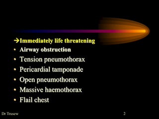
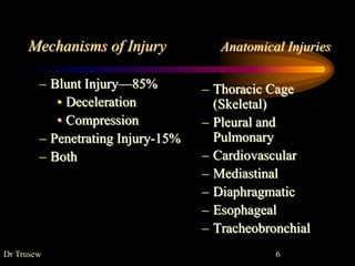
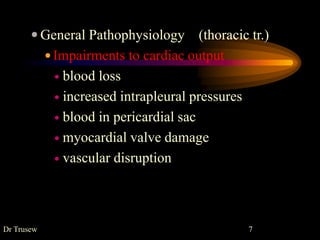
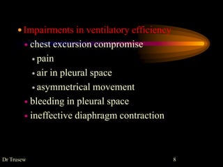
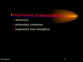
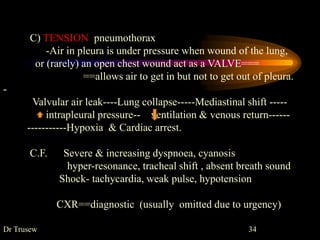
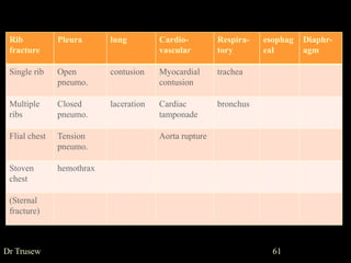
5. THORACIC TRAUMA
ď Second leading cause of trauma deaths after head
injury
ď Cause of about 10-20% of all trauma deaths
ď Many deaths due to thoracic trauma are preventable
Dr Trusew 5
6. Mechanisms of Injury Anatomical Injuries
â Blunt Injuryâ85%
⢠Deceleration
⢠Compression
â Penetrating Injury-15%
â Both
â Thoracic Cage
(Skeletal)
â Pleural and
Pulmonary
â Cardiovascular
â Mediastinal
â Diaphragmatic
â Esophageal
â Tracheobronchial
Dr Trusew 6
7. ď General Pathophysiology (thoracic tr.)
ď Impairments to cardiac output
ď blood loss
ď increased intrapleural pressures
ď blood in pericardial sac
ď myocardial valve damage
ď vascular disruption
Dr Trusew 7
8. ď Impairments in ventilatory efficiency
ď chest excursion compromise
ď pain
ď air in pleural space
ď asymmetrical movement
ď bleeding in pleural space
ď ineffective diaphragm contraction
Dr Trusew 8
9. ďImpairments in gas exchange
ď atelectasis
ď pulmonary contusion
ď respiratory tract disruption
Dr Trusew 9
10. ď Often result in:
ď Hypoxia
ď hypovolemia
ď pulmonary V/P mismatch
ď ď in intrathoracic pressure relationships
ď Hypercarbia
ď ď in intrathoracic pressure relationships
ď ďŻ level of consciousness
ď Acidosis
ď hypo perfusion of tissues (metabolic)
Dr Trusew 10
13. RIB FRACTURE
1).Single rib fracture
often trivial ,causeâdirect injury or excessive flexion
common site âat costal angle or middle of the shaft.
--5â9 th ribs ,not protected
ď§ 1 & 2 ribs fracture
requires high force
potentially serious chest injuryâ aorta, bronchi,
subclavian artery /vein
30% with this # may die
ď§ 10--12 th ribs fracture
may result in damage of LIVER/ SPLEEN/KIDNEYS
Dr Trusew 13
15. ď Assessment Findings ==rib #
ď Localized pain, tenderness
Increases on palpation or when patient:
Coughs
Moves
Breathes deeply
ď âSplintedâ Respirations
ď Instability in chest wall, Crepitus
ď Deformity and discoloration
ď Associated pneumo or hemothorax
Dr Trusew 15
16. Rxâ analgesics, intercostal nerve block,
assurance.
--strapping is may be needed
--elderly may require cough assistance &
intratracheal suction
CXR done initially and after 24 hrs to rule out
pneumothorax or hemothorax
Dr Trusew 16
17. 3).FLAIL CHEST
when several ribs are # in two places
either in one side of the chest (or both side of
sternum)
=result in segments of ribs with no chest wall
attachments (flail segment)
Paradoxical movement
flail segment âsucked in during Inspiration (-ve ITP)
--pushed out during Expiration
Dr Trusew 17
18. ⢠Flail chest
⢠A flail chest occurs when a segment of the
chest wall does not
⢠have bony continuity with the rest of the
thoracic cage.
⢠This condition usually results from blunt
trauma associated with multiple rib
fractures, i.e. three or more ribs fractured in
two or more places.
19. ⢠The blunt force required to disrupt the
integrity of the
⢠thoracic cage typically produces an
underlying pulmonary contusion
⢠as well. The diagnosis is made clinically,
not by radiography.
⢠On inspiration the loose segment of the
chest wall is displaced
⢠inwards and less air therefore moves into
the lungs.
20. ⢠To confirm
⢠the diagnosis the chest wall can be observed
for paradoxical
⢠motion of a chest wall segment for several
respiratory cycles and
⢠during coughing. Voluntary splinting as a
result of pain, mechanically
⢠impaired chest wall movement and the
associated lung
⢠contusion are all causes of the hypoxia.
21. ⢠The patient is also at high
⢠risk of developing a pneumothorax or
haemothorax.
⢠Traditionally, treatment consisted of
mechanical ventilation to
⢠âinternally splintâ the chest until fibrous
union of the broken ribs
⢠occurred. The price for this was
considerable in terms of intensive
⢠care unit resources and ventilation-
dependent morbidity
22. Currently, treatment consists of oxygen
administration, adequate
⢠analgesia (including opiates) and
physiotherapy. If a chest tube is
⢠in situ, intrapleural local analgesia can be
used as well. Ventilation
⢠is reserved for cases developing respiratory
failure despite adequate
⢠analgesia and oxygen.
23. ⢠Surgery to stabilise the flail chest is
⢠currently in use again; it may be useful in a
selected group with
⢠isolated or severe chest injury and
pulmonary contusion who
⢠have been shown to benefit from internal
operative fixation of
⢠the flail segment.
24. Paradoxical breathing
Air moves from one lung to other ( pendulum movement)
not being replaced from out side.
==hypoxia, hypercapnea & respiratory failure
this air movement cause mediastinum to shift to & fro
==inadequate circulation leading to shock.
Rxâpadding or strapping
- place him on affected side-chest splinted/normal lung up
- skeletal traction/surgical fixation ârarely done
-severe--- intubation & mechanical ventilation (IPPR)
Dr Trusew 24
25. 4).STOVE IN CHEST
by local crushing force cause depression of chest wall
Rx âstrapping
--elevation of depressed segment.
STERNAL FRACTURE
uncommonâlarge force is required
in steering wheel injury
high mortality âinjury to heart ,aorta, trachea...
Rx ânon-displaced #-analgesics, observe for cardiac injury
--displaced # --surgical fixation.
Dr Trusew 25
26. PLEURA
1. PNEUMOTHORAX
==Collection of air in the pleural cavity.
Incidence=20-30% in blunt trauma
~100% in penetrating
TYPES
A) Closed pneumothorax â(simple)
Cause = by a fractured rib, lacerate lung
Morbidity & Mortality dependent on
extent of atelectasis
associated injuries
Dr Trusew 26
27. ď Pathophysiology
ď Air enters pleural space causing partial lung collapse
ď Small tears self-seal
ď larger tears may progress
ď Usually well-tolerated in the young & healthy
ď Severe compromise can occur in the elderly or patients
with pulmonary disease
ď Degree of distress depends on amount and speed of
collapse
Dr Trusew 27
28. Rx=If air in pleura is smallâNo Rx ,(will be absorbed slowly)
only repeat CXR after few hours to see if it
increase.
If large air-(>3cm from apex) ==Intercostal tube
C.F. Little air---no symptom
Large air---dyspnoea, sign of rib #
CXR-air in pleural cavity.
Dr Trusew 28
29. B) Open pneumothorax (sucking wound of chest)
penetrating injury of chest wallâair enter
==lung collapse âmediastinal shift âhypoventilation
-- low cardiac out put
C.F. Chest wound, pain , audible air leak,
tachypnea, dyspnea,...
Tracheal shift, hyper-resonant
CXR-absent lung markings, pleura seen as faint line
Rx âseal chest wound == strapping ,proper wound repair
--intercostal tube drainage
Dr Trusew 29
30. ⢠Open pneumothorax (âsucking chest
woundâ)
⢠This is due to a large open defect in the
chest (> 3 cm), leading
⢠to equilibration between intrathoracic and
atmospheric pressure.
⢠;
31. ⢠Air accumulates in the hemithorax (rather
than in the lung) with
⢠each inspiration, leading to profound
hypoventilation on the
⢠affected side and hypoxia. Signs and
symptoms are usually proportionate
⢠to the size of the defect. If there is a
valvular effect,increasing amounts of air
will result in a tension pneumothorax
32. ⢠Initial management consists of promptly
closing the defect
⢠with a sterile occlusive plastic dressing (e.g.
Opsite), taped on
⢠three sides to act as a flutter-type valve. A
chest tube is inserted
⢠as soon as possible in a site remote from the
injury site.
33. ⢠Definitive treatment may warrant formal
debridement and closure, preferably
⢠in the operating room, and all such patients
should be referred early.
⢠The following points are important in the
management of an open pneumothorax:
⢠a common problem is using too small a tube
â a 28FG or larger tube should be used in an
adult
34. C) TENSION pneumothorax
-Air in pleura is under pressure when wound of the lung,
or (rarely) an open chest wound act as a VALVE===
==allows air to get in but not to get out of pleura.
-
Valvular air leak----Lung collapse-----Mediastinal shift -----
intrapleural pressure-- ventilation & venous return------
-----------Hypoxia & Cardiac arrest.
C.F. Severe & increasing dyspnoea, cyanosis
hyper-resonance, tracheal shift , absent breath sound
Shock- tachycardia, weak pulse, hypotension
CXR==diagnostic (usually omitted due to urgency)
Dr Trusew 34
35. Tension pneumothorax
⢠A tension pneumothorax develops when a
âone-way valveâ air
⢠leak occurs either from the lung or through
the chest wall. Air is
⢠forced into the thoracic cavity without any
means of escape, completely
⢠collapsing the affected lung.
36. ⢠The mediastinum is displaced to the
opposite side, decreasing venous return and
compressing opposite lung.
⢠The most common causes are penetrating
chest trauma, blunt chest trauma with
parenchymal lung injury and air leak that
did not spontaneously close, iatrogenic lung
punctures (e.g. due to subclavian central
venepuncture) and mechanical +pressure
ventilation.
37. ⢠The clinical presentation is dramatic. The
patient is panicky
⢠with tachypnoea, dyspnoea and distended
neck veins (similar to
⢠pericardial tamponade). Clinical
examination can reveal tracheal
⢠deviation (a late finding â not necessary to
clinically confirm diagnosis),
hyperresonance and absent breath sounds
over the affected hemithorax.
38. ⢠Tension pneumothorax is a clinical
diagnosis
⢠and treatment should not be delayed by
waiting for radiological confirmation
⢠Tx consists of immediate decompression
and is
⢠managed initially by rapid insertion of a
large-bore needle into
⢠the second intercostal space in the mid-
clavicular line of the affected hemithorax.
39. ⢠This is immediately followed by insertion
of
⢠a chest tube through the fifth intercostal
space in the anterior axillary line.
40. Rx urgentâ wide boar needle at 2nd intercostal space
later â proper intercostal tube drain
Needle Thoracostomy Review
Decompress with 14 g (large bore) needle
Midclavicular line: 2nd intercostals space
Midaxillary line: 4-5th intercostals space
Go over superior margin of rib to avoid blood
vessels
Be careful not to kink or bend needle or catheter
If available, attach a one-way valve
Dr Trusew 40
41. 2.HEMOTHORAX
Collection of Blood in the pleural cavity.
ď Pathophysiology
ď Most common result of major trauma to the chest wall
ď Present in 70 - 80% of penetrating and major non-
penetrating trauma cases
ď Associated with pneumothorax
ď Rib fractures are frequent cause
ď Blood from intercostal, lungs & bronchial blood
vessels or internal mammary artery
Dr Trusew 41
42. ⢠Massive haemothorax
⢠The most common cause of massive
haemothorax in blunt injury
⢠is continuing bleeding from torn intercostal
vessels or occasionally z internal mammary
artery.
⢠Accumulation of blood in a hemithorax can
significantly compromise
⢠respiratory efforts by compressing the lung
and preventing adequate ventilation.
43. ⢠Such massive accumulation of blood
⢠presents as haemorrhagic shock with flat
neck veins, unilateral
⢠absence of breath sounds and dullness to
percussion. The treatment
⢠consists of correcting the hypovolaemic
shock, insertion of
⢠an intercostal drain and, in some cases,
intubation.
44. ⢠Blood in the pleural space should be
removed as completely
⢠and rapidly as possible to prevent on-going
bleeding, empyema or
⢠a late fibrothorax. Clamping a chest drain to
tamponade a massive
⢠haemothorax is usually not helpful.
45. ⢠Initial drainage of more than 1500 ml of
blood or on-going
⢠haemorrhage of more than 200 ml hâ1 over
3â4 hours is generally
⢠considered an indication for urgent
thoracotomy.
⢠The following points are important in the
management of massive haemothorax:
46. ⢠Bleeding may vary from minor to
massive.the pleural space can accumulate
up to 3 liter. massive HT is generally result
from major pul vascular injuries or major
arterial wound , whereas minor lung injuries
cause a small HT.(grade 1 = <
1000ml,grade 2=1000_ 1500ml, and grade
3= >1500ml)
47. ď clinical examination may be misleading if
only done from the
⢠supine position, as the lung may âfloatâ on
the haemothorax and breath sounds
anteriorly may be normal;
⢠caution is required in a case that drains
more than 500 ml into the drainage bottle
but has persistent dullness or radiographic
opacification.
48. ď Accumulated blood can eventually produce
a tension hemothorax
ď Shifting the mediastinum producing
ventilatory impairment
cardiovascular collaps
-The blood in pleura doesn't clot for a long
time â b/e of chest movement
- high risk of pyothorax
Dr Trusew 48
49. ď Clinical Findings
ď Hx of violence & chest pain
ď Tachypnea & dyspneoa
ď Shock
ď Collapsed neck veins
ď Trachea & apex shifted to the other side
ď Dullness on percussion
ď Decreased breath sounds
Dr Trusew 49
50. Ixâ CXRâobliterated costophrenic angle
-- air-fluid level or meniscial sign
Rx
Needle thoracostomy if tension or unable to differentiate
it from tension pneumothorax
1.)Inter costal tube drain (Tube thoracostomy)
2.)THORACOTOMY
ďDrainage of blood is >1000ml or 100ml/hr for 4 hrs
ďClotted or infected hemothorax is present
Dr Trusew 50
51. Pulmonary injuries
1)Lung contusion
Dr Trusew 51
ď Pathophysiology
ď Blunt trauma to the chest
ď Deceleration injury cause lung to strike chest wall
ď Crush trauma
ď high energy shock wave from explosion
ď high velocity missile wound
ď§ Most common injury from blunt thoracic
trauma
ď Hemorrhage & interstitial edema occur ,
results in consolidation of lung tissue
53. Dr Trusew 53
Rx
Usually self-limiting, if there are no other conditions
Fluid restriction
Chest physiotherapy
rarely ventilation
2.Lung laceration
ď minor lacerationâHemopneumothorax
--usually intercostal tube is enough
ďMajor lacerationâHemopneumothorax
-intercostal tube/thoracotomy
54. Tracheobronchial Trauma
Rare injury ,High mortality
1.Tracheal Injury
-- common in cervical trachea , cut injuries
clinical pictureâAir way obstruction
--Emphysema (mediastinal & cervical)
--pneumothorax
--voice impairment
Rx==Intubation
Tracheostomy
Surgery and repair
Dr Trusew 54
55. Dr Trusew 55
2.Bronchial Injury
commonly(80%) occur at or near carina.
Rapid air leak into pleural cavity occur
Clinical picture
--Respiratory Distress ==dyspnea, tachypnea
--Subcutaneous emphysema
--Hemoptysis
ď Sign of tension pneumothorax unresponsive to needle
decompression
--Uncontrolled air leak
Rx ==Endotracheal intubation
==Surgery --bronchoplasty
56. Cardiovascular Trauma
⢠Any patient with significant blunt or penetrating trauma to chest
has heart/great vessel injury until proven otherwise
1. Myocardial contusion
ď most common blunt injury to the heart
ď usually anterior chest impact-steering wheel injury
ď Features=Arrhythmias, low COP, cardiac tamponade
ď May cause hypotension unresponsive to fluid or drug
therapy
Dr Trusew 56
57. Dr Trusew 57
2.Pericadial tamponade
is accumulation of blood in pericardiac sac.
ď Usually associated with penetrating trauma ,rare in blunt
ď GSW wounds have higher mortality than stab wounds
ď Lower mortality rate if isolated tamponade
Signs and Symptoms
Dyspnea;cyanosis
Resistant hypotension
Increased central venous pressure(distended neck/arm
veins in presence of decreased arterial BP)
Enlarged cardiac dullness &decreased heart sounds
58. Dr Trusew 58
Definite treatment is aspiration( pericardiocentesis)
followed by surgery
Pericardial Window
3.Rupture of Aorta
--85% die immediately
Complete ruptureâimmediate death Incomplete
ruptureâ shock , widening of mediastinum
Rx surgery and repair
Dx is usually difficult
ECG helps
Rx
59. Esophageal Injury
Most frequent causeâpenetrating trauma
-- Rare in blunt trauma can
perforate spontaneously -violent vomiting
(instrumentation) -carcinoma
Clinical finding
=pain ,hoarseness, dysphagia,respiratory distress
=subcutaneous emphysema in neck
=pleural effusion on one or both side
Rxâsurgery and repair
Dr Trusew 59
60. Diaphragmatic Rupture
Usually due to blunt trauma
but may occur with penetrating trauma.
Commonly on the left diaphragm (R-liver)
Abdominal compressionâcontents rupture through the
diaphragm-bowel obstructed ,lung compressed,
mediastinum shift.
C.F.
Respiratory distress, dullness, decrease breath sound
Dx by U/S , CXR
Rx â Surgical repair
Dr Trusew 60
62. EMPYEMA
⢠Is a collection of pus in the pleural cavity
cause: ~ is always secondary
a)Chest wall --wounds,osteomyelitis
b)Lungs --pneumonia,abscess,TB,....
c)postop.
d)Rupture of esophagus
e)Extension of sub phrenic ,hepatic abscess
f)Hematogenous spread â(septicemia)
Dr Trusew 62
63. Dr Trusew 63
(Bacteriology ,pathogenesis ,symptoms ,signs-local & general)
Ix Hct, wbc (anemia,leucocytosis)
sputumâgram`s stain AFS
pleural fluid analysis
CXR
Rx a)General: correct anemia ,high protein diet, vitamins.....
systemic antibiotics
b)Local : AIM
= eradication of infection
=removing dead space to allow expansion of lung
choice of Rx depend on
Pus-thickness & amount
Patient`s clinical state
64. Dr Trusew 64
1.Aspiration
2.Intercostal chest tube
3.Rib resection methods of Rx
4.Decortication
5.Thoracoplasty
1.)Aspiration
--used when the pus is thin and remain thin
--wide bore needle at 8/9th ICS posterior axillary line
done every 2--3rd day
success= pus remain thin ,less purulent, amount less,
lung expand, patient afebrile
65. Dr Trusew 65
2.)Intercostal tube drainage
--used when pus is thick or aspiration fails
--tube is inserted at 8/9th ICS posterior axillary/scapular line
(often the tube is blocked by fibrin ,so followed by (3)
3.)Rib resection
--used when . pus is thick & lots of fibrin is present
.<60ml/day pus drain of chest tube
--short tube is inserted after rib resection at lowest level of
empyema .Pus escape into dressing gauze which is
changed twice
66. Dr Trusew 66
5.)Thoracoplasty
is obliteration of the pleural space by mobilizing the
chest wall and making it fall against the underlying lung.
--used when (4) fails or after pneumonectomy
4.)Decortication
is surgical removal of fibrous walls of the empyema
cavity(visceral/parietal pleura)from lung, chest wall &
diaphragm.
LUNG expands as a result.
67. Dr Trusew 67
LUNG ABSCESS
~50% from aspiration pneumonia
--present like pneumonia, but sputum becomes fetid
( )
Rx âmost respond to medical Rx
~5% may need surgical intervention
--Drainageâ done u/s or CT guided
--Resection â eg. Lobectomy
68. Dr Trusew 68
BRONCHIECTASIS
is a chronic irreversible dilatation of the medium-
sized bronchi.
Rx-medical
surgery in localized lesion âsegmental resection
--lobectomy
--pneumonectomy
69. Dr Trusew 69
PULMONARY TUBERCULOSIS
The role of surgery in Mx of TB is gradually decreasing
Indications:
. suspicious lesion on CXR (CA)
.chronic tuberculous abscess ,resistant to
chemotherapy
. hemoptysis life âthreatening
TWO types:
ď Excision surgeryâsegment , lobe, lung.
ď Collapse therapy âartificial pneumothorax....
--thoracoplasty
70. Dr Trusew 70
Tumours of lung and bronchi
most common malignancy in men,
next to breast in female
Factors: smoking
irritant chemicalsâsulphur smokes, fumes
Types: . squamous carcinoma
. oat cell carcinoma
. adenocarcinoma
C.F: cough ,hemoptysis
bronchial obstructionâemphysema, atelectasis
secondary infectionâconsolidation, abscess
pressure effectâdysphagia, stridor
71. Dr Trusew 71
Ix = CXR
bronchoscopy
cytologyâsputum , exfoliative
bronchography ....
Rx
1.Excisionâlobectomy
--pneumonectomy
2.Radiation therapy
Editor's Notes
Condition which affect venous return and cardiac contractlity ,