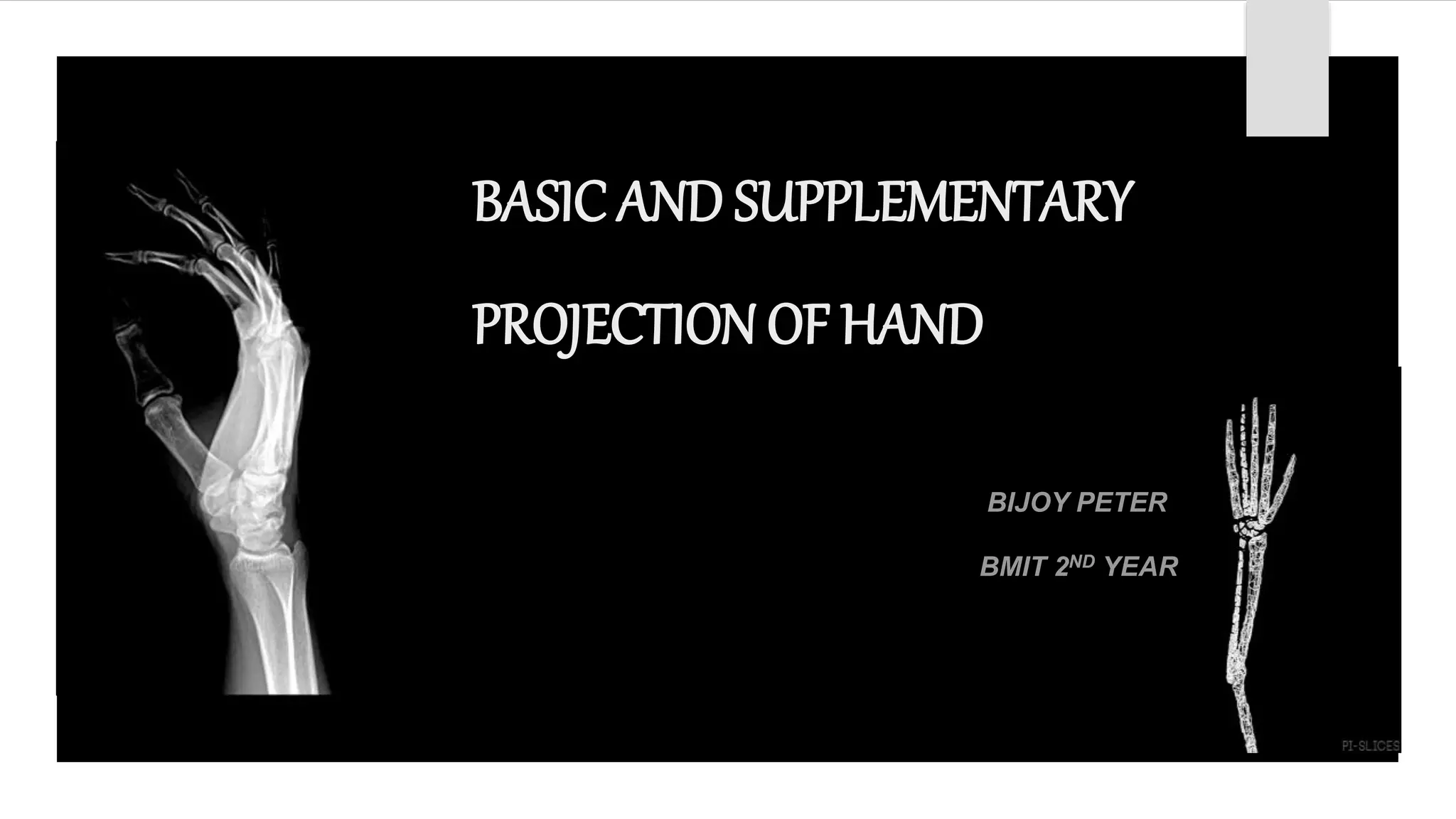
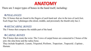
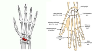
BASIC AND SUPPLEMENTARY
PROJECTION OF HAND
BIJOY PETER
BMIT 2ND YEAR
ANATOMY
There are 3 major types of bones in the hand itself, including:
PHALANGES
The 14 bones that are found in the fingers of each hand and also in the toes of each foot.
Each finger has 3 phalanges (the distal, middle, and proximal); the thumb only has 2.
METACARPAL BONES
The 5 bones that compose the middle part of the hand.
CARPAL BONES
The 8 bones that create the wrist. The 2 rows of carpal bones are connected to 2 bones of the
arm--the ulna bone and the radius bone.
They include Scaphoid , Lunate, Triquetral, Pisiform , Trapezium , Trapezoid , Capitate ,
Hamate
JOINTS :
Radiocarpal Joint
Its between radius and carpus.
 Carpometacarpal Joint
It connect the carpal bone to the metacarpus.
 Metacarpophalangeal joint
Its between metacarpal bones and phalanges.
INDICATIONS
 Trauma with suspected fracture
Suspected metacarpal dislocation
Foreign body detection and localisation
Investigation of joint pain and deformity
Rheumatoid arthritis
Osteoarthrosis
Projections
BASIC PROJECTIONS
POSTERO-ANTERIOR VIEW
OBLIQUE VIEW
LATERAL VIEW
SUPPLEMENTARY PROJECTIONS
BALL-CATCHER VIEW (NORGAARD PROJECTION)
The Hand PA view is apart of a two view series metacarpals, phalanges, carpal bones
and distal radial ulnar joint.
 Patient is seated alongside the table
 The forearm is pronated and placed on the table with palmer surface of the hand in contact
with the image receptor
 The fingers are separated and extended but relaxed to ensure that they remain in contact with
the image receptor.
 The wrist is adjusted so that the radial and ulna styloid processes are equidistant from the
image receptor.
The image should demonstrate all the phalanges, including the soft tissue of the
finger tips, the carpal and metacarpal bones and distal end of the radius and ulna.
The interphalangeal and metacarpo-phalangeal and carpometacarpal joints should be
demonstrated clearly.
No rotation of hand
A) POSTERO-ANTERIOR PROJECTION
1) CENTRING POINT
 THIRD METACARPAL HEAD
2) COLLIMATION
 LATERALLY TO THE SKIN MARGINS
 PROXIMAL TO INCLUDE DISTAL RADIO-ULNAR JOINT
 DISTAL TO THE TIPS OF THE DISTAL PHALANGES
3) ORIENTATION
 PORTRAIT
4) DETECTOR SIZE
 18cm x 24cm
TECHNICAL FACTORS
5) EXPOSURE
 50-60kVp
 1-5mAS
6) SID
 100 cm
7) GRID
 NO
POSITION OF PATIENT
 Patient is seated alongside the table
 From the basic postero-anterior position ,the hand is externally rotated 45 with the fingers
extended.
 The fingers should be separated slightly and the hand supported on a 45 non –opaque
pad.
 A sand bag is placed over the lower end of the forearm for immobilisation.
ESSENTIAL IMAGE CHARACTERISTICS
The image should demonstrate all the phalanges, including the soft tissue of the fingertips,
the carpal and metacarpal bones and the distal end of the radius and ulna.
The correct degree of rotation has been achieved when the heads of the 1st and the 2nd
metacarpals are seen separated whilst those of the 4th and 5th are just superimposed
A. POSTERO-ANTERIOR PROJECTION
1) CENTRING POINT
2) COLLIMATION
a) ORIENTATION
b) DETECTOR SIZE
 Third metacarpal head
 Laterally to the skin margins
 Proximal to include distal radioulnar joint
 Distal to the tips of the distal phalanges
 Portrait
 18cm X 24cm
Technical Factors
f) EXPOSURE
 50-60 KVp
 1-5 mAs
e) SID
 100 CM
f) GRID
 NO
LATERAL
POSITION OF PATIENT
 From the postero-anterior(DP) , the hand is externally rotated 90
 The palm of the hand is perpendicular to the image receptor with the fingers
extended and thumb abducted and supported parallel to the image receptor on a non
opaque pad.
 The radial and ulnar styloid processes are superimposed
DIRECTION AND LOCATION OF THE X-RAY BEAM
The collimated vertical beam is centred over the head of the 2nd metacarpal.
ESSENTIAL IMAGE CHARACTERISTICS
 The image should include the finger tips, including soft tissue , and radial and
ulnar styloid processes.
 The heads of the metacarpal should be super imposed . The thumb should be
demonstrated clearly without superimposition of other structures.
TECHNICAL FACTORS
A) LATERAL PROJECTION
1) CENTRING POINT
 Over the head of the second metacarpal
2) COLLIMATION
 Anteroposterior to the skin margins
 Distal to the tips of the fingers
 Proximal to include one-third of the distal radius
and ulna
3) ORIENTATION
 Portrait
4) DETECTOR SIZE
 18cmx24cm
5) EXPOSURE
 50-60 KVp
 1-5 mAs
6) SID
 100 CM
7) GRID
 NO
BALL CATCHER VIEW (NORGAARD PROJECTION)
POSITION OF PATIENT
 Patient may be seated alongside or facing the table
 Both hands are supinated with their dorsal surfaces are placed on the cassette/FPDs
 The hands are then rotated medially by 45 degree to assume a position likened to one
about to receive or catch a ball.
ESSENTIAL IMAGE CHARACTERISTICS
 The image should demonstrate all the phalanges, including the soft tissue of the
finger tips, the carpal and metacarpal bones and distal end of the radius and ulna.
 The exposure factors selected must produce an accurate EI and optically
demonstrate joint details.
 The heads of the metacarpals should not be superimposed
3) COLLIMATION
 Laterally to the skin margins
 Distal to the skin margins of the finger
 Proximal to the include one-third of the distal radius and ulna
4) ORIENTATION
 Landscape
TECHNICAL FACTORS
7) SID
 100CM
8) GRID
 NO
REFERENCE
CLARKS POSITIONING IN RADIOGRAPHY
Basic and supplementary projection of hand

Basic and supplementary projection of hand

  • 1.
    BASIC AND SUPPLEMENTARY PROJECTIONOF HAND BIJOY PETER BMIT 2ND YEAR
  • 2.
    ANATOMY There are 3major types of bones in the hand itself, including: PHALANGES The 14 bones that are found in the fingers of each hand and also in the toes of each foot. Each finger has 3 phalanges (the distal, middle, and proximal); the thumb only has 2. METACARPAL BONES The 5 bones that compose the middle part of the hand. CARPAL BONES The 8 bones that create the wrist. The 2 rows of carpal bones are connected to 2 bones of the arm--the ulna bone and the radius bone. They include Scaphoid , Lunate, Triquetral, Pisiform , Trapezium , Trapezoid , Capitate , Hamate
  • 3.
    JOINTS : Radiocarpal Joint Itsbetween radius and carpus.  Carpometacarpal Joint It connect the carpal bone to the metacarpus.  Metacarpophalangeal joint Its between metacarpal bones and phalanges.
  • 5.
    INDICATIONS  Trauma withsuspected fracture Suspected metacarpal dislocation Foreign body detection and localisation Investigation of joint pain and deformity Rheumatoid arthritis Osteoarthrosis
  • 6.
    Projections BASIC PROJECTIONS POSTERO-ANTERIOR VIEW OBLIQUEVIEW LATERAL VIEW SUPPLEMENTARY PROJECTIONS BALL-CATCHER VIEW (NORGAARD PROJECTION)
  • 7.
    The Hand PAview is apart of a two view series metacarpals, phalanges, carpal bones and distal radial ulnar joint.  Patient is seated alongside the table  The forearm is pronated and placed on the table with palmer surface of the hand in contact with the image receptor  The fingers are separated and extended but relaxed to ensure that they remain in contact with the image receptor.  The wrist is adjusted so that the radial and ulna styloid processes are equidistant from the image receptor.
  • 8.
    The image shoulddemonstrate all the phalanges, including the soft tissue of the finger tips, the carpal and metacarpal bones and distal end of the radius and ulna. The interphalangeal and metacarpo-phalangeal and carpometacarpal joints should be demonstrated clearly. No rotation of hand
  • 10.
    A) POSTERO-ANTERIOR PROJECTION 1)CENTRING POINT  THIRD METACARPAL HEAD 2) COLLIMATION  LATERALLY TO THE SKIN MARGINS  PROXIMAL TO INCLUDE DISTAL RADIO-ULNAR JOINT  DISTAL TO THE TIPS OF THE DISTAL PHALANGES 3) ORIENTATION  PORTRAIT 4) DETECTOR SIZE  18cm x 24cm TECHNICAL FACTORS 5) EXPOSURE  50-60kVp  1-5mAS 6) SID  100 cm 7) GRID  NO
  • 11.
    POSITION OF PATIENT Patient is seated alongside the table  From the basic postero-anterior position ,the hand is externally rotated 45 with the fingers extended.  The fingers should be separated slightly and the hand supported on a 45 non –opaque pad.  A sand bag is placed over the lower end of the forearm for immobilisation. ESSENTIAL IMAGE CHARACTERISTICS The image should demonstrate all the phalanges, including the soft tissue of the fingertips, the carpal and metacarpal bones and the distal end of the radius and ulna. The correct degree of rotation has been achieved when the heads of the 1st and the 2nd metacarpals are seen separated whilst those of the 4th and 5th are just superimposed
  • 13.
    A. POSTERO-ANTERIOR PROJECTION 1)CENTRING POINT 2) COLLIMATION a) ORIENTATION b) DETECTOR SIZE  Third metacarpal head  Laterally to the skin margins  Proximal to include distal radioulnar joint  Distal to the tips of the distal phalanges  Portrait  18cm X 24cm Technical Factors f) EXPOSURE  50-60 KVp  1-5 mAs e) SID  100 CM f) GRID  NO
  • 14.
    LATERAL POSITION OF PATIENT From the postero-anterior(DP) , the hand is externally rotated 90  The palm of the hand is perpendicular to the image receptor with the fingers extended and thumb abducted and supported parallel to the image receptor on a non opaque pad.  The radial and ulnar styloid processes are superimposed DIRECTION AND LOCATION OF THE X-RAY BEAM The collimated vertical beam is centred over the head of the 2nd metacarpal.
  • 15.
    ESSENTIAL IMAGE CHARACTERISTICS The image should include the finger tips, including soft tissue , and radial and ulnar styloid processes.  The heads of the metacarpal should be super imposed . The thumb should be demonstrated clearly without superimposition of other structures.
  • 17.
    TECHNICAL FACTORS A) LATERALPROJECTION 1) CENTRING POINT  Over the head of the second metacarpal 2) COLLIMATION  Anteroposterior to the skin margins  Distal to the tips of the fingers  Proximal to include one-third of the distal radius and ulna 3) ORIENTATION  Portrait 4) DETECTOR SIZE  18cmx24cm 5) EXPOSURE  50-60 KVp  1-5 mAs 6) SID  100 CM 7) GRID  NO
  • 18.
    BALL CATCHER VIEW(NORGAARD PROJECTION) POSITION OF PATIENT  Patient may be seated alongside or facing the table  Both hands are supinated with their dorsal surfaces are placed on the cassette/FPDs  The hands are then rotated medially by 45 degree to assume a position likened to one about to receive or catch a ball. ESSENTIAL IMAGE CHARACTERISTICS  The image should demonstrate all the phalanges, including the soft tissue of the finger tips, the carpal and metacarpal bones and distal end of the radius and ulna.  The exposure factors selected must produce an accurate EI and optically demonstrate joint details.  The heads of the metacarpals should not be superimposed
  • 20.
    3) COLLIMATION  Laterallyto the skin margins  Distal to the skin margins of the finger  Proximal to the include one-third of the distal radius and ulna 4) ORIENTATION  Landscape TECHNICAL FACTORS 7) SID  100CM 8) GRID  NO
  • 21.