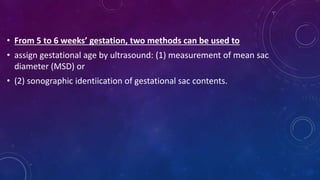
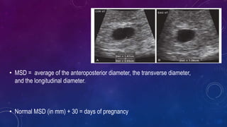
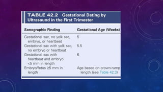
EARLY OG USG PROTOCOL
INDICATIONS
1. Confirm presence of intrauterine pregnancy
2. Evaluate:
a. Suspected ectopic pregnancy
b. Pelvic pain
c. Maternal pelvic masses and/or uterine abnormalities
d. Suspected hydatidiform mole
3. Identify cause of vaginal bleeding
4. Estimate gestational age
5. Diagnose or evaluate multiple gestations
6. Confirm cardiac activity
HISTORY
• Gravidity, Parity
• Any fertility treatment
• LMP
• Gynecological history
LOOK FOR
• Uterus –longitudinal and transverse
• Both ovaries and adnexa
• Cervix and POD
• Gestational sac – longitudinal and transverse
• From 5 to 6 weeks’ gestation, two methods can be used to
• assign gestational age by ultrasound: (1) measurement of mean sac
diameter (MSD) or
• (2) sonographic identiication of gestational sac contents.
• MSD = average of the anteroposterior diameter, the transverse diameter,
and the longitudinal diameter.
• Normal MSD (in mm) + 30 = days of pregnancy
SONOGRAPHIC TIMELINE (TVS)
• The earliest sonographic finding of pregnancy is a small rounded intrauterine fluid collection in the
central portion of the uterus.
• The fluid collection may or may not demonstrate the intradecidual sign or the double sac sign. When
ultrasound demonstrates an intrauterine fluid collection with no visible yolk sac or embryo, the
pregnancy can be accurately assigned a gestational age of 5.0 weeks.
•
• When the fluid collection contains a yolk sac but no embryo, the
pregnancy can be accurately assigned a gestational age of 5.5
weeks.
• Yolk sac can be visualized at 4 weeks
• The demonstration of a yolk sac may be critical in diferentiating an
early intrauterine gestational sac from a pseudosac.
• Although the double-decidual sign is not 100% specific for
presence of an IUP, the identiication of a yolk sac within the early
gestational sac is diagnostic of IUP.
• The number of yolk sacs present can be helpful in determining
amnionicity of a multifetal pregnancy
• Yolk sac should be round with an anechoic centre.
• It should not be calcified, misshapen or >6mm from the inner to
inner diameter.
• Yolk sacs larger than 6 mm are usually indicative of an abnormal
pregnancy.
• The presence of a small embryo, less than 2 mm in size, with the embryonic
heartbeat visible adjacent to the yolk sac, can be accurately dated as 6.0 weeks’
gestation .
• The accuracy of these early sonographic milestones for dating pregnancy is ±0.5
week
• From 6 weeks until the end of the first trimester, gestational age correlates closely
with the crown-rump length (CRL) of the embryo or fetus
• The term embryo is commonly used up to 10 weeks’ gestation, and the term fetus
applies thereafter.
CRL( CROWN RUMP LENGHTH
• The CRL is the length of the embryo or fetus from the top of its head to the bottom of its torso.
• It is measured as the longest dimension of the embryo, excluding the yolk sac and extremities
• The CRL can be used to assign gestational age accurately up to 14 weeks because there is little
biologic variability in fetal length up until that age
HEART RATE
• Confirm heart beat and rate with M –Mode evaluation
• Doppler traces is not recommended in the 1st trimester
• The very early embryonic heart will be a subtle flicker
• Initially heart rate maybe slow It may be below 100 beats per
minute but this will increase to between 120- 180 beats per
minute by 7 weeks.
SUBCHORIONIC HEMATOMA
• A subchorionic hematoma appears on
ultrasound as an irregular hypoechoic
space, sometimes crescent-shaped,
around part of the gestational sac.
• The hematoma usually has internal
echoes and is separated from the fluid
inside the gestational sac by a thick
band of tissue
REFERENCES
• Rumacks
• Callens USG in obstetrics
Early OG USG Protocol (1).pptx

Early OG USG Protocol (1).pptx

  • 1.
    EARLY OG USGPROTOCOL
  • 2.
    INDICATIONS 1. Confirm presenceof intrauterine pregnancy 2. Evaluate: a. Suspected ectopic pregnancy b. Pelvic pain c. Maternal pelvic masses and/or uterine abnormalities d. Suspected hydatidiform mole 3. Identify cause of vaginal bleeding 4. Estimate gestational age 5. Diagnose or evaluate multiple gestations 6. Confirm cardiac activity
  • 3.
    HISTORY • Gravidity, Parity •Any fertility treatment • LMP • Gynecological history
  • 4.
    LOOK FOR • Uterus–longitudinal and transverse • Both ovaries and adnexa • Cervix and POD • Gestational sac – longitudinal and transverse
  • 5.
    • From 5to 6 weeks’ gestation, two methods can be used to • assign gestational age by ultrasound: (1) measurement of mean sac diameter (MSD) or • (2) sonographic identiication of gestational sac contents.
  • 6.
    • MSD =average of the anteroposterior diameter, the transverse diameter, and the longitudinal diameter. • Normal MSD (in mm) + 30 = days of pregnancy
  • 7.
    SONOGRAPHIC TIMELINE (TVS) •The earliest sonographic finding of pregnancy is a small rounded intrauterine fluid collection in the central portion of the uterus. • The fluid collection may or may not demonstrate the intradecidual sign or the double sac sign. When ultrasound demonstrates an intrauterine fluid collection with no visible yolk sac or embryo, the pregnancy can be accurately assigned a gestational age of 5.0 weeks. •
  • 8.
    • When thefluid collection contains a yolk sac but no embryo, the pregnancy can be accurately assigned a gestational age of 5.5 weeks. • Yolk sac can be visualized at 4 weeks • The demonstration of a yolk sac may be critical in diferentiating an early intrauterine gestational sac from a pseudosac. • Although the double-decidual sign is not 100% specific for presence of an IUP, the identiication of a yolk sac within the early gestational sac is diagnostic of IUP. • The number of yolk sacs present can be helpful in determining amnionicity of a multifetal pregnancy • Yolk sac should be round with an anechoic centre. • It should not be calcified, misshapen or >6mm from the inner to inner diameter. • Yolk sacs larger than 6 mm are usually indicative of an abnormal pregnancy.
  • 9.
    • The presenceof a small embryo, less than 2 mm in size, with the embryonic heartbeat visible adjacent to the yolk sac, can be accurately dated as 6.0 weeks’ gestation . • The accuracy of these early sonographic milestones for dating pregnancy is ±0.5 week • From 6 weeks until the end of the first trimester, gestational age correlates closely with the crown-rump length (CRL) of the embryo or fetus • The term embryo is commonly used up to 10 weeks’ gestation, and the term fetus applies thereafter.
  • 10.
    CRL( CROWN RUMPLENGHTH • The CRL is the length of the embryo or fetus from the top of its head to the bottom of its torso. • It is measured as the longest dimension of the embryo, excluding the yolk sac and extremities • The CRL can be used to assign gestational age accurately up to 14 weeks because there is little biologic variability in fetal length up until that age
  • 11.
    HEART RATE • Confirmheart beat and rate with M –Mode evaluation • Doppler traces is not recommended in the 1st trimester • The very early embryonic heart will be a subtle flicker • Initially heart rate maybe slow It may be below 100 beats per minute but this will increase to between 120- 180 beats per minute by 7 weeks.
  • 12.
    SUBCHORIONIC HEMATOMA • Asubchorionic hematoma appears on ultrasound as an irregular hypoechoic space, sometimes crescent-shaped, around part of the gestational sac. • The hematoma usually has internal echoes and is separated from the fluid inside the gestational sac by a thick band of tissue
  • 15.