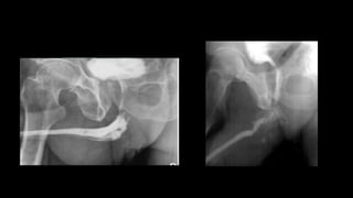
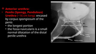
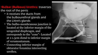
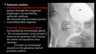
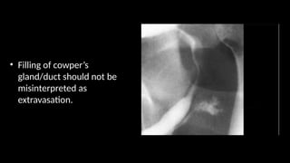
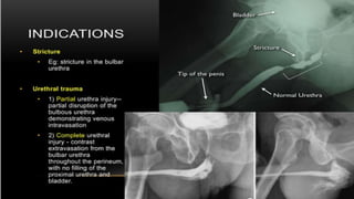
AUG IMAGING
BY
DR.ANJANA AUGUSTINE
Indications
1. Stricture
2. Urethral trauma
3. Fistulae or false passage
4. Congenital abnormalities
Contraindications
1. Acute urinary tract infection
2. Recent instrumentation
Complications Due to the Technique
1. Acute urinary tract infection
2. Urethral trauma
3. Intravasation of contrast medium, especially if excessive pressure is
used to overcome a stricture
Contrast Medium
LOCM (urograffin).
Prewarming the contrast medium
will help reduce the incidence of spasm of the external sphincter.
Preliminary Image
supine posteroanterior (PA) of the bladder base and urethra.
Technique
1. Patient supine with slightly tilted legs.
2. Using aseptic technique, the tip of the catheter is inserted so that the balloon lies
in the fossa navicularis and its balloon is inflated with 2–3mL
3. Contrast medium is injected and steep (30–45°) oblique films are taken.
• Gentle traction on the catheter is used to straighten the penis over the
ipsilateral leg and prevent urethral overlap
 Anterior urethra:
• Penile (Spongy, Pendulous)
Urethra (~16 cm long): encased
by corpus spongiosum of the
penis
• the longest portion
• the fossa navicularis is a small
normal dilatation of the distal
penile urethra
•Bulbar (Bulbous) Urethra: traverses
the root of the penis
• it receives the ducts from
the bulbourethral glands and
the Littre’s glands
• The bulbo-membranous junction is
located at the inferior margin of the
urogenital diaphragm, and
corresponds to the “cone”- Located
at 1-1.5cm distal to inferior margin
of verumontanum.
• Connecting inferior margin of
obturator foramina intersecting
urethra.
 Posterior urethra-
• Membranous Urethra (1 cm long):
passes through the urogenital
diaphragm, surrounded by
sphincter urethrae
the shortest and narrowest portion
o Least distensible segment
• Prostatic Urethra (3 cm long):
surrounded by the prostate gland;
o The verumontanum -ovoid mound of
tissue on the posterior wall, housing
the utricle and ejaculatory duct
openings.
o it is seen as a triangular
structre on the posterior wall of
prostatic urethra
• Filling of cowper’s
gland/duct should not be
misinterpreted as
extravasation.
• Fistula extending from
bulbous urethra to perineum
• PENILE FRACTURE-Contrast
extravasation in penile
urethra
• Urethral diverticulum
• MC at peno scrotal
junction.
• Condyloma accuminata-
multiple small filling defects
• MALIGNANT TUMORS:
• MC location-bulbomembranous
urethra.
• SCC-Irregular narrowing of
bulbar urethra with multiple
sinus tracts.
references
• Chapman’s guide to radiological procedures.
• APA. Gray, H. (2013). Grays Anatomy. Arcturus
Publishing.
THANKYOU

Anterior urethrogram,RETROGRADE URETHROGRAM.pptx

  • 1.
  • 3.
    Indications 1. Stricture 2. Urethraltrauma 3. Fistulae or false passage 4. Congenital abnormalities Contraindications 1. Acute urinary tract infection 2. Recent instrumentation Complications Due to the Technique 1. Acute urinary tract infection 2. Urethral trauma 3. Intravasation of contrast medium, especially if excessive pressure is used to overcome a stricture
  • 4.
    Contrast Medium LOCM (urograffin). Prewarmingthe contrast medium will help reduce the incidence of spasm of the external sphincter. Preliminary Image supine posteroanterior (PA) of the bladder base and urethra. Technique 1. Patient supine with slightly tilted legs. 2. Using aseptic technique, the tip of the catheter is inserted so that the balloon lies in the fossa navicularis and its balloon is inflated with 2–3mL 3. Contrast medium is injected and steep (30–45°) oblique films are taken. • Gentle traction on the catheter is used to straighten the penis over the ipsilateral leg and prevent urethral overlap
  • 5.
     Anterior urethra: •Penile (Spongy, Pendulous) Urethra (~16 cm long): encased by corpus spongiosum of the penis • the longest portion • the fossa navicularis is a small normal dilatation of the distal penile urethra
  • 6.
    •Bulbar (Bulbous) Urethra:traverses the root of the penis • it receives the ducts from the bulbourethral glands and the Littre’s glands • The bulbo-membranous junction is located at the inferior margin of the urogenital diaphragm, and corresponds to the “cone”- Located at 1-1.5cm distal to inferior margin of verumontanum. • Connecting inferior margin of obturator foramina intersecting urethra.
  • 7.
     Posterior urethra- •Membranous Urethra (1 cm long): passes through the urogenital diaphragm, surrounded by sphincter urethrae the shortest and narrowest portion o Least distensible segment • Prostatic Urethra (3 cm long): surrounded by the prostate gland; o The verumontanum -ovoid mound of tissue on the posterior wall, housing the utricle and ejaculatory duct openings. o it is seen as a triangular structre on the posterior wall of prostatic urethra
  • 8.
    • Filling ofcowper’s gland/duct should not be misinterpreted as extravasation.
  • 10.
    • Fistula extendingfrom bulbous urethra to perineum
  • 11.
  • 12.
    • Urethral diverticulum •MC at peno scrotal junction.
  • 13.
  • 14.
    • MALIGNANT TUMORS: •MC location-bulbomembranous urethra. • SCC-Irregular narrowing of bulbar urethra with multiple sinus tracts.
  • 15.
    references • Chapman’s guideto radiological procedures. • APA. Gray, H. (2013). Grays Anatomy. Arcturus Publishing.
  • 16.