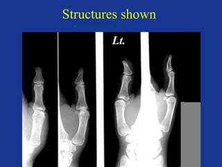
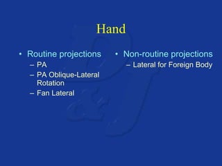
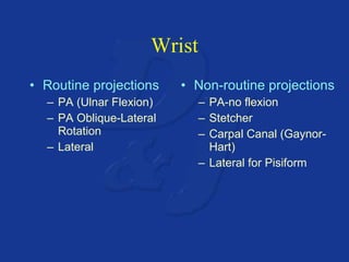
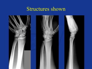
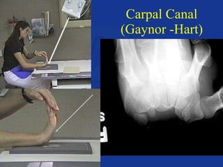
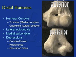
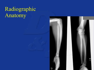
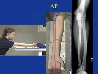
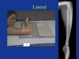
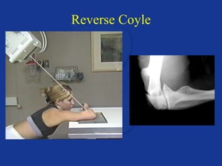
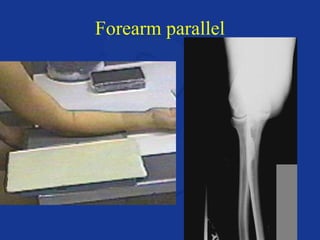
Radiographic Anatomy and Positioning of the Upper Extremity Dennis Winders Jeff Ahrendsen
Anatomy
Upper extremity consists of: Phalanges Metacarpals Carpals Radius Ulna Humerus
Anatomy of the Hand & Wrist
The hand & wrist consists of : 27 Bones Phalanges  - 14 Metacarpals - 5 Carpals  - 8
Phalanges Fingers & thumb 3 separate bones Digits 2-5 Proximal Middle Distal Tuft Thumb Proximal Distal
Naming of Digits 1 2 3 4 5 Thumb Index Middle Ring Little
Joints Interphalangeal Metacarpophalangeal Distal Interphalangeal Proximal Interphalangeal
Metacarpals Palm Numbering Three parts Head Shaft Base Joints MP Carpometacarpal Head Base Shaft
Carpals (Wrist) 8 bones Proximal row Navicular - Scaphoid Lunate  - Semilunar Triquetral - Cuneiform Pisiform a b c d
Carpals (continued) Distal row Greater Multangular - Trapezium Lesser Multangular  -  Trapezoid Capitate  - Os Magnum Hamate  - Unciform a b c d
Mnemonic Never Lower Tillies Pants Grandma Might  Come Home Some Sassy Children Play Through Their Old Underwear Some Lovers Try  Positions  That They  Can’t  Handle Alternative mnemonic
Carpal Joints Radiocarpal Intercarpal
Distal Radius & Ulna Radial Styloid Process Ulnar Styloid Process Distal Radioulnar Jt.
Radiographic  Anatomy
A. B. C. D. E. F. G. H.
Tuft 2nd DIP Jt. 2nd PIP Jt. 2nd MP Jt. IP Jt. 1st MP Jt. CM Jt. Radiocarpal Jt.
A. B. C. D.
Trapezium Trapezoid Scaphoid Pisiform
A. B. C. D.
Os Magnum Semilunar Unciform Cuneiform
Carpal Canal Trapezium Os Magnum Unciform Pisiform
Motions of the Hand & Wrist Radial Flexion (Ulnar Deviation) Ulnar Flexion (Radial Deviation)
Positioning of the Hand & Wrist
Finger Routine projections PA Medial Oblique Lateral Oblique Lateral Film size SID CR
PA
Lateral
Medial oblique
Lateral oblique
Parallel vs. Not Parallel
Structures shown
Technique
Prevention of Immobilize Sandbags Tape Short exposure time 10mAs = 200mA x .05s 10mAs = 400mA x .025s
Thumb Routine projections AP PA Oblique AP Oblique Lateral Film size SID CR
AP
Lateral
PA Oblique
AP Oblique
Structures shown
Technique
Hand Routine projections PA PA Oblique-Lateral Rotation Fan Lateral Non-routine projections Lateral for Foreign Body
Routine Hand Projections Routine projections PA PA Oblique-Lateral Rotation Fan Lateral Film size SID CR
PA
PA Oblique-Lateral Rotation
Fingers Down vs. Fingers Straight
Fan Lateral
Structures Shown
Technique
Non-routine projections of the Hand
Lateral for Foreign Body
Wrist Routine projections PA (Ulnar Flexion) PA Oblique-Lateral Rotation Lateral Non-routine projections PA-no flexion Stetcher Carpal Canal (Gaynor-Hart) Lateral for Pisiform
Routine Wrist Projections Routine projections PA (Ulnar Flexion) PA Oblique-Lateral Rotation Lateral Film size SID CR
PA (Ulnar Flexion)
PA Oblique-Lateral Rotation
Lateral
Structures shown
Technique
Non-routine projections of the Wrist
PA-no flexion
Scaphoid views (Stetcher)
Carpal Canal (Gaynor -Hart)
10° Supination Lateral
Casted Extremities
Changes in technique  No cast Extremity film 10mAs @ 60kV Casted Regular film Lower mAs 10 times Wet cast Add 15 to the kV Dry cast Add 10 to the kV Fiberglass Add 5 to the kV
Anatomy of the Forearm & Elbow
Radius Distal Styloid Process Ulnar Notch Proximal Head Neck Tuberosity Shaft
Ulna Distal Head Styloid Process Proximal Olecranon process Coronoid process Trochlear notch Radial notch Shaft
Effects of pronation on the forearm
Distal Humerus Humeral Condyle Trochlea (Medial condyle) Capitulum (Lateral condyle) Lateral epicondyle Medial epicondyle Depressions Coronoid fossa Radial fossa Olecranon fossa  posterior anterior
Classification of Joints Radioulnar Proximal Distal Elbow
Radiographic Anatomy
A. B. C. D. E. F. G. H. I. J. (Jt.) K.
Medial Epicondyle Coronoid Process Shaft (Ulna) Ulnar Head Ulnar Styloid Process Lateral Epicondyle Radial Head Radial Tuberosity Shaft (Radius) Distal Radioulnar Jt. Radial Styloid Process
A. B. C. (Jt.) D. E. F.  G. Post. H. I. J.
Lateral epicondyle Capitulum Proximal radioulnar jt. Radial head Radial neck Radial tuberosity Olecranon fossa Medial epicondyle Trochlea Coronoid process
C.  B.  A.  D. E. (not the jt.)  F.  G. (depression)
Coronoid Process Radial head Radial neck Condyles Trochlear notch Olecranon  process Radial notch
Positioning of the Forearm & Elbow
Forearm Routine projections AP Lateral Film size SID CR
AP
Lateral
Structures shown
Technique
Elbow Routine projections AP Lateral Coyle Non-routine Obliques Reverse Coyle Partial Flexion Rotational views of the radial head
Routine Elbow Routine projections AP Lateral Coyle Film size SID CR
AP
Lateral
Coyle
Structures shown
Technique
Non-routine projections of the Elbow
Obliques
Reverse Coyle
Partial Flexion
Humerus parallel
Forearm parallel
Rotational views of the Radial Head
Hand supinated
Hand AP oblique
Hand lateral
Hand pronated
Lateral positioning

Upper extremity anatomy & positioning