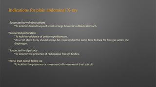
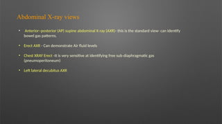
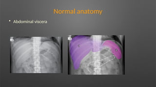
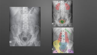
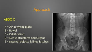
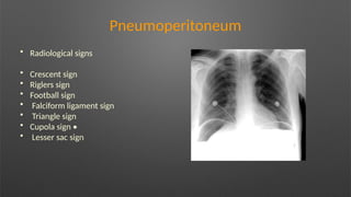
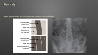
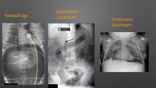
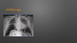
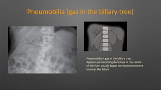
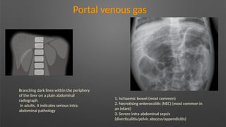
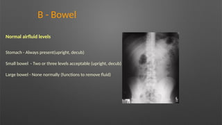
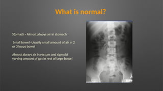
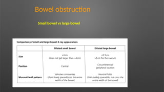
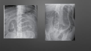
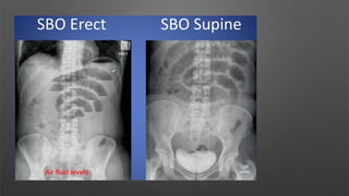
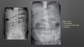
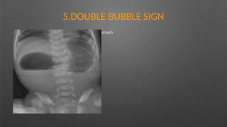
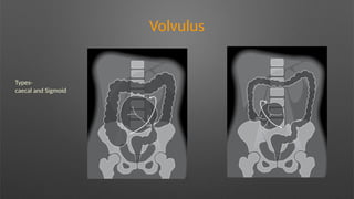
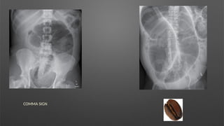
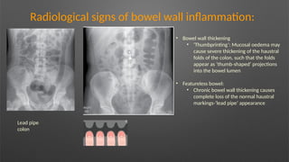
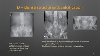
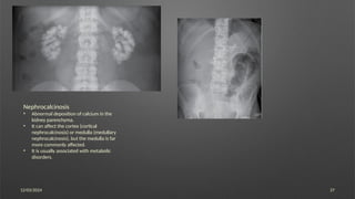
Abdominal x-ray
Dr Swaroop Sankar
Junior resident RD
Indications for plain abdominal X ray
‐
Suspected bowel obstructions
To look for dilated loops of small or large bowel or a dilated stomach.
Suspected perforation
To look for evidence of pneumoperitoneum.
An erect chest X ray should always be requested at the same time to look for free gas under the
‐
diaphragm.
Suspected foreign body
To look for the presence of radiopaque foreign bodies.
Renal tract calculi follow up
‐
To look for the presence or movement of known renal tract calculi.
Abdominal X ray views
‐
• Anterior–posterior (AP) supine abdominal X ray (AXR)
‐ - this is the standard view- can Identify
bowel gas patterns.
• Erect AXR - Can demonstrate Air fluid levels
• Chest XRAY Erect -It is very sensitive at identifying free sub diaphragmatic gas
‐
(pneumoperitoneum)
• Left lateral decubitus AXR
Inclusion
The entire anatomy should be included from the
hemi diaphragms to the symphysis pubis.
‐
• The superior aspect of the liver (1) and spleen (2)
should be included at the top of the radiograph.
• The lateral abdominal walls (3) should be seen
on either side of the radiograph.
• The pubic symphysis (4) should be clearly
visualised at the bottom of the radiograph.
Normal anatomy
• Abdominal viscera
Approach
ABDO X
A = Air in wrong place
B = Bowel
C = Calcification
D = Dense structeres and Organs
E = external objects & lines & tubes
A is for Air in the wrong place
• Look for pneumoperitoneum and pneumoretroperitoneum
• Look for gas in the biliary tree and portal vein
B is for Bowel
• Look for dilated small and large bowel
• Look for a volvulus
• Look for a distended stomach
• Look for a hernia
C = Calcification
• Look for clinically significant calcified structures such as calcified gallstones, renal calculus,
nephrocalcinosis, pancreatic calcification and an abdominal aortic aneurysm (AAA)
D = Dense structeres and Organs
Look at the bony skeleton for fractures and sclerotic/lytic bone lesions
Look at the spine for vertebral body height, alignment, pedicles
Organomegaly
Look for a foetus (females)
E -external objects & lines & tubes
• Look for evidence of previous surgery and other medical devices
• Look for foreign bodies
• Look at the lung bases
Pneumoperitoneum
• Radiological signs
• Crescent sign
• Riglers sign
• Football sign
• Falciform ligament sign
• Triangle sign
• Cupola sign •
• Lesser sac sign
Rigler's sign
Bowel wall visualised on both sides due to intra and extraluminal air .
Football sign
Football sign
FALCIFORM
LIGAMENT Continuous
diaphragm
CUPOLA sign
Pneumobilia (gas in the biliary tree)
Pneumobilia is gas in the biliary tree.
Appears as branching dark lines in the centre
of the liver, usually larger and more prominent
towards the hilum
Portal venous gas
Branching dark lines within the periphery
of the liver on a plain abdominal
radiograph.
In adults, it indicates serious intra‐
abdominal pathology
1. Ischaemic bowel (most common)
2. Necrotising enterocolitis (NEC) (most common in
an infant)
3. Severe intra abdominal sepsis
‐
(diverticulitis/pelvic abscess/appendicitis)
B - Bowel
Normal airfluid levels
Stomach - Always present(upright, decub)
Small bowel - Two or three levels acceptable (upright, decub)
Large bowel - None normally (functions to remove fluid)
What is normal?
Stomach - Almost always air in stomach
Small bowel -Usually small amount of air in 2
or 3 loops bowel
Almost always air in rectum and sigmoid
varying amount of gas in rest of large bowel
Bowel obstruction
Small bowel vs large bowel
Rigler’s triad:
1. Pneumobilia
2. Small bowel obstruction
3. Gallstone
4. SINGLE BUBBLE SIGN
• Abdominal radiographs classically show a gas filled distended stomach with absence
of distal bowel gas, giving rise to single bubble sign.
• Pyeloric atresia is most common cause ---Leading to complete obstruction of the
pyloric lumen of stomach.
5.DOUBLE BUBBLE SIGN
Findings -Dilatation of the proximal duodenum and stomach
Causes :
– Duodenal atresia
Volvulus
Types-
caecal and Sigmoid
COMMA SIGN
Radiological signs of bowel wall inflammation:
Lead pipe
colon
• Bowel wall thickening
• ‘Thumbprinting’: Mucosal oedema may
cause severe thickening of the haustral
folds of the colon, such that the folds
appear as ‘thumb shaped’ projections
‐
into the bowel lumen
• Featureless bowel:
• Chronic bowel wall thickening causes
complete loss of the normal haustral
markings-‘lead pipe’ appearance
26
D = Dense structures & calcification
Only around 15% of
gallstones contain enough
calcium to be visible on a
plain radiograph.
Most renal stones (90%) contain enough calcium to be visible
on a plain radiograph
Radiolucent stones -uric acid stones are not visualised.
12/03/2024 27
Nephrocalcinosis
• Abnormal deposition of calcium in the
kidney parenchyma.
• It can affect the cortex (cortical
nephrocalcinosis) or medulla (medullary
nephrocalcinosis), but the medulla is far
more commonly affected.
• It is usually associated with metabolic
disorders.
12/03/2024 28
29
Spine
Ossification of the interspinous and supraspinous
ligaments, and marginal syndesmophytes causing
fusion (ankylosis)
12/03/2024 30
12/03/2024 31
TEST
This is a supine AP abdominal radiograph.
The pubic symphysis is included on this radiograph,
however the hemi diaphragms are not visualised. Ideally I
‐
would like to see both hemi diaphragms”
‐
A: “There is no evidence of free gas.”
B: “The bowel gas pattern is within normal limits.”
C: “There is no abnormal calcification.”
D: “There is no fracture or bony abnormality.”
E: “There is no evidence of previous surgery, medical
devices or any foreign body.” “
Impression
Normal abdominal radiograph.”
12/03/2024 32
This is a supine AP abdominal radiograph
The pubic symphysis is included on this radiograph,
however the hemi diaphragms are not visualised.
‐
Ideally I would like to see both hemi diaphragms.”
‐
A: “There is no evidence of free gas.”
B: “There are multiple centrally located gas filled
‐
loops of bowel
Valvulae conniventes are seen in many of the loops
and they measure >3cm in diameter in keeping
with dilated loops of small bowel.”
C: “There is no abnormal calcification.”
D: “There is no fracture or bony abnormality.”
E: “There is no evidence of previous surgery,
medical devices or any foreign body.”
Impression
This is an abnormal abdominal radiograph showing
dilated loops of small bowel.

Abdominal xray RADIOLOGY final.pptx .pptx

  • 1.
    Abdominal x-ray Dr SwaroopSankar Junior resident RD
  • 2.
    Indications for plainabdominal X ray ‐ Suspected bowel obstructions To look for dilated loops of small or large bowel or a dilated stomach. Suspected perforation To look for evidence of pneumoperitoneum. An erect chest X ray should always be requested at the same time to look for free gas under the ‐ diaphragm. Suspected foreign body To look for the presence of radiopaque foreign bodies. Renal tract calculi follow up ‐ To look for the presence or movement of known renal tract calculi.
  • 3.
    Abdominal X rayviews ‐ • Anterior–posterior (AP) supine abdominal X ray (AXR) ‐ - this is the standard view- can Identify bowel gas patterns. • Erect AXR - Can demonstrate Air fluid levels • Chest XRAY Erect -It is very sensitive at identifying free sub diaphragmatic gas ‐ (pneumoperitoneum) • Left lateral decubitus AXR
  • 4.
    Inclusion The entire anatomyshould be included from the hemi diaphragms to the symphysis pubis. ‐ • The superior aspect of the liver (1) and spleen (2) should be included at the top of the radiograph. • The lateral abdominal walls (3) should be seen on either side of the radiograph. • The pubic symphysis (4) should be clearly visualised at the bottom of the radiograph.
  • 5.
  • 7.
    Approach ABDO X A =Air in wrong place B = Bowel C = Calcification D = Dense structeres and Organs E = external objects & lines & tubes
  • 8.
    A is forAir in the wrong place • Look for pneumoperitoneum and pneumoretroperitoneum • Look for gas in the biliary tree and portal vein B is for Bowel • Look for dilated small and large bowel • Look for a volvulus • Look for a distended stomach • Look for a hernia C = Calcification • Look for clinically significant calcified structures such as calcified gallstones, renal calculus, nephrocalcinosis, pancreatic calcification and an abdominal aortic aneurysm (AAA) D = Dense structeres and Organs Look at the bony skeleton for fractures and sclerotic/lytic bone lesions Look at the spine for vertebral body height, alignment, pedicles Organomegaly Look for a foetus (females) E -external objects & lines & tubes • Look for evidence of previous surgery and other medical devices • Look for foreign bodies • Look at the lung bases
  • 9.
    Pneumoperitoneum • Radiological signs •Crescent sign • Riglers sign • Football sign • Falciform ligament sign • Triangle sign • Cupola sign • • Lesser sac sign
  • 10.
    Rigler's sign Bowel wallvisualised on both sides due to intra and extraluminal air .
  • 11.
  • 12.
  • 13.
    Pneumobilia (gas inthe biliary tree) Pneumobilia is gas in the biliary tree. Appears as branching dark lines in the centre of the liver, usually larger and more prominent towards the hilum
  • 14.
    Portal venous gas Branchingdark lines within the periphery of the liver on a plain abdominal radiograph. In adults, it indicates serious intra‐ abdominal pathology 1. Ischaemic bowel (most common) 2. Necrotising enterocolitis (NEC) (most common in an infant) 3. Severe intra abdominal sepsis ‐ (diverticulitis/pelvic abscess/appendicitis)
  • 15.
    B - Bowel Normalairfluid levels Stomach - Always present(upright, decub) Small bowel - Two or three levels acceptable (upright, decub) Large bowel - None normally (functions to remove fluid)
  • 16.
    What is normal? Stomach- Almost always air in stomach Small bowel -Usually small amount of air in 2 or 3 loops bowel Almost always air in rectum and sigmoid varying amount of gas in rest of large bowel
  • 17.
  • 20.
    Rigler’s triad: 1. Pneumobilia 2.Small bowel obstruction 3. Gallstone
  • 21.
    4. SINGLE BUBBLESIGN • Abdominal radiographs classically show a gas filled distended stomach with absence of distal bowel gas, giving rise to single bubble sign. • Pyeloric atresia is most common cause ---Leading to complete obstruction of the pyloric lumen of stomach.
  • 22.
    5.DOUBLE BUBBLE SIGN Findings-Dilatation of the proximal duodenum and stomach Causes : – Duodenal atresia
  • 23.
  • 24.
  • 25.
    Radiological signs ofbowel wall inflammation: Lead pipe colon • Bowel wall thickening • ‘Thumbprinting’: Mucosal oedema may cause severe thickening of the haustral folds of the colon, such that the folds appear as ‘thumb shaped’ projections ‐ into the bowel lumen • Featureless bowel: • Chronic bowel wall thickening causes complete loss of the normal haustral markings-‘lead pipe’ appearance
  • 26.
    26 D = Densestructures & calcification Only around 15% of gallstones contain enough calcium to be visible on a plain radiograph. Most renal stones (90%) contain enough calcium to be visible on a plain radiograph Radiolucent stones -uric acid stones are not visualised.
  • 27.
    12/03/2024 27 Nephrocalcinosis • Abnormaldeposition of calcium in the kidney parenchyma. • It can affect the cortex (cortical nephrocalcinosis) or medulla (medullary nephrocalcinosis), but the medulla is far more commonly affected. • It is usually associated with metabolic disorders.
  • 28.
  • 29.
    29 Spine Ossification of theinterspinous and supraspinous ligaments, and marginal syndesmophytes causing fusion (ankylosis)
  • 30.
  • 31.
    12/03/2024 31 TEST This isa supine AP abdominal radiograph. The pubic symphysis is included on this radiograph, however the hemi diaphragms are not visualised. Ideally I ‐ would like to see both hemi diaphragms” ‐ A: “There is no evidence of free gas.” B: “The bowel gas pattern is within normal limits.” C: “There is no abnormal calcification.” D: “There is no fracture or bony abnormality.” E: “There is no evidence of previous surgery, medical devices or any foreign body.” “ Impression Normal abdominal radiograph.”
  • 32.
    12/03/2024 32 This isa supine AP abdominal radiograph The pubic symphysis is included on this radiograph, however the hemi diaphragms are not visualised. ‐ Ideally I would like to see both hemi diaphragms.” ‐ A: “There is no evidence of free gas.” B: “There are multiple centrally located gas filled ‐ loops of bowel Valvulae conniventes are seen in many of the loops and they measure >3cm in diameter in keeping with dilated loops of small bowel.” C: “There is no abnormal calcification.” D: “There is no fracture or bony abnormality.” E: “There is no evidence of previous surgery, medical devices or any foreign body.” Impression This is an abnormal abdominal radiograph showing dilated loops of small bowel.