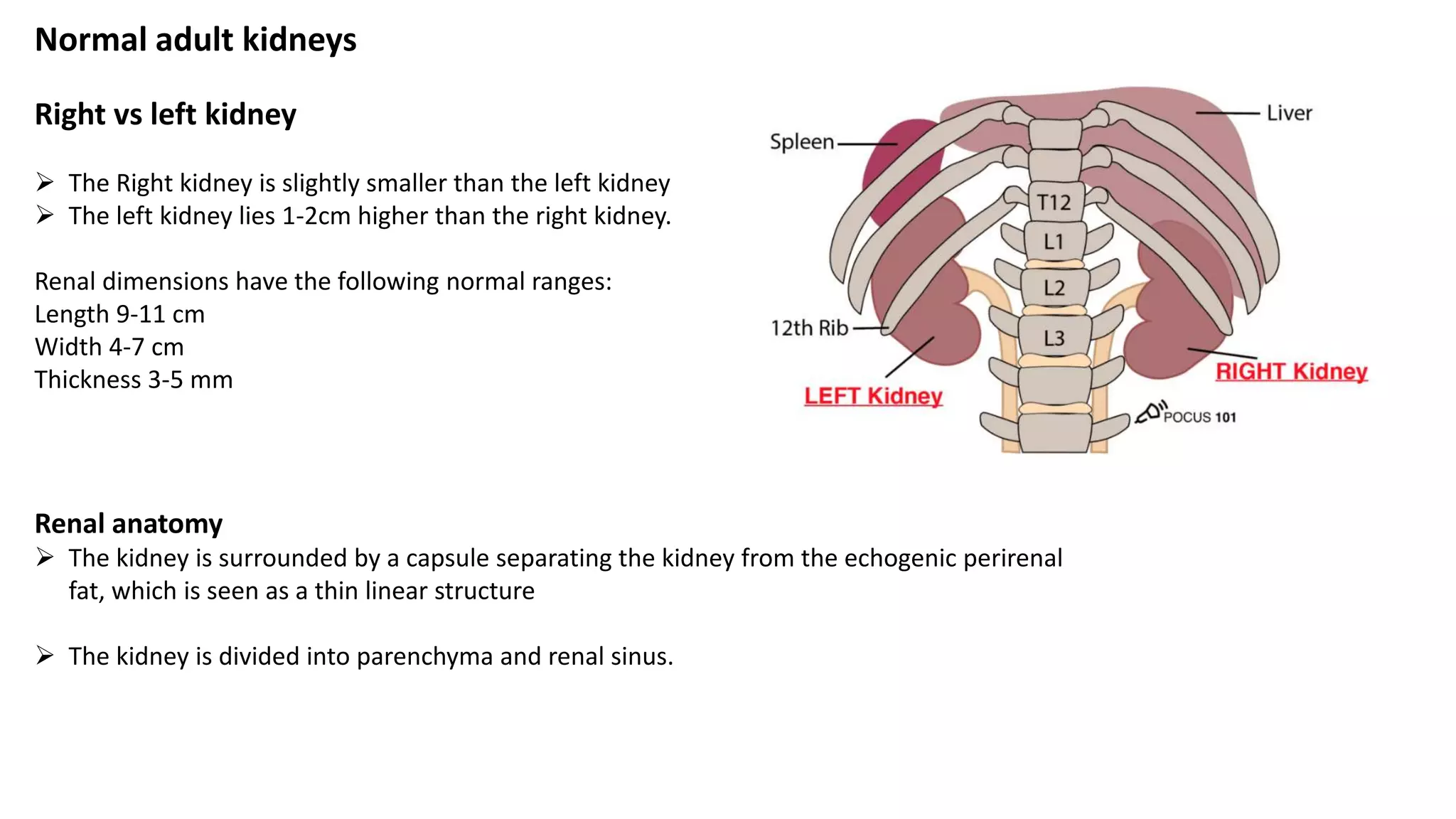
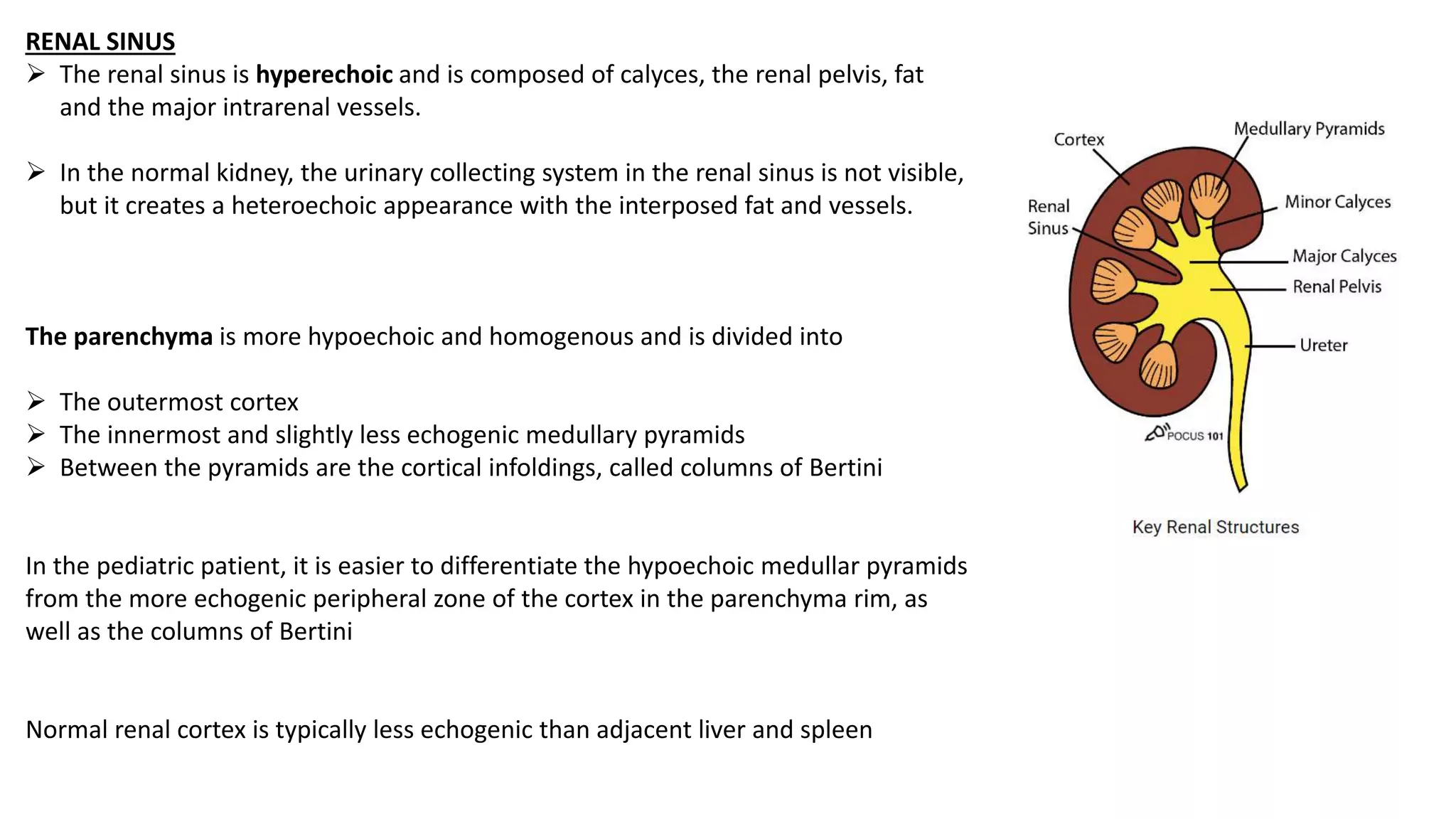
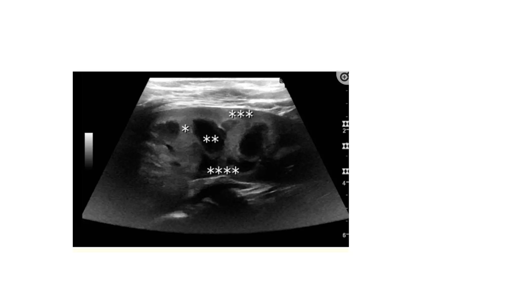
The document summarizes the technique for performing an ultrasound examination of the kidneys. It outlines the proper probe selection, patient positioning, and scanning approaches. It describes how to obtain longitudinal and transverse views of both kidneys and minimum images that should be included. Normal kidney anatomy is defined including dimensions, cortex thickness and echogenicity compared to liver. Causes of non-pathological renal pelvis dilation are listed.
Introducing Diagnostic Ultrasound in General Practiceupstatevet
Chris Ryan, DVM, DACVR
This lecture will begin by reviewing the basic operation of ultrasound equipment with a focus on hardware and software features common to almost all machines. The various settings and controls will be reviewed, along with the effects that these have on overall image quality, and how to utilize these settings to optimize image quality. A roadmap will then be developed for applying ultrasound in everyday general practice, beginning with the basics of evaluation for abdominal or pleural cavity fluid, and proceeding to perform a complete basic abdominal ultrasound exam. Normal sonographic anatomy and measurements will be reviewed, along with a recommended acquisition protocol for submission to teleradiology services.
Excretionurography
Also known as intravenous urography (IVU).
Most frequently employed radiologic investigation of renal rainage.
The contrast material is administered intravenously.
Best method for adults unless use of other methods is specified and is used in examinations of upper urinary tracts of infants and children.
New Drug Discovery and Development .....NEHA GUPTA
The "New Drug Discovery and Development" process involves the identification, design, testing, and manufacturing of novel pharmaceutical compounds with the aim of introducing new and improved treatments for various medical conditions. This comprehensive endeavor encompasses various stages, including target identification, preclinical studies, clinical trials, regulatory approval, and post-market surveillance. It involves multidisciplinary collaboration among scientists, researchers, clinicians, regulatory experts, and pharmaceutical companies to bring innovative therapies to market and address unmet medical needs.
Report Back from SGO 2024: What’s the Latest in Cervical Cancer?bkling
Are you curious about what’s new in cervical cancer research or unsure what the findings mean? Join Dr. Emily Ko, a gynecologic oncologist at Penn Medicine, to learn about the latest updates from the Society of Gynecologic Oncology (SGO) 2024 Annual Meeting on Women’s Cancer. Dr. Ko will discuss what the research presented at the conference means for you and answer your questions about the new developments.
New Directions in Targeted Therapeutic Approaches for Older Adults With Mantl...i3 Health
i3 Health is pleased to make the speaker slides from this activity available for use as a non-accredited self-study or teaching resource.
This slide deck presented by Dr. Kami Maddocks, Professor-Clinical in the Division of Hematology and
Associate Division Director for Ambulatory Operations
The Ohio State University Comprehensive Cancer Center, will provide insight into new directions in targeted therapeutic approaches for older adults with mantle cell lymphoma.
STATEMENT OF NEED
Mantle cell lymphoma (MCL) is a rare, aggressive B-cell non-Hodgkin lymphoma (NHL) accounting for 5% to 7% of all lymphomas. Its prognosis ranges from indolent disease that does not require treatment for years to very aggressive disease, which is associated with poor survival (Silkenstedt et al, 2021). Typically, MCL is diagnosed at advanced stage and in older patients who cannot tolerate intensive therapy (NCCN, 2022). Although recent advances have slightly increased remission rates, recurrence and relapse remain very common, leading to a median overall survival between 3 and 6 years (LLS, 2021). Though there are several effective options, progress is still needed towards establishing an accepted frontline approach for MCL (Castellino et al, 2022). Treatment selection and management of MCL are complicated by the heterogeneity of prognosis, advanced age and comorbidities of patients, and lack of an established standard approach for treatment, making it vital that clinicians be familiar with the latest research and advances in this area. In this activity chaired by Michael Wang, MD, Professor in the Department of Lymphoma & Myeloma at MD Anderson Cancer Center, expert faculty will discuss prognostic factors informing treatment, the promising results of recent trials in new therapeutic approaches, and the implications of treatment resistance in therapeutic selection for MCL.
Target Audience
Hematology/oncology fellows, attending faculty, and other health care professionals involved in the treatment of patients with mantle cell lymphoma (MCL).
Learning Objectives
1.) Identify clinical and biological prognostic factors that can guide treatment decision making for older adults with MCL
2.) Evaluate emerging data on targeted therapeutic approaches for treatment-naive and relapsed/refractory MCL and their applicability to older adults
3.) Assess mechanisms of resistance to targeted therapies for MCL and their implications for treatment selection
Knee anatomy and clinical tests 2024.pdfvimalpl1234
This includes all relevant anatomy and clinical tests compiled from standard textbooks, Campbell,netter etc..It is comprehensive and best suited for orthopaedicians and orthopaedic residents.
micro teaching on communication m.sc nursing.pdfAnurag Sharma
Microteaching is a unique model of practice teaching. It is a viable instrument for the. desired change in the teaching behavior or the behavior potential which, in specified types of real. classroom situations, tends to facilitate the achievement of specified types of objectives.
Basavarajeeyam is an important text for ayurvedic physician belonging to andhra pradehs. It is a popular compendium in various parts of our country as well as in andhra pradesh. The content of the text was presented in sanskrit and telugu language (Bilingual). One of the most famous book in ayurvedic pharmaceutics and therapeutics. This book contains 25 chapters called as prakaranas. Many rasaoushadis were explained, pioneer of dhatu druti, nadi pareeksha, mutra pareeksha etc. Belongs to the period of 15-16 century. New diseases like upadamsha, phiranga rogas are explained.
263778731218 Abortion Clinic /Pills In Harare ,sisternakatoto
263778731218 Abortion Clinic /Pills In Harare ,ABORTION WOMEN’S CLINIC +27730423979 IN women clinic we believe that every woman should be able to make choices in her pregnancy. Our job is to provide compassionate care, safety,affordable and confidential services. That’s why we have won the trust from all generations of women all over the world. we use non surgical method(Abortion pills) to terminate…Dr.LISA +27730423979women Clinic is committed to providing the highest quality of obstetrical and gynecological care to women of all ages. Our dedicated staff aim to treat each patient and her health concerns with compassion and respect.Our dedicated group ABORTION WOMEN’S CLINIC +27730423979 IN women clinic we believe that every woman should be able to make choices in her pregnancy. Our job is to provide compassionate care, safety,affordable and confidential services. That’s why we have won the trust from all generations of women all over the world. we use non surgical method(Abortion pills) to terminate…Dr.LISA +27730423979women Clinic is committed to providing the highest quality of obstetrical and gynecological care to women of all ages. Our dedicated staff aim to treat each patient and her health concerns with compassion and respect.Our dedicated group of receptionists, nurses, and physicians have worked together as a teamof receptionists, nurses, and physicians have worked together as a team wwww.lisywomensclinic.co.za/
- Video recording of this lecture in English language: https://youtu.be/lK81BzxMqdo
- Video recording of this lecture in Arabic language: https://youtu.be/Ve4P0COk9OI
- Link to download the book free: https://nephrotube.blogspot.com/p/nephrotube-nephrology-books.html
- Link to NephroTube website: www.NephroTube.com
- Link to NephroTube social media accounts: https://nephrotube.blogspot.com/p/join-nephrotube-on-social-media.html
2. Probe selection
• In the adult patient- a curved array transducer
• In the pediatric patient -a linear array transducer
Patient positioning
Patient is usually placed in supine position.
Best position is lateral decubitus
Kidneys are examined in longitudinal and transverse scan planes with the transducer placed in the flanks
Examination technique and positioning
Probe maybe
kept at a oblique
plane
corresponding
to the lie of the
kidney on the
psoas muscle.
3. Slowly tilt/fan the probe anteriorly and posteriorly to assess the entire
kidney
Step 1: RIGHT Kidney Ultrasound Longitudinal View
4. Step 2: RIGHT Kidney Ultrasound Transverse View
Tilt the probe superiorly and inferiorly
5. Scanning apporach
A combination of subcostal and intercostal approaches is required to evaluate the kidneys fully; the upper
pole of the let kidney may be particularly difficult to image without a combination of approaches.
Barriers to scanning
1)Artifacts of the lowest ribs always shadow the upper poles of the kidneys.
Ask the patient to take a deep breath and hold.
Ipsilateral arms can be raised to widen rib spaces
2)When insonation of the kidney is obscured by intestinal air
The supine scan position is combined with the lateral decubitus position and the transducer moved dorsally.
In obese patients
A prone position scanning approach can be used
6. A renal examination series should include the following minimum images;
•Both kidneys with length measurements
•Right kidney longitudinal section with liver for comparison
•Both kidneys longitudinal, anterior and posterior sections
•Both kidneys transverse at level of
• Superior pole
• midpole
• Inferior pole
•Left kidney longitudinal with spleen for comparison
•Document the normal anatomy. Any pathology found in 2 planes, including measurements and any vascularity
7. What to Check
•Kidney size (should not be >1cm difference between sides)
•Cortical thickness(not <10mm)
•Cortico-medullary differentiation
•Cortex at least as hypoechoic as the liver
•Pyramids slightly hypoechoic relative to the cortex
•No hydronephrosis
•Renal scarring(beware mistaking prominant lobulations as scars)
8. Normal adult kidneys
Right vs left kidney
The Right kidney is slightly smaller than the left kidney
The left kidney lies 1-2cm higher than the right kidney.
Renal dimensions have the following normal ranges:
Length 9-11 cm
Width 4-7 cm
Thickness 3-5 mm
Renal anatomy
The kidney is surrounded by a capsule separating the kidney from the echogenic perirenal
fat, which is seen as a thin linear structure
The kidney is divided into parenchyma and renal sinus.
9. RENAL SINUS
The renal sinus is hyperechoic and is composed of calyces, the renal pelvis, fat
and the major intrarenal vessels.
In the normal kidney, the urinary collecting system in the renal sinus is not visible,
but it creates a heteroechoic appearance with the interposed fat and vessels.
The parenchyma is more hypoechoic and homogenous and is divided into
The outermost cortex
The innermost and slightly less echogenic medullary pyramids
Between the pyramids are the cortical infoldings, called columns of Bertini
In the pediatric patient, it is easier to differentiate the hypoechoic medullar pyramids
from the more echogenic peripheral zone of the cortex in the parenchyma rim, as
well as the columns of Bertini
Normal renal cortex is typically less echogenic than adjacent liver and spleen
10. Cortical thickness measurement and echogenicity
Cortical thickness should be estimated from the base of the pyramid and is
generally 7–10 mm.
If the pyramids are difficult to differentiate, the parenchymal thickness can be
measured instead and should be 15–20 mm
The echogenicity of the cortex decreases with age and is less echogenic than or
equal to the liver and spleen at the same depth in individuals older than six
months
In neonates and children up to six months of age, the cortex is more echogenic
than the liver and spleen when compared at the same depth
11. Non pathological causes of Dilated Renal pelvis
With increased fluid intake the renal pelvis can distend and be visualized as a more echo free structure.
A similar finding can be seen in a developmental variant of an extrarenal pelvis.
In both conditions ,the dilation does not involve the calyces and infundibula
12.
13. An enlarged adrenal gland should be looked for within the
perirenal fat above the upper pole of the kidney, where it
can appear as a hypoechoic mass within the echogenic
perirenal fat.