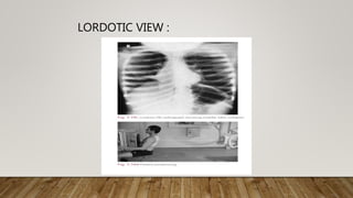
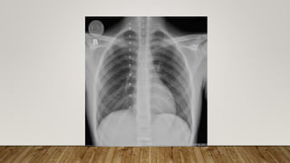
NORMAL CHEST X
RAY
BY DR.AKHIL ROHAN
• INTRODUCTION
• MAJOR VIEWS OF CHEST RADIOGRAPH
• TECHNICAL ASPECTS
• APPROACH
• SUMMARY
INTRODUCTION
• Radiographic examination of the lungs is
performed for a wide variety of medical conditions
providing an easily accessible,cheap and effective
diagnostic tool.
• Typical effective dose of chest X ray is around
0.2mSv
MAJOR VIEWS OF CHEST RADIOGRAPH
• Postero anterior
• Antero posterior
• Lateral
• Oblique
• Lateral Decubitus
• Lordotic
POSTERO ANTERIOR POSITION
• Patient stands upright with the anterior wall of chest placed
against the front of film.
• The shoulders are rotated forward enough to touch the film
or ask the patient to place hands on the waist ensuring that
the scapulae do not obscure a portion of lung fields
• Exposure usually taken with patient in full inspiration.
• Focus to film distance of 6 feet
ANTERO POSTERIOR POSITION
• Used when the patient is debilitated, immobilizer,
or unable to co operate.
• Film is placed behind the patient’s back with the
patient in a supine position
Scapula Seen in the periphery of
thorax
Seen over the lung fields
Clavicles Project over the lung Above the apex of lung
fields
Ribs Posterior ribs distinct Anterior ribs distinct
LATERAL POSITION
• Patient stands upright with the left side of chest
against the film and arms raised over the head
• Typically used in conjunction with other views
• esophagus(A),
trachea (B), lung hili
(C), heart silhouette
(D), lung apices (E),
scapulae (F), thoracic
vertebra (G), thoracic
intervertebral
foramen (H),
superimposed
posterior ribs (I),
costophrenic angles
(J), and diaphragm
(yellow arrows).
LATERAL DECUBITUS POSITION
• The patient lies on either side.The radiograph is
labeled according to the side that is placed against
the film .
• Useful in detecting small amount of pleural
effusion
OBLIQUE VIEW
LORDOTIC VIEW :
APICAL VIEW
FACTORS DETERMINING THE TECHNICAL QUALITY
OF THE RADIOGRAPH
• Inspiration
• Penetration
• Rotation
INSPIRATION
• The chest radiograph should be obtained with the
patient in full inspiration to help assess
intrapulmonary abnormalities.
• At full inspiration,the diaphragm should be
observed at the level of the 8-10 ribs posteriorly or
the 5-6 rib anteriorly.
PENETRATION
• Vertebral bodies and disc spaces should be just
visible through the cardiac shadow .
• Over penetration leads to loss of visibility of low
density lesions such as early consolidation.
ROTATION
• Medial aspects of clavicles equidistant from
vertebral spinous processes.
APPROACH
• Patient identication details , marker labeling.
• X ray view –PA ,AP, Lateral
• Breath : Inspiration or Expiration
• X ray penetration : Under or over penetrated
• Rotation
SYSTEMATIC SEARCH
• TRACHEA
Check whether the trachea is midline or
deviated.The carina lies at the level of T6 on
inspiration and at the level of T4 on expiration.In
adults the right main bronchus has a steeper angle
than the left , but the angles are symmetrical in
children.
HILUM
• Left hilum is usually higher than the right hilum
• The hilar shadows in both projections are produced
mainly by the right and left pulmonary arteries
MEDIASTINUM
HEART
• Position
• Cardiothoracic ratio :ratio between the maximum
transverse diameter of heart and max width of
thorax above costophrenic angles.
RIGHT HEART BORDER
1. SVC
2. RIGHT ATRIUM
3. IVC
LEFT HEART BORDER
1. AORTIC KNUCKLE
2. PULMONARY TRUNK
3. LEFT VENTRICLE
LUNG
• On a PA view , for descriptive purposes the lungs
are divided into three zones
ACCESSORY FISSURES
JUNCTION LINES:
HIDDEN AREAS
PLEURA
• Trace around the entire edge of lung to look for
pleural
abnormalities/thickening/calcifications/pneumotho
rax.
• B/l costophrenic angles are well defined in a
normal chest radiograph.
DIAPHRAGM
• Right hemi diaphragm is higher than the left.
• Assess the curvature of b/l hemidiaphragms to
identify diaphragmatic flattening or bulge .
• Assess b/l costophrenic angles .
BONES
• Clavicles
• Each rib
• Scapulae
• Vertebra
SOFT TISSUES
• Breast shadows
• Skin folds
• Muscles
Normal chest X ray radiography interpretation

Normal chest X ray radiography interpretation

  • 1.
    NORMAL CHEST X RAY BYDR.AKHIL ROHAN
  • 2.
    • INTRODUCTION • MAJORVIEWS OF CHEST RADIOGRAPH • TECHNICAL ASPECTS • APPROACH • SUMMARY
  • 3.
    INTRODUCTION • Radiographic examinationof the lungs is performed for a wide variety of medical conditions providing an easily accessible,cheap and effective diagnostic tool. • Typical effective dose of chest X ray is around 0.2mSv
  • 4.
    MAJOR VIEWS OFCHEST RADIOGRAPH • Postero anterior • Antero posterior • Lateral • Oblique • Lateral Decubitus • Lordotic
  • 5.
    POSTERO ANTERIOR POSITION •Patient stands upright with the anterior wall of chest placed against the front of film. • The shoulders are rotated forward enough to touch the film or ask the patient to place hands on the waist ensuring that the scapulae do not obscure a portion of lung fields • Exposure usually taken with patient in full inspiration. • Focus to film distance of 6 feet
  • 7.
    ANTERO POSTERIOR POSITION •Used when the patient is debilitated, immobilizer, or unable to co operate. • Film is placed behind the patient’s back with the patient in a supine position
  • 9.
    Scapula Seen inthe periphery of thorax Seen over the lung fields Clavicles Project over the lung Above the apex of lung fields Ribs Posterior ribs distinct Anterior ribs distinct
  • 10.
    LATERAL POSITION • Patientstands upright with the left side of chest against the film and arms raised over the head • Typically used in conjunction with other views
  • 12.
    • esophagus(A), trachea (B),lung hili (C), heart silhouette (D), lung apices (E), scapulae (F), thoracic vertebra (G), thoracic intervertebral foramen (H), superimposed posterior ribs (I), costophrenic angles (J), and diaphragm (yellow arrows).
  • 13.
    LATERAL DECUBITUS POSITION •The patient lies on either side.The radiograph is labeled according to the side that is placed against the film . • Useful in detecting small amount of pleural effusion
  • 15.
  • 16.
  • 17.
  • 18.
    FACTORS DETERMINING THETECHNICAL QUALITY OF THE RADIOGRAPH • Inspiration • Penetration • Rotation
  • 19.
    INSPIRATION • The chestradiograph should be obtained with the patient in full inspiration to help assess intrapulmonary abnormalities. • At full inspiration,the diaphragm should be observed at the level of the 8-10 ribs posteriorly or the 5-6 rib anteriorly.
  • 21.
    PENETRATION • Vertebral bodiesand disc spaces should be just visible through the cardiac shadow . • Over penetration leads to loss of visibility of low density lesions such as early consolidation.
  • 23.
    ROTATION • Medial aspectsof clavicles equidistant from vertebral spinous processes.
  • 25.
    APPROACH • Patient identicationdetails , marker labeling. • X ray view –PA ,AP, Lateral • Breath : Inspiration or Expiration • X ray penetration : Under or over penetrated • Rotation
  • 26.
    SYSTEMATIC SEARCH • TRACHEA Checkwhether the trachea is midline or deviated.The carina lies at the level of T6 on inspiration and at the level of T4 on expiration.In adults the right main bronchus has a steeper angle than the left , but the angles are symmetrical in children.
  • 28.
    HILUM • Left hilumis usually higher than the right hilum • The hilar shadows in both projections are produced mainly by the right and left pulmonary arteries
  • 30.
  • 31.
    HEART • Position • Cardiothoracicratio :ratio between the maximum transverse diameter of heart and max width of thorax above costophrenic angles.
  • 32.
    RIGHT HEART BORDER 1.SVC 2. RIGHT ATRIUM 3. IVC LEFT HEART BORDER 1. AORTIC KNUCKLE 2. PULMONARY TRUNK 3. LEFT VENTRICLE
  • 35.
    LUNG • On aPA view , for descriptive purposes the lungs are divided into three zones
  • 40.
  • 41.
  • 42.
  • 43.
    PLEURA • Trace aroundthe entire edge of lung to look for pleural abnormalities/thickening/calcifications/pneumotho rax. • B/l costophrenic angles are well defined in a normal chest radiograph.
  • 45.
    DIAPHRAGM • Right hemidiaphragm is higher than the left. • Assess the curvature of b/l hemidiaphragms to identify diaphragmatic flattening or bulge . • Assess b/l costophrenic angles .
  • 47.
    BONES • Clavicles • Eachrib • Scapulae • Vertebra
  • 48.
    SOFT TISSUES • Breastshadows • Skin folds • Muscles