Downloaded 115 times

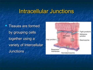


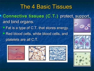


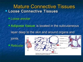


Tissues are groups of cells that work together to perform specialized functions. There are four basic tissue types: epithelial, connective, muscular, and nervous. Epithelial tissues line body surfaces and form glands. Connective tissues support and bind organs. Muscular tissues generate force for movement. Nervous tissues detect changes and generate nerve impulses. Tissues are named based on cell shape and layer arrangement, with the most common epithelia being simple squamous, simple columnar, and pseudostratified columnar. Connective tissues perform many functions and contain sparse cells in an extracellular matrix.


![06 [chapter 6 the skeletal system bone tissue]](https://cdn.slidesharecdn.com/ss_thumbnails/06chapter6theskeletalsystem-bonetissue-170828035633-thumbnail.jpg?width=640&height=640&fit=bounds)


![22 [chapter 22 the lymphatic system and immunity]](https://cdn.slidesharecdn.com/ss_thumbnails/22chapter22thelymphaticsystemandimmunity-170828153258-thumbnail.jpg?width=640&height=640&fit=bounds)

![05 [chapter 5 the integumentary system]](https://cdn.slidesharecdn.com/ss_thumbnails/05chapter5theintegumentarysystem-170828035624-thumbnail.jpg?width=640&height=640&fit=bounds)

![02 [chapter 2 the chemical level of organization]](https://cdn.slidesharecdn.com/ss_thumbnails/02chapter2thechemicalleveloforganization-170828035601-thumbnail.jpg?width=640&height=640&fit=bounds)


![Body_Tissues fully detailed notes [1].pptx](https://cdn.slidesharecdn.com/ss_thumbnails/bodytissues1-250917032454-2d23aff7-thumbnail.jpg?width=640&height=640&fit=bounds)


![EPITHELIA_BASIC_HISTOLTOLOGY[2] pro.ppt](https://cdn.slidesharecdn.com/ss_thumbnails/epitheliabasichistoltology2pro-250325063041-f58decb8-thumbnail.jpg?width=640&height=640&fit=bounds)


