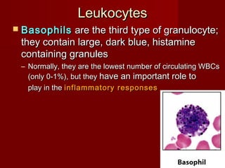
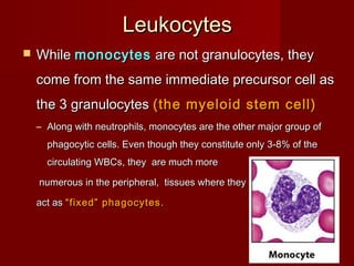
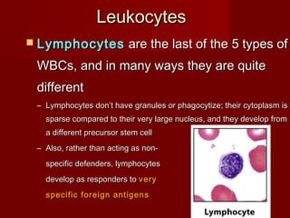
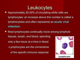
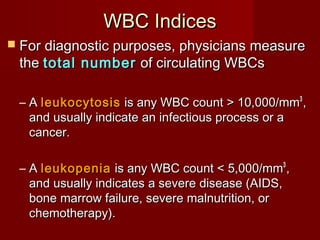
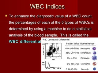
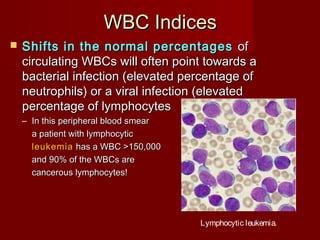
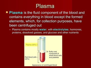
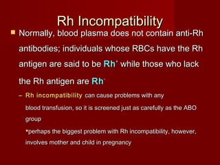
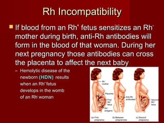
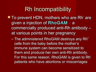
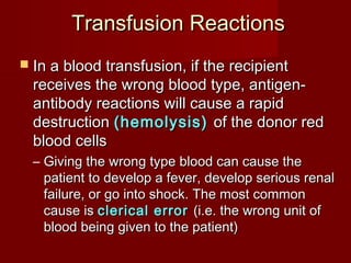
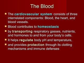
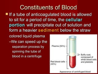
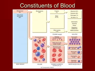
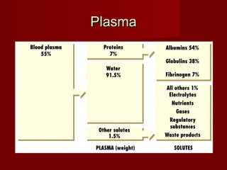
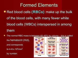
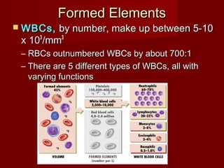
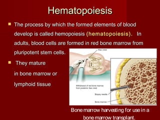
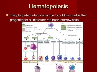
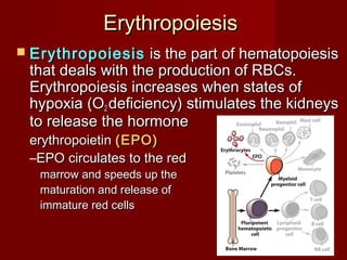
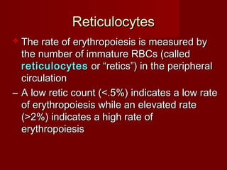
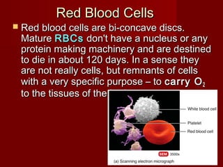
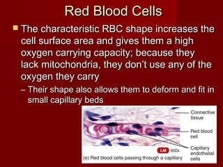
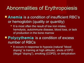
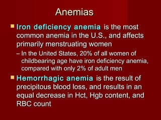
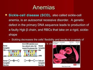
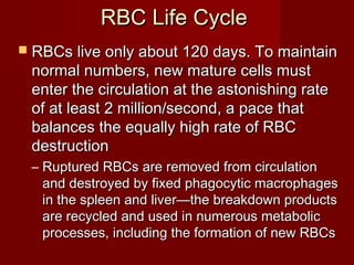
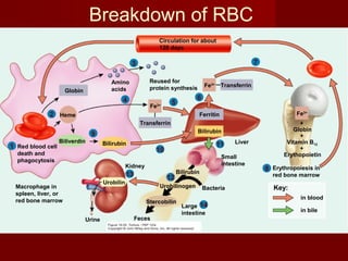
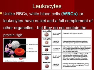
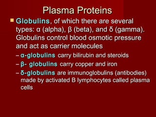
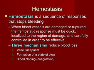
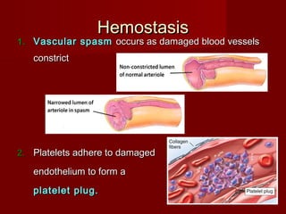
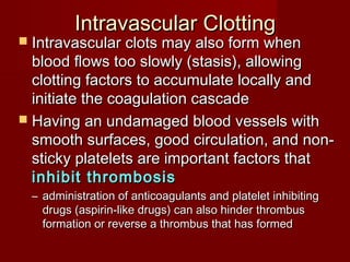
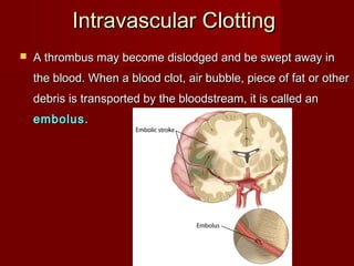
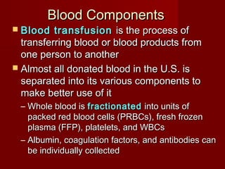
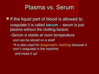
2. The BloodThe Blood
TheThe cardiovascular systemcardiovascular system consists of threeconsists of three
interrelated components: Blood, the heart, andinterrelated components: Blood, the heart, and
blood vessels.blood vessels.
Blood contributes toBlood contributes to homeostasishomeostasis
byby transportingtransporting respiratory gasses, nutrients,respiratory gasses, nutrients,
and hormones to and from your body’s cells.and hormones to and from your body’s cells.
It helpsIt helps regulateregulate body pH and temperature,body pH and temperature,
and providesand provides protectionprotection through its clottingthrough its clotting
mechanisms and immune defensesmechanisms and immune defenses
3. Physical characteristicsPhysical characteristics
Blood is denser and more viscous.Blood is denser and more viscous.
The temperature is 100.4The temperature is 100.4oo
F.F.
Alkaline pH ranging from 7.35 to 7.45.Alkaline pH ranging from 7.35 to 7.45.
The color of blood varies with its oxygenThe color of blood varies with its oxygen
content.content.
The blood volume is 5 to 6 liters in anThe blood volume is 5 to 6 liters in an
average adult male and 4 to 5 liters inaverage adult male and 4 to 5 liters in
female.female.
4. Constituents of BloodConstituents of Blood
If a tube of anticoagulated blood is allowedIf a tube of anticoagulated blood is allowed
to sit for a period of time, theto sit for a period of time, the cellularcellular
portionportion will precipitate out of solution andwill precipitate out of solution and
form a heavierform a heavier sedimentsediment below the strawbelow the straw
colored liquid plasmacolored liquid plasma
–We can speed up theWe can speed up the
separation process byseparation process by
spinning the tube ofspinning the tube of
blood in a centrifugeblood in a centrifuge
7. Formed ElementsFormed Elements
Red blood cellsRed blood cells (RBCs)(RBCs) make up the bulkmake up the bulk
of the blood cells, with many fewer whiteof the blood cells, with many fewer white
blood cells (WBCs) interspersed in amongblood cells (WBCs) interspersed in among
themthem
The normal RBC mass is between 40–45% by volume – this is calledThe normal RBC mass is between 40–45% by volume – this is called
thethe hematocrit (Hct),hematocrit (Hct),
and correspondsand corresponds
to 4–6to 4–6 xx 101066
/mm/mm33
by number.by number.
8. Formed ElementsFormed Elements
WBCs,WBCs, by number, make up between 5-10by number, make up between 5-10
x 10x 1033
/mm/mm33
– RBCs outnumbered WBCs by about 700:1RBCs outnumbered WBCs by about 700:1
– There are 5 different types of WBCs, all withThere are 5 different types of WBCs, all with
varying functionsvarying functions
9. MegakaryocytesMegakaryocytes are huge cells thatare huge cells that
splinter into 2000 to 3000 fragmentssplinter into 2000 to 3000 fragments
while still in the red bonewhile still in the red bone
marrowmarrow
– Each fragment, enclosed by a pieceEach fragment, enclosed by a piece
of the plasma membrane, is aof the plasma membrane, is a plateletplatelet
Formed ElementsFormed Elements
10. Formed ElementsFormed Elements
PlateletsPlatelets are more numerous than WBCsare more numerous than WBCs
(150-400 x 103/mm3 ), but they have a(150-400 x 103/mm3 ), but they have a
short life span (5 to 9 days) and they don’tshort life span (5 to 9 days) and they don’t
have much mass. They appear as littlehave much mass. They appear as little
specks interspersed among thespecks interspersed among the
many red cellsmany red cells
–Their granulesTheir granules
contain chemicals that,contain chemicals that,
once released, promoteonce released, promote
blood clottingblood clotting
11. HematopoiesisHematopoiesis
The process by which the formed elements of bloodThe process by which the formed elements of blood
develop is called hemopoiesisdevelop is called hemopoiesis (hematopoiesis).(hematopoiesis). InIn
adults, blood cells are formed in red bone marrow fromadults, blood cells are formed in red bone marrow from
pluripotent stem cells.pluripotent stem cells.
They matureThey mature
in bone marrow orin bone marrow or
lymphoid tissuelymphoid tissue
Bonemarrow harvesting for usein a
bonemarrow transplant.
12. HematopoiesisHematopoiesis
The pluripotent stem cell at the top of this chart is theThe pluripotent stem cell at the top of this chart is the
progenitor of all the other red bone marrow cellsprogenitor of all the other red bone marrow cells
13. ErythropoiesisErythropoiesis
ErythropoiesisErythropoiesis is the part of hematopoiesisis the part of hematopoiesis
that deals with the production of RBCs.that deals with the production of RBCs.
Erythropoiesis increases when states ofErythropoiesis increases when states of
hypoxia (Ohypoxia (O22 deficiency) stimulates the kidneysdeficiency) stimulates the kidneys
to release the hormoneto release the hormone
erythropoietinerythropoietin (EPO)(EPO)
–EPO circulates to the redEPO circulates to the red
marrow and speeds up themarrow and speeds up the
maturation and release ofmaturation and release of
immature red cellsimmature red cells
14. ReticulocytesReticulocytes
The rate of erythropoiesis is measured byThe rate of erythropoiesis is measured by
the number of immature RBCs (calledthe number of immature RBCs (called
reticulocytesreticulocytes or “retics”) in the peripheralor “retics”) in the peripheral
circulationcirculation
– A low retic count (<.5%) indicates a low rateA low retic count (<.5%) indicates a low rate
of erythropoiesis while an elevated rateof erythropoiesis while an elevated rate
(>2%) indicates a high rate of(>2%) indicates a high rate of
erythropoiesiserythropoiesis
15. Red Blood CellsRed Blood Cells
Red blood cells are bi-concave discs.Red blood cells are bi-concave discs.
MatureMature RBCsRBCs don't have a nucleus or anydon't have a nucleus or any
protein making machinery and are destinedprotein making machinery and are destined
to die in about 120 days. In a sense theyto die in about 120 days. In a sense they
are not really cells, but remnants of cellsare not really cells, but remnants of cells
with a very specific purpose – towith a very specific purpose – to carry Ocarry O22
to the tissues of the body.to the tissues of the body.
16. Red Blood CellsRed Blood Cells
The characteristic RBC shape increases theThe characteristic RBC shape increases the
cell surface area and gives them a highcell surface area and gives them a high
oxygen carrying capacity; because theyoxygen carrying capacity; because they
lack mitochondria, they don’t use any of thelack mitochondria, they don’t use any of the
oxygen they carryoxygen they carry
– Their shape also allows them to deform and fit inTheir shape also allows them to deform and fit in
small capillary bedssmall capillary beds
17. ReticulocytesReticulocytes
As cells mature in the bone marrow, they become smaller, theAs cells mature in the bone marrow, they become smaller, the
nucleus disappears, and the amount of Hgb increasesnucleus disappears, and the amount of Hgb increases
18. HemoglobinHemoglobin (Hgb)(Hgb) is a protein moleculeis a protein molecule
adapted to carry Oadapted to carry O22 (and CO(and CO22 as well), andas well), and
each RBC contains 280 million molecules ofeach RBC contains 280 million molecules of
Hgb.Hgb.
HemoglobinHemoglobin
19. Abnormalities of ErythropoiesisAbnormalities of Erythropoiesis
AnemiaAnemia is a condition of insufficient RBC’sis a condition of insufficient RBC’s
or hemoglobin (quality or quantity)or hemoglobin (quality or quantity)
It is most often the result of low iron intake,It is most often the result of low iron intake,
hemolysis, autoimmune disease, blood loss, or lackhemolysis, autoimmune disease, blood loss, or lack
of production in the bone marrowof production in the bone marrow
PolycythemiaPolycythemia is a condition of excessis a condition of excess
number of RBCsnumber of RBCs
It occurs in response to hypoxia (natural “bloodIt occurs in response to hypoxia (natural “blood
doping” is training at high altitude), shots of EPOdoping” is training at high altitude), shots of EPO
(illegal “doping”), smoking (COPD), or dehydration(illegal “doping”), smoking (COPD), or dehydration
20. AnemiasAnemias
Iron deficiency anemiaIron deficiency anemia is the mostis the most
common anemia in the U.S., and affectscommon anemia in the U.S., and affects
primarily menstruating womenprimarily menstruating women
– In the United States, 20% of all women ofIn the United States, 20% of all women of
childbearing age have iron deficiency anemia,childbearing age have iron deficiency anemia,
compared with only 2% of adult mencompared with only 2% of adult men
Hemorrhagic anemiaHemorrhagic anemia is the result ofis the result of
precipitous blood loss, and results in anprecipitous blood loss, and results in an
equal decrease in Hct, Hgb content, andequal decrease in Hct, Hgb content, and
RBC countRBC count
21. AnemiasAnemias
Sickle-cell disease (SCD),Sickle-cell disease (SCD), also called sickle-cellalso called sickle-cell
anemia, is an autosomal recessive disorder. A geneticanemia, is an autosomal recessive disorder. A genetic
defect in the primary DNA sequence leads to production ofdefect in the primary DNA sequence leads to production of
a faulty Hgba faulty Hgb ββ chain, and RBCs that take on a rigid, sickle-chain, and RBCs that take on a rigid, sickle-
shapeshape
– Sickling decreases the cells' flexibility and results in a variety ofSickling decreases the cells' flexibility and results in a variety of
complications; life expectancy is shortenedcomplications; life expectancy is shortened
22. RBC Life CycleRBC Life Cycle
RBCs live only about 120 days. To maintainRBCs live only about 120 days. To maintain
normal numbers, new mature cells mustnormal numbers, new mature cells must
enter the circulation at the astonishing rateenter the circulation at the astonishing rate
of at least 2 million/second, a pace thatof at least 2 million/second, a pace that
balances the equally high rate of RBCbalances the equally high rate of RBC
destructiondestruction
– Ruptured RBCs are removed from circulationRuptured RBCs are removed from circulation
and destroyed by fixed phagocytic macrophagesand destroyed by fixed phagocytic macrophages
in the spleen and liver—the breakdown productsin the spleen and liver—the breakdown products
are recycled and used in numerous metabolicare recycled and used in numerous metabolic
processes, including the formation of new RBCsprocesses, including the formation of new RBCs
23. Red blood cell
death and
phagocytosis
Key:
in blood
in bile
Macrophage in
spleen, liver, or
red bone marrow
1
Globin
Red blood cell
death and
phagocytosis
Key:
in blood
in bile
Macrophage in
spleen, liver, or
red bone marrow
Heme2
1
Amino
acids
Reused for
protein synthesisGlobin
Red blood cell
death and
phagocytosis
Key:
in blood
in bile
Macrophage in
spleen, liver, or
red bone marrow
Heme
3
2
1
Amino
acids
Reused for
protein synthesisGlobin
Red blood cell
death and
phagocytosis
Transferrin
Fe3+
Key:
in blood
in bile
Macrophage in
spleen, liver, or
red bone marrow
Heme
4
3
2
1
Amino
acids
Reused for
protein synthesisGlobin
Red blood cell
death and
phagocytosis
Transferrin
Fe3+
Liver
Key:
in blood
in bile
Macrophage in
spleen, liver, or
red bone marrow
FerritinHeme
54
3
2
1
Amino
acids
Reused for
protein synthesisGlobin
Red blood cell
death and
phagocytosis
Transferrin
Fe3+
Fe3+ Transferrin
Liver
Key:
in blood
in bile
Macrophage in
spleen, liver, or
red bone marrow
FerritinHeme
6
54
3
2
1
Amino
acids
Reused for
protein synthesisGlobin
Red blood cell
death and
phagocytosis
Transferrin
Fe3+
Fe3+ Transferrin
Liver
+
Globin
+
Vitamin B12
+
Erythopoietin
Key:
in blood
in bile
Macrophage in
spleen, liver, or
red bone marrow
FerritinHeme Fe3+
7
6
54
3
2
1
Amino
acids
Reused for
protein synthesisGlobin
Circulation for about
120 days
Red blood cell
death and
phagocytosis
Transferrin
Fe3+
Fe3+ Transferrin
Liver
+
Globin
+
Vitamin B12
+
Erythopoietin
Key:
in blood
in bile
Erythropoiesis in
red bone marrow
Macrophage in
spleen, liver, or
red bone marrow
FerritinHeme Fe3+
8
7
6
54
3
2
1
Amino
acids
Reused for
protein synthesisGlobin
Circulation for about
120 days
Red blood cell
death and
phagocytosis
Transferrin
Fe3+
Fe3+ Transferrin
Liver
+
Globin
+
Vitamin B12
+
Erythopoietin
Key:
in blood
in bile
Erythropoiesis in
red bone marrow
Macrophage in
spleen, liver, or
red bone marrow
FerritinHeme
Biliverdin Bilirubin
Fe3+
9
8
7
6
54
3
2
1
Amino
acids
Reused for
protein synthesisGlobin
Circulation for about
120 days
Bilirubin
Red blood cell
death and
phagocytosis
Transferrin
Fe3+
Fe3+ Transferrin
Liver
+
Globin
+
Vitamin B12
+
Erythopoietin
Key:
in blood
in bile
Erythropoiesis in
red bone marrow
Macrophage in
spleen, liver, or
red bone marrow
FerritinHeme
Biliverdin Bilirubin
Fe3+
10
9
8
7
6
54
3
2
1
Amino
acids
Reused for
protein synthesisGlobin
Stercobilin
Bilirubin
Urobilinogen
Feces
Small
intestine
Circulation for about
120 days
Bacteria
Bilirubin
Red blood cell
death and
phagocytosis
Transferrin
Fe3+
Fe3+ Transferrin
Liver
+
Globin
+
Vitamin B12
+
Erythopoietin
Key:
in blood
in bile
Erythropoiesis in
red bone marrow
Macrophage in
spleen, liver, or
red bone marrow
FerritinHeme
Biliverdin Bilirubin
Fe3+
12
11
10
9
8
7
6
54
3
2
1
Amino
acids
Reused for
protein synthesisGlobin
Urine
Stercobilin
Bilirubin
Urobilinogen
Feces
Small
intestine
Circulation for about
120 days
Bacteria
Bilirubin
Red blood cell
death and
phagocytosis
Transferrin
Fe3+
Fe3+ Transferrin
Liver
+
Globin
+
Vitamin B12
+
Erythopoietin
Key:
in blood
in bile
Erythropoiesis in
red bone marrow
Kidney
Macrophage in
spleen, liver, or
red bone marrow
Ferritin
Urobilin
Heme
Biliverdin Bilirubin
Fe3+
13 12
11
10
9
8
7
6
54
3
2
1
Amino
acids
Reused for
protein synthesisGlobin
Urine
Stercobilin
Bilirubin
Urobilinogen
Feces
Large
intestine
Small
intestine
Circulation for about
120 days
Bacteria
Bilirubin
Red blood cell
death and
phagocytosis
Transferrin
Fe3+
Fe3+ Transferrin
Liver
+
Globin
+
Vitamin B12
+
Erythopoietin
Key:
in blood
in bile
Erythropoiesis in
red bone marrow
Kidney
Macrophage in
spleen, liver, or
red bone marrow
Ferritin
Urobilin
Heme
Biliverdin Bilirubin
Fe3+
14
13 12
11
10
9
8
7
6
54
3
2
1
Breakdown of RBC
24. LeukocytesLeukocytes
Unlike RBCs, white blood cellsUnlike RBCs, white blood cells (WBCs)(WBCs) oror
leukocytesleukocytes have nuclei and a full complement ofhave nuclei and a full complement of
other organelles - but they do not contain theother organelles - but they do not contain the
protein Hgb.protein Hgb.
25. LeukocytesLeukocytes
Leukocytes are divided intoLeukocytes are divided into two groupstwo groups depending ondepending on
whether they contain conspicuous chemical-filledwhether they contain conspicuous chemical-filled
cytoplasmic granules (when stained)cytoplasmic granules (when stained)
– GranulocytesGranulocytes include the neutrophils, eosinophils,include the neutrophils, eosinophils,
and basophilsand basophils
– AgranulocytesAgranulocytes are the monocytes and lymphocytesare the monocytes and lymphocytes
26. LeukocytesLeukocytes
The most numerous WBC in normal blood (60-The most numerous WBC in normal blood (60-
70% of circulating white cells) is the70% of circulating white cells) is the neutrophil,neutrophil,
or polymorphonucleocyte (PMN)or polymorphonucleocyte (PMN)
– PMNs are granulocytes with a pinkish cytoplasm,PMNs are granulocytes with a pinkish cytoplasm,
and they are one of the two majorand they are one of the two major
phagocytesphagocytes in the bodyin the body
their principal role is to fighttheir principal role is to fight
bacterial infectionsbacterial infections
27. LeukocytesLeukocytes
Chemicals released by microbes and inflamedChemicals released by microbes and inflamed
tissues attract phagocytes, a phenomenontissues attract phagocytes, a phenomenon
called chemotaxis.called chemotaxis.
– This graphic shows a PMN phagocytizing a microbeThis graphic shows a PMN phagocytizing a microbe
for internalfor internal
digestion anddigestion and
destructiondestruction
28. LeukocytesLeukocytes
EosinophilsEosinophils are characterized by theirare characterized by their
large red granuleslarge red granules
– They are much less numerous than neutrophilsThey are much less numerous than neutrophils
(2-4% of circulating WBCs), but their numbers(2-4% of circulating WBCs), but their numbers
increase slightly withincrease slightly with parasitic infectionparasitic infection
they have also beenthey have also been
associated with theassociated with the
development ofdevelopment of allergiesallergies
29. LeukocytesLeukocytes
BasophilsBasophils are the third type of granulocyte;are the third type of granulocyte;
they contain large, dark blue, histaminethey contain large, dark blue, histamine
containing granulescontaining granules
– Normally, they are the lowest number of circulating WBCsNormally, they are the lowest number of circulating WBCs
(only 0-1%), but they(only 0-1%), but they have an important role tohave an important role to
play in theplay in the inflammatory responsesinflammatory responses
30. LeukocytesLeukocytes
WhileWhile monocytesmonocytes are not granulocytes, theyare not granulocytes, they
come from the same immediate precursor cell ascome from the same immediate precursor cell as
the 3 granulocytesthe 3 granulocytes (the myeloid stem cell)(the myeloid stem cell)
– Along with neutrophils, monocytes are the other major group ofAlong with neutrophils, monocytes are the other major group of
phagocytic cells. Even though they constitute only 3-8% of thephagocytic cells. Even though they constitute only 3-8% of the
circulating WBCs, they are much morecirculating WBCs, they are much more
numerous in the peripheral, tissues where theynumerous in the peripheral, tissues where they
act asact as “fixed” phagocytes.“fixed” phagocytes.
31. LeukocytesLeukocytes
LymphocytesLymphocytes are the last of the 5 types ofare the last of the 5 types of
WBCs, and in many ways they are quiteWBCs, and in many ways they are quite
differentdifferent
– Lymphocytes don’t have granules or phagocytize; their cytoplasm isLymphocytes don’t have granules or phagocytize; their cytoplasm is
sparse compared to their very large nucleus, and they develop fromsparse compared to their very large nucleus, and they develop from
a different precursor stem cella different precursor stem cell
– Also, rather than acting as non-Also, rather than acting as non-
specific defenders, lymphocytesspecific defenders, lymphocytes
develop as responders todevelop as responders to veryvery
specific foreign antigensspecific foreign antigens
32. LeukocytesLeukocytes
Approximately 20-30% of circulating white cells areApproximately 20-30% of circulating white cells are
lymphocytes: an increase above this number is called alymphocytes: an increase above this number is called a
lymphocytosis and often represents anlymphocytosis and often represents an acute viralacute viral
infection.infection.
Most lymphocytes continually move among lymphoidMost lymphocytes continually move among lymphoid
tissues, lymph, and blood, spendingtissues, lymph, and blood, spending
only a few hours at a time in bloodonly a few hours at a time in blood
–Lymphocytes are the cornerstoneLymphocytes are the cornerstone
of the specific immune responseof the specific immune response
33. WBC IndicesWBC Indices
For diagnostic purposes, physicians measureFor diagnostic purposes, physicians measure
thethe total numbertotal number of circulating WBCsof circulating WBCs
– AA leukocytosisleukocytosis is any WBC count > 10,000/mmis any WBC count > 10,000/mm33
,,
and usually indicate an infectious process or aand usually indicate an infectious process or a
cancer.cancer.
– AA leukopenialeukopenia is any WBC count < 5,000/mmis any WBC count < 5,000/mm33
,,
and usually indicates a severe disease (AIDS,and usually indicates a severe disease (AIDS,
bone marrow failure, severe malnutrition, orbone marrow failure, severe malnutrition, or
chemotherapy).chemotherapy).
34. WBC IndicesWBC Indices
To enhance the diagnostic value of a WBC count,To enhance the diagnostic value of a WBC count,
the percentages of each of the 5 types of WBCs isthe percentages of each of the 5 types of WBCs is
determined by using a machine to do a statisticaldetermined by using a machine to do a statistical
analysis of the blood sample. This is called theanalysis of the blood sample. This is called the
WBC differentialWBC differential
35. WBC IndicesWBC Indices
Shifts in the normal percentagesShifts in the normal percentages ofof
circulating WBCs will often point towards acirculating WBCs will often point towards a
bacterial infection (elevated percentage ofbacterial infection (elevated percentage of
neutrophils) or a viral infection (elevatedneutrophils) or a viral infection (elevated
percentage of lymphocytespercentage of lymphocytes
– In this peripheral blood smearIn this peripheral blood smear
a patient with lymphocytica patient with lymphocytic
leukemialeukemia has a WBC >150,000has a WBC >150,000
and 90% of the WBCs areand 90% of the WBCs are
cancerous lymphocytes!cancerous lymphocytes!
Lymphocytic leukemia.
36. PlasmaPlasma is the fluid component of the blood andis the fluid component of the blood and
contains everything in blood except the formedcontains everything in blood except the formed
elements, which, for collection purposes, haveelements, which, for collection purposes, have
been centrifuged outbeen centrifuged out
– Plasma contains mostlyPlasma contains mostly waterwater ,, withwith electrolyteselectrolytes ,, hormones,hormones,
proteins, dissolved gasses, and glucose and other nutrientsproteins, dissolved gasses, and glucose and other nutrients
PlasmaPlasma
37. Plasma ProteinsPlasma Proteins
The major protein in plasma isThe major protein in plasma is albuminalbumin; it also; it also
has many clotting proteins, antibodies, andhas many clotting proteins, antibodies, and
enzymes.enzymes.
Albumin is synthesized in the liver and contributesAlbumin is synthesized in the liver and contributes
significantly to the blood viscosity and the body’ssignificantly to the blood viscosity and the body’s
ability to maintain blood pressure.ability to maintain blood pressure.
It also plays an important role as a carrierIt also plays an important role as a carrier
molecule.molecule.
38. Plasma ProteinsPlasma Proteins
GlobulinsGlobulins, of which there are several, of which there are several
types:types: αα (alpha),(alpha), ββ (beta), and(beta), and δδ (gamma).(gamma).
Globulins control blood osmotic pressureGlobulins control blood osmotic pressure
and act as carrier moleculesand act as carrier molecules
– αα-globulins-globulins carry bilirubin and steroidscarry bilirubin and steroids
– ββ-- globulinsglobulins carry copper and ironcarry copper and iron
– δδ-globulins-globulins are immunoglobulins (antibodies)are immunoglobulins (antibodies)
made by activated B lymphocytes called plasmamade by activated B lymphocytes called plasma
cellscells
39. HemostasisHemostasis
HemostasisHemostasis is a sequence of responsesis a sequence of responses
that stops bleedingthat stops bleeding
– When blood vessels are damaged or ruptured,When blood vessels are damaged or ruptured,
the hemostatic response must be quick,the hemostatic response must be quick,
localized to the region of damage, and carefullylocalized to the region of damage, and carefully
controlled in order to be effectivecontrolled in order to be effective
– Three mechanismsThree mechanisms reduce blood lossreduce blood loss
1.1. Vascular spasmVascular spasm
2.2. Formation of a platelet plugFormation of a platelet plug
3.3. Blood clotting (coagulation)Blood clotting (coagulation)
40. HemostasisHemostasis
1.1. Vascular spasmVascular spasm occurs as damaged blood vesselsoccurs as damaged blood vessels
constrictconstrict
2.2. Platelets adhere to damagedPlatelets adhere to damaged
endothelium to form aendothelium to form a
platelet plug.platelet plug.
41. 1
Red blood cell
Platelet
Collagen fibers
and damaged
endothelium
Platelet adhesion11
2
Red blood cell
Platelet
Collagen fibers
and damaged
endothelium
Liberated ADP,
serotonin, and
thromboxane A2
Platelet adhesion1
Platelet release reaction2
1
2
3
Red blood cell
Platelet
Collagen fibers
and damaged
endothelium
Liberated ADP,
serotonin, and
thromboxane A2
Platelet plug
Platelet adhesion1
Platelet release reaction2
Platelet aggregation3
Platelet
Plug
Formation
42. HemostasisHemostasis
3.3. ClottingClotting (coagulation)(coagulation) is possible because ofis possible because of
the presence of several clotting proteinsthe presence of several clotting proteins
normally dissolved (soluble) in the blood. Therenormally dissolved (soluble) in the blood. There
areare 2 pathways to activate the system2 pathways to activate the system
43. HemostasisHemostasis
TheThe extrinsic pathwayextrinsic pathway has fewhas few
steps and occurs rapidly, often withinsteps and occurs rapidly, often within
seconds, once the protein “tissueseconds, once the protein “tissue
factor”factor” (TF)(TF) leaks into the bloodleaks into the blood
TheThe intrinsic pathwayintrinsic pathway is moreis more
complex and occurs more slowly incomplex and occurs more slowly in
response to damage toresponse to damage to endothelialendothelial
cellscells or phospholipids released byor phospholipids released by
activated platelets.activated platelets.
44. Both the extrinsic and intrinsic clottingBoth the extrinsic and intrinsic clotting
pathways converge at apathways converge at a common pointcommon point
(pathway)(pathway) where factor X becomeswhere factor X becomes
activated (Xa)activated (Xa)
– In thisIn this second stagesecond stage ofof
blood clotting prothrombinblood clotting prothrombin
is converted to thrombinis converted to thrombin
which in turn convertswhich in turn converts
soluble fibrinogen tosoluble fibrinogen to
insoluble fibrin threadsinsoluble fibrin threads
HemostasisHemostasis
45. Tissue trauma
Tissue
factor
(TF)
Blood trauma
Damaged
endothelial cells
expose collagen
fibers
(a) Extrinsic pathway (b) Intrinsic pathway
Activated XII
Ca2+
Damaged
platelets
Ca2+
Platelet
phospholipids
Activated X
Activated
platelets
Activated X
PROTHROMBINASE
Ca2+
V
Ca2+
V
1
Tissue trauma
Tissue
factor
(TF)
Blood trauma
Damaged
endothelial cells
expose collagen
fibers
(a) Extrinsic pathway (b) Intrinsic pathway
Activated XII
Ca2+
Damaged
platelets
Ca2+
Platelet
phospholipids
Activated X
Activated
platelets
Activated X
PROTHROMBINASE
Ca2+
V
Ca2+
Prothrombin
(II)
Ca2+
THROMBIN
(c) Common
pathway
V
1
2
+
+
Tissue trauma
Tissue
factor
(TF)
Blood trauma
Damaged
endothelial cells
expose collagen
fibers
(a) Extrinsic pathway (b) Intrinsic pathway
Activated XII
Ca2+
Damaged
platelets
Ca2+
Platelet
phospholipids
Activated X
Activated
platelets
Activated X
PROTHROMBINASE
Ca2+
V
Ca2+
Prothrombin
(II)
Ca2+
THROMBIN
Ca2+
Loose fibrin
threads
STRENGTHENED
FIBRIN THREADS
Activated XIII
Fibrinogen
(I)
XIII
(c) Common
pathway
V
1
2
3
+
+
Stages of
Clotting
46. The mineralThe mineral CaCa22++
plays an important role throughout theplays an important role throughout the
clotting system, and many steps have positive or negativeclotting system, and many steps have positive or negative
feedback on various other steps to propagate the process,feedback on various other steps to propagate the process,
yet maintain controlyet maintain control
Clot retractionClot retraction is the consolidation of the fibrin clot. Asis the consolidation of the fibrin clot. As
the clot retracts, it pulls the edges ofthe clot retracts, it pulls the edges of the damagedthe damaged
vessel closer together,vessel closer together,
decreasing the risk of furtherdecreasing the risk of further
damage – new endothelial cells candamage – new endothelial cells can
then repair the vessel liningthen repair the vessel lining
HemostasisHemostasis
47. FibrinolysisFibrinolysis
Because blood clotting involves amplification andBecause blood clotting involves amplification and
positive feedback cycles, a clot has a tendency topositive feedback cycles, a clot has a tendency to
enlarge, creating the potential for impairment ofenlarge, creating the potential for impairment of
blood flow through undamaged vesselsblood flow through undamaged vessels
– TheThe fibrinolytic systemfibrinolytic system dissolves small, inappropriate clots; itdissolves small, inappropriate clots; it
also dissolves clots at a site of damage once the damage isalso dissolves clots at a site of damage once the damage is
repairedrepaired
both body tissues and blood contain substances that can activateboth body tissues and blood contain substances that can activate
plasminogen to becomeplasminogen to become plasmin,plasmin, (the enzyme that actively(the enzyme that actively
dissolves clots)dissolves clots)
48. Intravascular ClottingIntravascular Clotting
Blood clots sometimes form unexpectedlyBlood clots sometimes form unexpectedly
within the cardiovascular system. Clotting inwithin the cardiovascular system. Clotting in
an unbroken blood vessel (usually a vein) isan unbroken blood vessel (usually a vein) is
calledcalled thrombosis;thrombosis; the clot itself, called athe clot itself, called a
thrombusthrombus
– Such clots may be initiated by roughenedSuch clots may be initiated by roughened
endothelial surfaces of a blood vessel resultingendothelial surfaces of a blood vessel resulting
from atherosclerosis, trauma, or infectionfrom atherosclerosis, trauma, or infection
49. Intravascular ClottingIntravascular Clotting
Intravascular clots may also form whenIntravascular clots may also form when
blood flows too slowly (stasis), allowingblood flows too slowly (stasis), allowing
clotting factors to accumulate locally andclotting factors to accumulate locally and
initiate the coagulation cascadeinitiate the coagulation cascade
Having an undamaged blood vessels withHaving an undamaged blood vessels with
smooth surfaces, good circulation, and non-smooth surfaces, good circulation, and non-
sticky platelets are important factors thatsticky platelets are important factors that
inhibit thrombosisinhibit thrombosis
– administration of anticoagulants and platelet inhibitingadministration of anticoagulants and platelet inhibiting
drugs (aspirin-like drugs) can also hinder thrombusdrugs (aspirin-like drugs) can also hinder thrombus
formation or reverse a thrombus that has formedformation or reverse a thrombus that has formed
50. Intravascular ClottingIntravascular Clotting
A thrombus may become dislodged and be swept away inA thrombus may become dislodged and be swept away in
the blood. When a blood clot, air bubble, piece of fat or otherthe blood. When a blood clot, air bubble, piece of fat or other
debris is transported by the bloodstream, it is called andebris is transported by the bloodstream, it is called an
embolus.embolus.
51. Blood ComponentsBlood Components
Blood transfusionBlood transfusion is the process ofis the process of
transferring blood or blood products fromtransferring blood or blood products from
one person to anotherone person to another
Almost all donated blood in the U.S. isAlmost all donated blood in the U.S. is
separated into its various components toseparated into its various components to
make better use of itmake better use of it
– Whole blood isWhole blood is fractionatedfractionated into units ofinto units of
packed red blood cells (PRBCs), fresh frozenpacked red blood cells (PRBCs), fresh frozen
plasma (FFP), platelets, and WBCsplasma (FFP), platelets, and WBCs
– Albumin, coagulation factors, and antibodies canAlbumin, coagulation factors, and antibodies can
be individually collectedbe individually collected
52. Plasma vs. SerumPlasma vs. Serum
If the liquid part of blood is allowed toIf the liquid part of blood is allowed to
coagulate it is calledcoagulate it is called serum -serum - serum is justserum is just
plasma without the clotting factorsplasma without the clotting factors
–Serum is stable at room temperatureSerum is stable at room temperature
and can be stored on a shelfand can be stored on a shelf
it is also used forit is also used for diagnostic testingdiagnostic testing because itbecause it
won’t coagulate in the machinewon’t coagulate in the machine
and mess it up!and mess it up!
53. In transfusion medicine the presence orIn transfusion medicine the presence or
absence of theabsence of the
A and B red cell antigens forms the basis of theA and B red cell antigens forms the basis of the
ABO blood group systemABO blood group system
Blood GroupsBlood Groups
Another major red cell antigen istheRh antigen, which 85% of the
population have, and comprisesanother important blood grouping
54. Blood GroupsBlood Groups
For reason that are not totally clear, serum contains anti-For reason that are not totally clear, serum contains anti-
ABO antibodies of a type opposite to the ABO antigen onABO antibodies of a type opposite to the ABO antigen on
the red cell surfacethe red cell surface
– For instance, those with A antigens on their red cellsFor instance, those with A antigens on their red cells
have anti-B antibodies in their serumhave anti-B antibodies in their serum
55. Blood GroupsBlood Groups
By knowing the status of the A antigen, BBy knowing the status of the A antigen, B
antigen, and Rh antigen, most of the major bloodantigen, and Rh antigen, most of the major blood
incompatibility issues can be avoidedincompatibility issues can be avoided
– Type AB individuals areType AB individuals are “universal recipients”“universal recipients”
because they has neither anti-A nor anti-Bbecause they has neither anti-A nor anti-B
antibodies in their serum that would destroyantibodies in their serum that would destroy
transfused RBCstransfused RBCs
– Type O individuals areType O individuals are “universal donors”“universal donors”
because their RBCs have no antigens on the cellbecause their RBCs have no antigens on the cell
surface that can potentially react with thesurface that can potentially react with the
recipients serumrecipients serum
56. Blood GroupsBlood Groups
Blood typingBlood typing forfor
ABO status is doneABO status is done
using single drops ofusing single drops of
blood mixed withblood mixed with
different antiseradifferent antisera
– AgglutinationAgglutination with anwith an
antisera indicates theantisera indicates the
presence of that antigenpresence of that antigen
on the RBCon the RBC
57. Rh IncompatibilityRh Incompatibility
Normally, blood plasma does not contain anti-RhNormally, blood plasma does not contain anti-Rh
antibodies; individuals whose RBCs have the Rhantibodies; individuals whose RBCs have the Rh
antigen are said to beantigen are said to be RhRh++
while those who lackwhile those who lack
the Rh antigen arethe Rh antigen are RhRh--
– Rh incompatibilityRh incompatibility can cause problems with anycan cause problems with any
blood transfusion, so it is screened just as carefully as the ABOblood transfusion, so it is screened just as carefully as the ABO
groupgroup
perhaps the biggest problem with Rh incompatibility, however,perhaps the biggest problem with Rh incompatibility, however,
involves mother and child in pregnancyinvolves mother and child in pregnancy
58. Rh IncompatibilityRh Incompatibility
If blood from an RhIf blood from an Rh++
fetus sensitizes an Rhfetus sensitizes an Rh--
mother during birth, anti-Rh antibodies willmother during birth, anti-Rh antibodies will
form in the blood of that woman. During herform in the blood of that woman. During her
next pregnancy those antibodies can crossnext pregnancy those antibodies can cross
the placenta to affect the next babythe placenta to affect the next baby
– Hemolytic disease of theHemolytic disease of the
newbornnewborn (HDN)(HDN) resultsresults
when an Rhwhen an Rh++
fetusfetus
develops in the wombdevelops in the womb
of an Rhof an Rh--
womanwoman
59. Rh IncompatibilityRh Incompatibility
To prevent HDN, mothers who are RhTo prevent HDN, mothers who are Rh--
areare
given a injection ofgiven a injection of RhoGAMRhoGAM -- aa
commercially produced anti-Rh antibody –commercially produced anti-Rh antibody –
at various points in her pregnancyat various points in her pregnancy
– The administered RhoGAM destroys any RhThe administered RhoGAM destroys any Rh++
cells from the baby before the mother’scells from the baby before the mother’s
immune system can become sensitized toimmune system can become sensitized to
them and produce her own anti-Rh antibody.them and produce her own anti-Rh antibody.
For this same reason, RhoGAM is given to RhFor this same reason, RhoGAM is given to Rh--
patients who have abortions or miscarriagespatients who have abortions or miscarriages
60. Transfusion ReactionsTransfusion Reactions
In a blood transfusion, if the recipientIn a blood transfusion, if the recipient
receives the wrong blood type, antigen-receives the wrong blood type, antigen-
antibody reactions will cause a rapidantibody reactions will cause a rapid
destructiondestruction (hemolysis)(hemolysis) of the donor redof the donor red
blood cellsblood cells
– Giving the wrong type blood can cause theGiving the wrong type blood can cause the
patient to develop a fever, develop serious renalpatient to develop a fever, develop serious renal
failure, or go into shock. The most commonfailure, or go into shock. The most common
cause iscause is clerical errorclerical error (i.e. the wrong unit of(i.e. the wrong unit of
blood being given to the patient)blood being given to the patient)