Histology of basic tissues (Connective tissues).pptx
1.
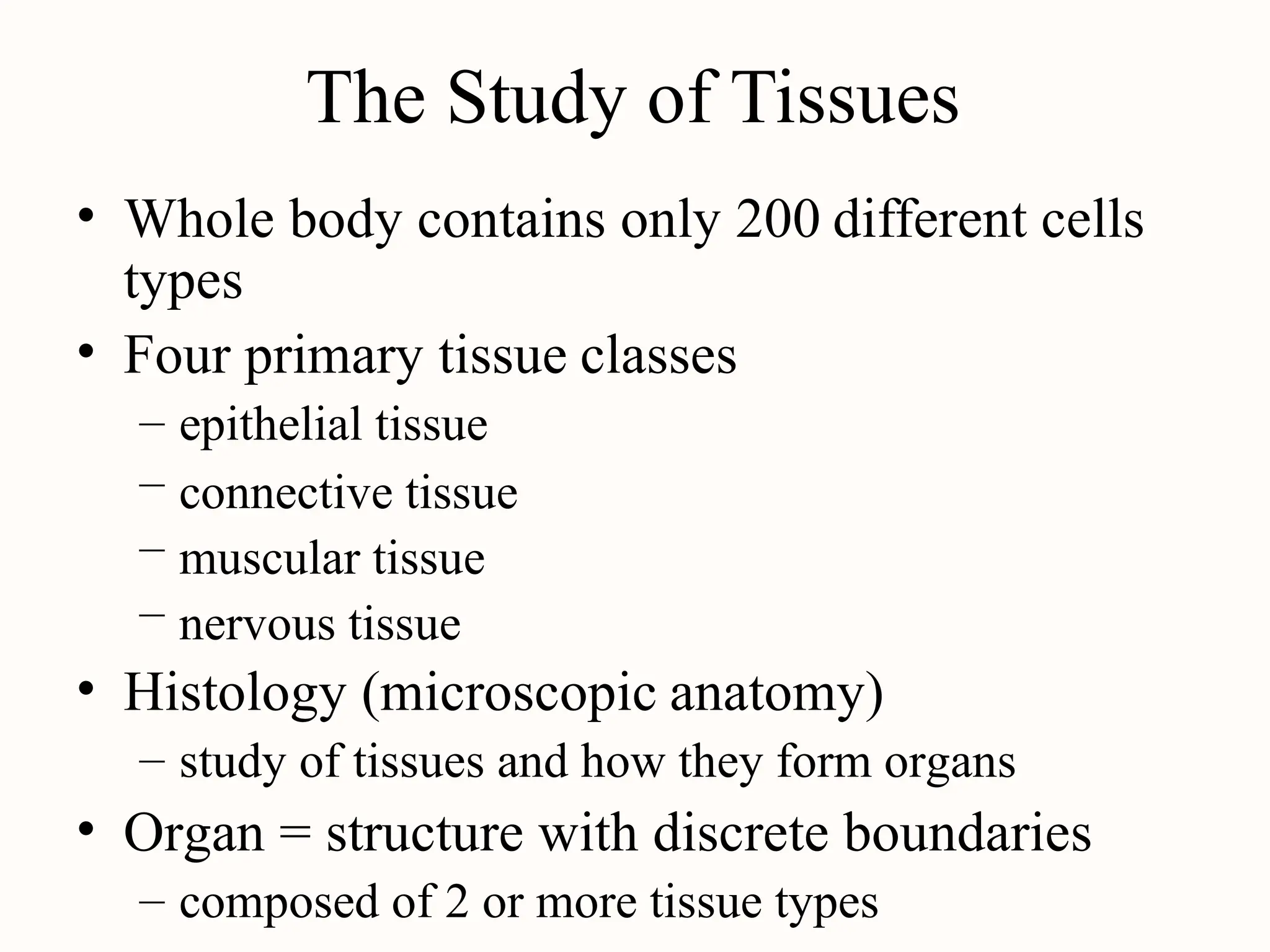
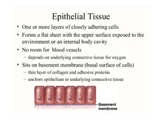
The Study ofTissues
• Whole body contains only 200
types
different cells
• Four primary tissue classes
–
–
–
–
epithelial tissue
connective tissue
muscular tissue
nervous tissue
• Histology (microscopic anatomy)
– study of tissues and how they form organs
• Organ = structure with discrete boundaries
– composed of 2 or more tissue types
2.
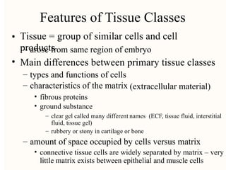
Features of TissueClasses
Tissue = group of similar cells and cell
products
•
– arose from same region of embryo
• Main differences between primary tissue classes
–
–
types and functions of cells
characteristics of the matrix (extracellular material)
•
•
fibrous proteins
ground substance
– clear gel called many different
fluid, tissue gel)
names (ECF, tissue fluid, interstitial
– rubbery or stony in cartilage or bone
– amount of space occupied by cells versus matrix
• connective tissue cells are widely separated by matrix – very
little matrix exists between epithelial and muscle cells
3.
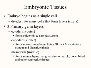
Embryonic Tissues
Embryo beginsas a single cell
•
– divides into many cells that form layers (strata)
• 3 Primary germ layers
– ectoderm (outer)
• forms epidermis & nervous system
– endoderm (inner)
• forms mucous membrane lining GI
system and digestive glands
tract & respiratory
– mesoderm (middle)
• forms mesenchyme that gives rise to muscle, bone, blood
and other connective tissues
4.
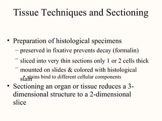
Tissue Techniques andSectioning
• Preparation of histological specimens
–
–
–
preserved in fixative prevents decay (formalin)
sliced into very thin sections only 1 or 2 cells thick
mounted on slides & colored with histological
stain
• stains bind to different cellular components
• Sectioning an organ or tissue reduces a 3-
dimensional structure to a 2-dimensional
slice
6.
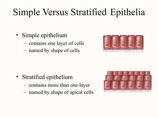
Simple Versus StratifiedEpithelia
• Simple epithelium
–
–
contains one layer of cells
named by shape of cells
• Stratified epithelium
–
–
contains more than one layer
named by shape of apical cells
7.
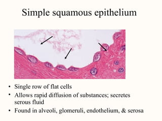
imp e SquamousEpithelium
Simple squamous epithelium
•
•
Single row of flat cells
Allows rapid diffusion of substances; secretes
serous fluid
• Found in alveoli, glomeruli, endothelium, & serosa
9.
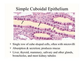
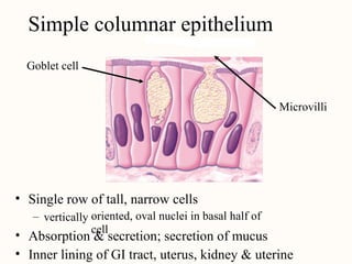
Simple Columnar Epithe
Simplecolumnar epithelium
Goblet cell
Microvilli
• Single row of tall, narrow cells
oriented, oval nuclei in basal half of
cell
– vertically
•
•
Absorption & secretion; secretion of mucus
Inner lining of GI tract, uterus, kidney & uterine
10.
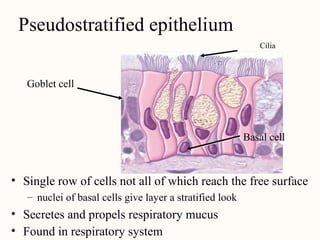
Pseudostratified Epithe
Cilia
Pseudostratified epithelium
Gobletcell
Basal cell
• Single row of cells not all of which reach the free surface
– nuclei of basal cells give layer a stratified look
•
•
Secretes and propels respiratory mucus
Found in respiratory system
11.
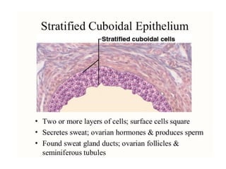
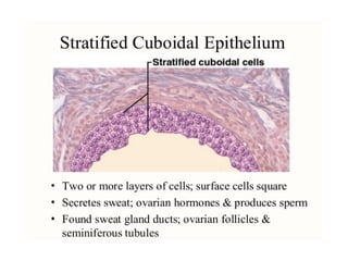
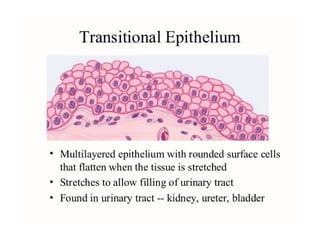
Stratified Epithelia
• Composedof more than one layer of
named for shape of surface cells
cells &
– exception is transitional epithelium
•
•
Deepest cells sit on basement membrane
Variations
–
–
keratinized epithelium has surface layer of dead cells
nonkeratinized epithelium lacks the layer of dead
cells
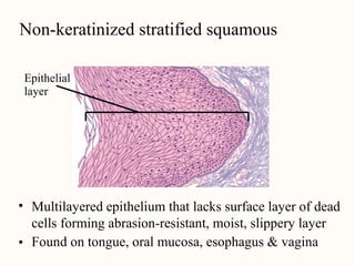
14.
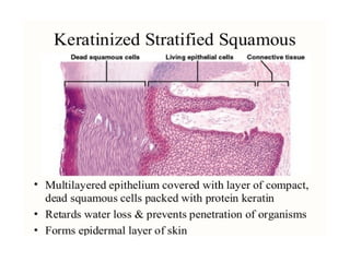
eratinized Stratified Squa
Non-keratinizedstratified squamous
Epithelial
layer
• Multilayered epithelium that lacks surface layer of dead
cells forming abrasion-resistant, moist, slippery layer
Found on tongue, oral mucosa, esophagus & vagina
•
17.
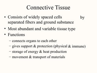
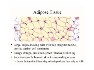
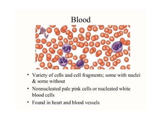
Connective Tissue
Consists ofwidely spaced cells
separated fibers and ground substance
• by
•
•
Most abundant and variable tissue
Functions
type
–
–
–
–
connects organs to each other
gives support & protection (physical
storage of energy & heat production
movement & transport of materials
& immune)
18.
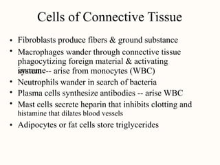
Cells of ConnectiveTissue
Fibroblasts produce fibers & ground substance
Macrophages wander through connective tissue
phagocytizing foreign material & activating
immune
•
•
system -- arise from monocytes (WBC)
•
•
•
Neutrophils wander in search of bacteria
Plasma cells synthesize antibodies -- arise WBC
Mast cells secrete heparin that inhibits clotting and
histamine that dilates blood vessels
Adipocytes or fat cells store triglycerides
•
19.
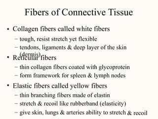
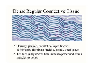
Fibers of ConnectiveTissue
Collagen fibers called white fibers
•
–
–
tough, resist stretch yet flexible
tendons, ligaments & deep layer of the skin
(dermis)
• Reticular fibers
–
–
thin collagen fibers coated with glycoprotein
form framework for spleen & lymph nodes
• Elastic fibers called yellow fibers
–
–
–
thin branching fibers made of elastin
stretch & recoil like rubberband (elasticity)
give skin, lungs & arteries ability to stretch & recoil
20.
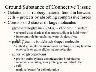
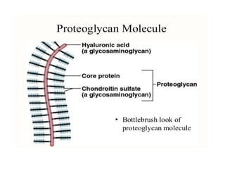
Ground Substance ofConnective Tissue
• Gelatinous or rubbery material found in between
cells – protects by absorbing compressive forces
Consists of 3 classes of large molecules
•
– glycosaminoglycans (GAGs) – chondroitin sulfate
•
•
unusual disaccharides that attract sodium & hold water
important role in regulating water & electrolyte
balance
– proteoglycan is bottlebrush-shaped molecule
• embedded in plasma membranes creating a strong bond to
other cells or extracellular macromolecules
– adhesive glycoproteins
• protein-carbohydrate complexes that bind plasma
membrane to collagen or proteoglycans outside the
cells
mark pathways for cell migration
•
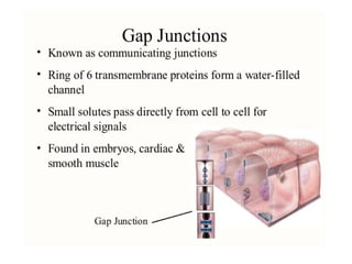
22.
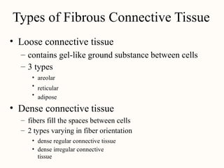
Types of FibrousConnective Tissue
• Loose connective tissue
–
–
contains gel-like ground
3 types
substance between cells
•
•
•
areolar
reticular
adipose
• Dense connective tissue
–
–
fibers fill the spaces between cells
2 types varying in fiber orientation
•
•
dense regular connective tissue
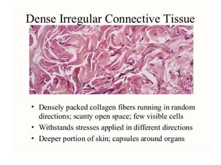
dense irregular connective
tissue
28.
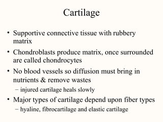
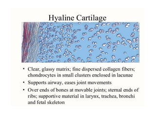
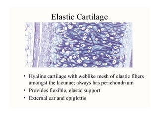
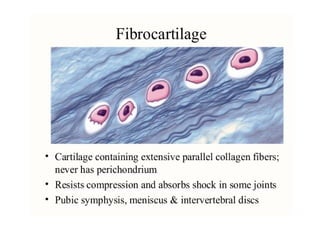
Cartilage
• Supportive connectivetissue with
matrix
rubbery
• Chondroblasts produce matrix, once surrounded
are called chondrocytes
No blood vessels so diffusion must bring in
nutrients & remove wastes
•
– injured cartilage heals slowly
• Major types of cartilage depend upon fiber types
– hyaline, fibrocartilage and elastic cartilage
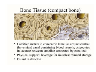
32.
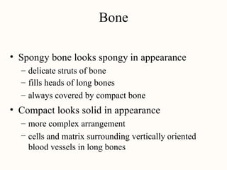
Bone
• Spongy bonelooks spongy in appearance
–
–
–
delicate struts of bone
fills heads of long bones
always covered by compact bone
• Compact looks solid in appearance
–
–
more complex arrangement
cells and matrix surrounding vertically
blood vessels in long bones
oriented
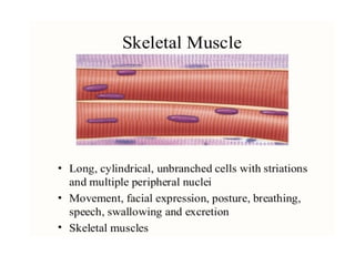
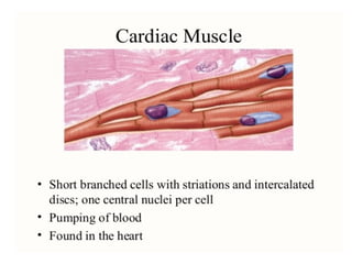
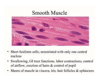
36.
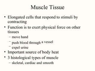
Muscle Tissue
• Elongatedcells that respond
contracting
to stimuli by
• Function is to exert
tissues
physical force on other
–
–
–
move hand
push blood through
expel urine
a vessel
•
•
Important source of body heat
3 histological types of muscle
– skeletal, cardiac and smooth
41.
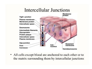
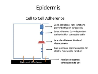
Epidermis
Basement Membrane
Cell toCell Adherence
Zona adherens: Ca++ dependent
cadherins that connect to actin
Zona occludens: tight junctions
prevent diffusion across cells
Macula adherens: Made of
desmosomes
Gap junctions: communication for
electric / metabolic function
Hemidesmosomes:
connect cells to BM
43.
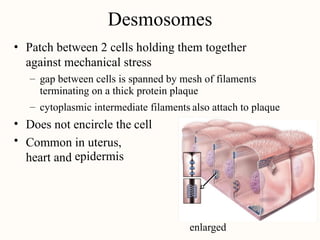
Desmosome
Desmosomes
Patch between 2cells holding them together
against mechanical stress
•
– gap between cells is spanned by mesh of filaments
terminating on a thick protein plaque
– cytoplasmic intermediate filaments also attach to plaque
•
•
Does not encircle the cell
Common in uterus,
epidermis
heart and
enlarged
45.
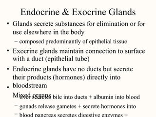
Endocrine & ExocrineGlands
Glands secrete substances for elimination or
use elsewhere in the body
• for
– composed predominantly of epithelial tissue
• Exocrine glands maintain connection to surface
with a duct (epithelial tube)
Endocrine glands have no ducts but secrete
their products (hormones) directly into
bloodstream
Mixed organs
•
•
–
–
–
liver secretes bile into ducts + albumin into blood
gonads release gametes + secrete hormones into
48.
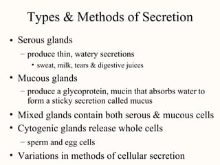
Types & Methodsof Secretion
Serous glands
•
– produce thin, watery secretions
• sweat, milk, tears & digestive juices
• Mucous glands
– produce a glycoprotein, mucin that absorbs water
form a sticky secretion called mucus
to
•
•
Mixed glands contain both serous & mucous cells
Cytogenic glands release whole cells
– sperm and egg cells
• Variations in methods of cellular secretion
50.
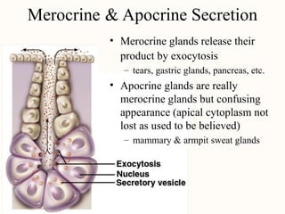
Merocrine &
•
Apocrine Secretion
Merocrineglands release their
product by exocytosis
– tears, gastric glands, pancreas, etc.
• Apocrine glands are really
merocrine glands but confusing
appearance (apical cytoplasm not
lost as used to be believed)
– mammary & armpit sweat glands
Editor's Notes
#41 The keratinocytes of the epidermis are connected to one another and the basement membrane by specialized attachement proteins. Some of the most clinically important are the desmosomes which are found inbetween keratinocytes. Desmosomes are composed of various types of desmolgliens. When the desmogleins, specifically desmoglien type 3, are attacked by a persons own immune system in diseases such as pemphigus vulgaris the cells will separate from one another and roll up in balls. This process is called acantholysis and results in crusted and blistered skin lesions. Also clinically important are the hemidesmosomes which anchor the cells of the basal layer to a protein structure called the basement membrane. If the hemidesomosomes are not working correctly the epidermis will lift off the dermis forming a subepidermal blister. This is seen in bullous pemphigoid which creates tense skin blisters most common in the elderly.


![Chapt05 Holes Lecture[1]](https://cdn.slidesharecdn.com/ss_thumbnails/chapt05holeslecture1-091122121913-phpapp02-thumbnail.jpg?width=640&height=640&fit=bounds)


