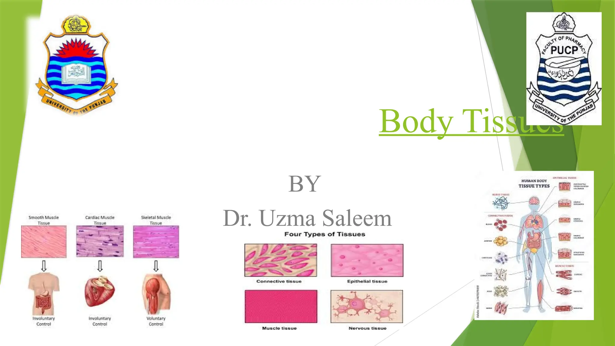
Tissue
The wordtissue comes from an old French verb meaning “to weave”.
Tissue is a group of similar cells, having same origin and performing a
specific function.
The term tissue was given by Bichat. The study of tissue is known as
histology.
The term histology was introduced by Mayer.
Xavier Bichat ("the father of histology") establishes the systematic
study of tissues as a discipline within anatomy by describing twenty-
one basic tissue types. Karl Mayer applies the word "histology" as
the name for the new discipline founded by Bichat.
3.
History of Bichat
XavierBichat, who lived a short life
(1771–1802), was prominent French anatomist
and physiologist during the time of revolution.
He played a key role in the creation of the science of histology. Indeed, he
was the first to see the organs of the body as being formed through the
specialization of simple, functional units (tissues). Bichat is also known as
one of the last of the major theorists of vitalism. [Vitalism is a scientific
theory that living organisms have a vital force, or life-force,
that is distinct from physical and chemical forces, and that this
force controls the development and activities of living
organisms].
4.
History of KarlMayer
Karl Mayer (1787–1865) was a German
anatomist and physiologist who coined the
term "histology" in 1819. The term comes from
the Greek words histos (tissue) and logia (science). Mayer used the term in his book on histology and a new classification
of tissues of the human body.
5.
Types of tissues
Theorgans in your body are composed of four basic types of tissue,
including:
1. Epithelial
2. Connective
3. Muscular
4. Nervous
6.
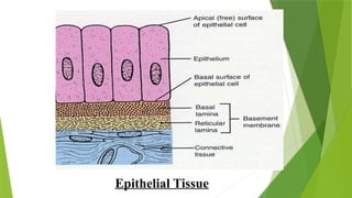
Epithelial Tissue
Epithelial tissuesare essentially large sheets of cells covering all
the surfaces of the body exposed to the outside world and lining the
outside of organs. Epithelial tissues covers body surface, lines body
cavities & ducts and form glands.
The epithelium is a type of body tissue that forms the covering on all
internal and external surfaces of the body, lines body cavities and
hollow organs and is the major tissue in glands. Epithelial tissue has a
variety of functions depending on where it’s located in your body,
including protection, secretion and absorption.
7.
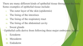
There are manydifferent kinds of epithelial tissue throughout the body.
Some examples of epithelial tissue include:
i. The outer layer of the skin (epidermis)
ii. The lining of the intestines
iii. The lining of the respiratory tract
iv. The lining of the abdominal cavity
v. Sweat glands
• Epithelial cells derive from following three major embryonic layers:
I. Ectoderm
II. Mesoderm
III. Endoderm
8.
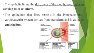
• The epithelialining the skin, parts of the mouth, nose and anus
develop from ectoderm.
• The epithelium that lines vessels in the lymphatic and
cardiovascular system derives from mesoderm and is called an
endothelium.
9.
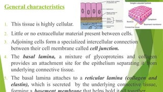
General characteristics
1. Thistissue is highly cellular.
2. Little or no extracellular material present between cells.
3. Adjoining cells form a specialized intercellular connection
between their cell membrane called cell junction.
4. The basal lamina, a mixture of glycoproteins and collagen
provides an attachment site for the epithelium separating it from
underlying connective tissue.
5. The basal lamina attaches to a reticular lamina (collagen and
elastin), which is secreted by the underlying connective tissue,
11.
6. Epithelial tissuesare nearly completely avascular. For instance no
blood vessels cross the basement membrane to enter the tissue.
7. Nutrients must come by diffusion or absorption from
underlying tissues or surface.
8. Many epithelial tissues are capable of rapidly replacing damaged
and dead cells.
9. Sloughing off of damaged or dead cells is a characteristic of
surface epithelium and allows our airways and digestive tracts to
rapidly replace damaged cells with new cells.
12.
Generalized function ofEpithelial tissues
1. Epithelial tissues provide the body’s first line of protection from:
• Physical
• Chemical, and Biological wear and tear.
2. The cells of an epithelium act as gatekeepers of the body
controlling permeability and allowing selective transfer of
materials across a physical barrier.
3. Some epithelia often include structural features that allow the
selective transport of molecules and ions across their cell
membranes.
13.
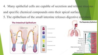
4. Many epithelialcells are capable of secretion and release mucous
and specific chemical compounds onto their apical surface.
5. The epithelium of the small intestine releases digestive enzymes.
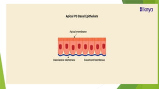
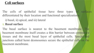
Cell surfaces
The cellsof epithelial tissue have three types of surfaces
differentiated by their location and functional specializations:
i) basal, ii) apical, and iii) lateral
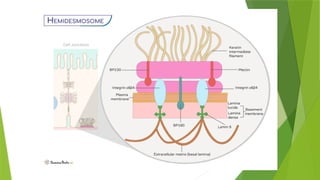
1. Basal surface
The basal surface is nearest to the basement membrane. The
basement membrane itself creates a thin barrier between connective
tissues and the most basal layer of epithelial cells. Specialized
junctions called hemi desmosomes secure the epithelial cells on the
basement membrane.
16.
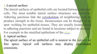
2. Lateral surface
Thelateral surfaces of epithelial cells are located between adjacent
cells. The most notable lateral surface structures are junctions.
Adhering junctions link the cytoskeleton of neighboring cells to
produce strength in the tissue. Desmosomes can be thought of as
spot- welding for epithelial tissues. They are usually located deep
to adhering junctions and are found in locations subject to stresses.
For example in the stratified epithelium of the skin.
3. Apical surface
The apical surface of an epithelial cell is nearest to the luminal or
free space. Apical cell surfaces may display specialized
extensions.
17.
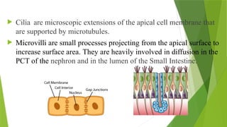
Cilia aremicroscopic extensions of the apical cell membrane that
are supported by microtubules.
Microvilli are small processes projecting from the apical surface to
increase surface area. They are heavily involved in diffusion in the
PCT of the nephron and in the lumen of the Small Intestine.
18.
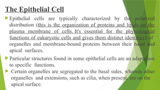
The Epithelial Cell
Epithelial cells are typically characterized by the polarized
distribution (this is the organization of proteins and lipids on the
plasma membrane of cells. It's essential for the physiological
functions of eukaryotic cells and gives them distinct identities) of
organelles and membrane-bound proteins between their basal and
apical surfaces.
Particular structures found in some epithelial cells are an adaptation
to specific functions.
Certain organelles are segregated to the basal sides, whereas other
organelles and extensions, such as cilia, when present, are on the
apical surface.
19.
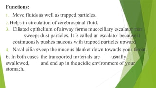
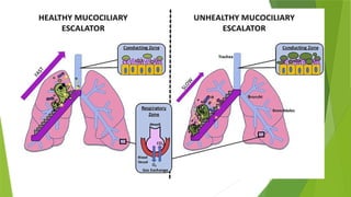
Functions:
1. Move fluidsas well as trapped particles.
2. Helps in circulation of cerebrospinal fluid.
3. Ciliated epithelium of airway forms mucociliary escalator that
sweeps dust particles. It is called an escalator because it
continuously pushes mucous with trapped particles upward.
4. Nasal cilia sweep the mucous blanket down towards your throat.
6. In both cases, the transported materials are usually
swallowed, and end up in the acidic environment of your
stomach.
21.
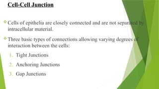
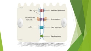
Cell-Cell Junction
Cells ofepithelia are closely connected and are not separated by
intracellular material.
Three basic types of connections allowing varying degrees of
interaction between the cells:
1. Tight Junctions
2. Anchoring Junctions
3. Gap Junctions
22.
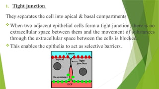
1. Tight junction
Theyseparates the cell into apical & basal compartments.
When two adjacent epithelial cells form a tight junction, there is no
extracellular space between them and the movement of substances
through the extracellular space between the cells is blocked.
This enables the epithelia to act as selective barriers.
23.
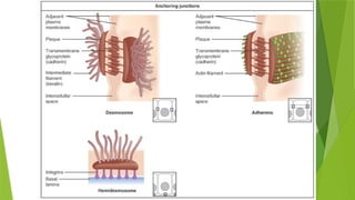
2. Anchoring junction
Ananchoring junction includes severaltypesof cell junctions that
help stabilize epithelial tissues.
Anchoring junctions are common on the lateral and basal surfaces
of cells where they provide strong and flexible connections.
There are three types of anchoring junctions:
i. Desmosomes
ii. Hemidesmosomes
iii. Adherens
25.
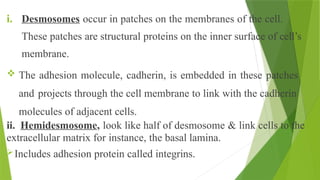
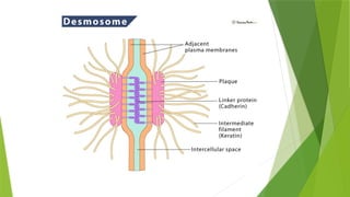
i. Desmosomes occurin patches on the membranes of the cell.
These patches are structural proteins on the inner surface of cell’s
membrane.
The adhesion molecule, cadherin, is embedded in these patches
and projects through the cell membrane to link with the cadherin
molecules of adjacent cells.
ii. Hemidesmosome, look like half of desmosome & link cells to the
extracellular matrix for instance, the basal lamina.
Includes adhesion protein called integrins.
28.
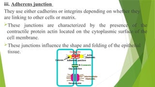
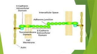
iii. Adherens junction
Theyuse either cadherins or integrins depending on whether they
are linking to other cells or matrix.
These junctions are characterized by the presence of the
contractile protein actin located on the cytoplasmic surface of the
cell membrane.
These junctions influence the shape and folding of the epithelial
tissue.
31.
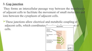
3. Gap junction
Theyforms an intercellular passage way between the membranes
of adjacent cells to facilitate the movement of small molecules and
ions between the cytoplasm of adjacent cells.
These junctions allow electrical and metabolic coupling of
adjacent cells, which coordinates function in large groups of
cells.
32.
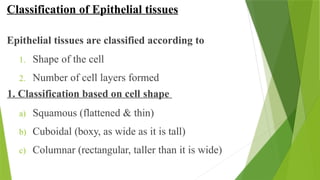
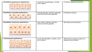
Classification of Epithelialtissues
Epithelial tissues are classified according to
1. Shape of the cell
2. Number of cell layers formed
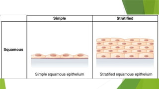
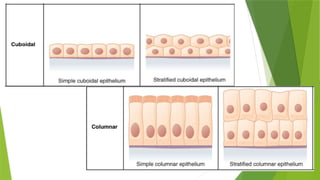
1. Classification based on cell shape
a) Squamous (flattened & thin)
b) Cuboidal (boxy, as wide as it is tall)
c) Columnar (rectangular, taller than it is wide)
34.
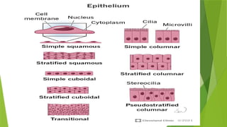
2. Classification basedon number of cell layers
Similarly, the number of cell layer in the tissue can be one, where every
cell rest on basal lamina, it is called simple epithelium.
If number of cell layer is more than one. It is called stratified
epithelium & only the basal layer of the cell rests on basal lamina.
2.1. Simple epithelialium
I. Squamous epithelium (flattened)
II. Cuboidal epithelium (cube shaped)
III. Columnar epithelium (elongated)
IV. Ciliated epithelium
V. Pseudo-stratified epithelium
35.
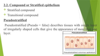
2.2. Compound orStratified epithelium
Stratified compound
Transitional compound
Pseudostratified
Pseudostratified (Pseudo = false) describes tissues with single layer
of irregularly shaped cells that give the appearance of more than one
layer.
38.
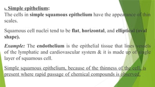
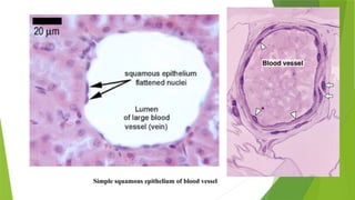
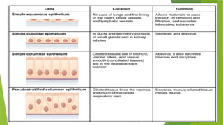
I. Simple epithelium:
Thecells in simple squamous epithelium have the appearance of thin
scales.
Squamous cell nuclei tend to be flat, horizontal, and elliptical (oval
shape).
Example: The endothelium is the epithelial tissue that lines vessels
of the lymphatic and cardiovascular system & it is made up of single
layer of squamous cell.
Simple squamous epithelium, because of the thinness of the cell, is
present where rapid passage of chemical compounds is observed.
39.
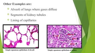
Other Examples are:
Alveoli of lungs where gases diffuse
Segments of kidney tubules
Lining of capillaries
Simple squamous epithelium of alveoli Simple squamous epithelium of kidney
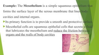
Example: The Mesotheliumis a simple squamous epithelium that
forms the surface layer of the serous membrane that lines body
cavities and internal organs.
Its primary function is to provide a smooth and protective surface.
Mesothelial cells are squamous epithelial cells that secrete a fluid
that lubricates the mesothelium and reduce the friction between
organs and the walls of body cavities
.
42.
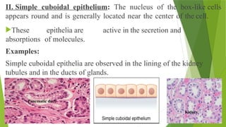
II. Simple cuboidalepithelium: The nucleus of the box-like cells
appears round and is generally located near the center of the cell.
These epithelia are active in the secretion and
absorptions of molecules.
Examples:
Simple cuboidal epithelia are observed in the lining of the kidney
tubules and in the ducts of glands.
Kidney
Pancreatic duct
43.
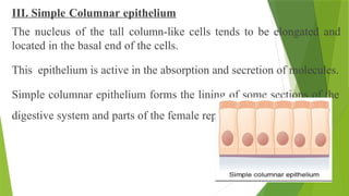
III. Simple Columnarepithelium
The nucleus of the tall column-like cells tends to be elongated and
located in the basal end of the cells.
This epithelium is active in the absorption and secretion of molecules.
Simple columnar epithelium forms the lining of some sections of the
digestive system and parts of the female reproductive tract.
44.
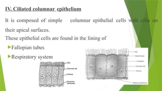
IV. Ciliated columnarepithelium
It is composed of simple columnar epithelial cells with cilia on
their apical surfaces.
These epithelial cells are found in the lining of
Fallopian tubes
Respiratory system
45.
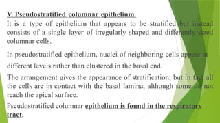
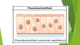
V. Pseudostratified columnarepithelium
It is a type of epithelium that appears to be stratified but instead
consists of a single layer of irregularly shaped and differently sized
columnar cells.
In pseudostratified epithelium, nuclei of neighboring cells appear at
different levels rather than clustered in the basal end.
The arrangement gives the appearance of stratification; but in fact all
the cells are in contact with the basal lamina, although some do not
reach the apical surface.
Pseudostratified columnar epithelium is found in the respiratory
tract.
47.
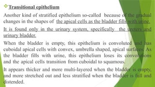
Transitional epithelium
Another kindof stratified epithelium so-called because of the gradual
changes in the shapes of the apical cells as the bladder fills with urine.
It is found only in the urinary system, specifically the ureters and
urinary bladder.
When the bladder is empty, this epithelium is convoluted and has
cuboidal apical cells with convex, umbrella shaped, apical surfaces. As
the bladder fills with urine, this epithelium loses its convolutions
and the apical cells transition from cuboidal to squamous.
It appears thicker and more multi-layered when the bladder is empty,
and more stretched out and less stratified when the bladder is full and
distended.
Introduction
As obviousfrom its name, one of the major function of
connective tissue is to connect tissue and organs.
Connective tissue cells are dispersed in a matrix.
The matrix usually includes a large amount of extracellular
material produced by the connective tissue cells that are
embedded within it.
The matrix plays a major role in the functioning of this
tissue.
52.
Two major componentsof the matrix are:
I. Ground substance
II. Protein fibers
I. Ground substance: It is usually a fluid (water), but it can also be
mineralized and solid, as in bones.
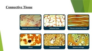
Connective tissues come in a vast variety of forms, yet they typically
have in common three characteristic components:
Cells
Large amount of ground substance
Protein fibers
53.
The amount andstructure of each component correlates with the
function of the tissue. Like from the rigid ground substance in
bones supporting the body to the inclusion of specialized cells; for
example, a phagocytic cell that engulfs pathogens and also rids tissue
of cellular debris.
Fibroblast: The most common cell found within connective tissue.
Polysaccharides and proteins secreted by fibroblasts combine with
extra-cellular fluids to produce a viscous ground substance that with
embedded fibrous proteins, forms the extracellular matrix.
54.
Types of fiberssecreted by fibroblasts:
Collagen
Elastic
Reticular
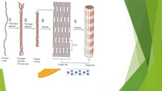
Collagen fiber: It is made from fibrous protein subunits linked together to
form a long and straight fiber.
Flexible
Great tensile strength
Resist stretching, and give ligaments and tendons their characteristic
resilience and strength.
These fibers hold connective tissues together, even during the
movement of the body.
56.
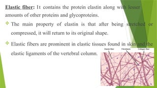
Elastic fiber: Itcontains the protein elastin along with lesser
amounts of other proteins and glycoproteins.
The main property of elastin is that after being stretched or
compressed, it will return to its original shape.
Elastic fibers are prominent in elastic tissues found in skin and the
elastic ligaments of the vertebral column.
58.
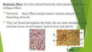
Reticular fiber: Itis also formed from the same protein subunits as
collagen fibers.
However, these fibers remain narrow and are arrayed in a
branching network.
They are found throughout the body, but are most abundant in the
reticular tissue of soft organs, such as liver and spleen.
Liver
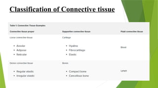
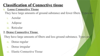
Classification of Connectivetissue
1. Loose Connective Tissue
They have large amounts of ground substance and fewer fibers. Types are:
i. Aerolar
ii. Adipose
iii. Reticular
2. Dense Connective Tissue
They have large amounts of fibers and less ground substance. Types are:
iv. Dense regular
v. Dense irregular
vi. Elastic Connective Tissue
61.
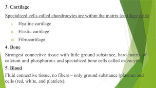
3. Cartilage
Specialized cellscalled chondrocytes are within the matrix (cartilage cells)
i. Hyaline cartilage
ii. Elastic cartilage
iii. Fibrocartilage
4. Bone
Strongest connective tissue with little ground substance, hard matrix of
calcium and phosphorous and specialized bone cells called osteocytes.
5. Blood
Fluid connective tissue, no fibers – only ground substance (plasma) and
cells (red, white, and platelets).
62.
1. Loose connectivetissue
Loose connective tissue is found between many organs where it
acts both to absorb shock and bind tissues together.
It allows water, salts, and various nutrients to diffuse through to
adjacent or imbedded cells and tissues.
Fat contributes mostly to lipid storage, can serve as insulation from
cold temperatures and mechanical injuries.
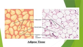
Example: Adipose tissue consists mostly of fat storage cells called
adipocytes that store lipids as droplets that fill most of the cytoplasm.
A large number of capillaries allow rapid storage and mobilization of
lipid molecules.
1.1. Areolar tissue
Thisis the most generalized type of connective tissue. The matrix is
semisolid with many fibroblasts and some fat cells (adipocytes), mast
cells and macrophages, widely separated by elastic and collagen fibers.
It is found in almost every part of the body, providing elasticity and
tensile strength. It connects and supports other tissues, for example:
under the skin
between muscles
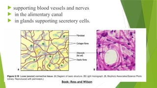
65.
supporting bloodvessels and nerves
in the alimentary canal
in glands supporting secretory cells.
Book: Ross and Wilson
66.
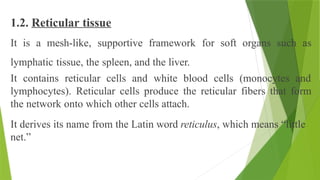
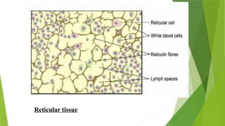
1.2. Reticular tissue
Itis a mesh-like, supportive framework for soft organs such as
lymphatic tissue, the spleen, and the liver.
It contains reticular cells and white blood cells (monocytes and
lymphocytes). Reticular cells produce the reticular fibers that form
the network onto which other cells attach.
It derives its name from the Latin word reticulus, which means “little
net.”
2. Dense connectivetissue
Dense connective tissue contains more collagen fibers than does loose
connective tissue.
As a consequence, it displays greater resistance to stretching.
There are three major categories of dense connective tissue: regular,
irregular, and elastic.
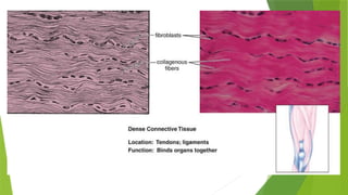
2.1. Dense regular connective tissue fibers are parallel to each other,
enhancing tensile strength and resistance to stretching in the direction of
the fiber orientations.
Example: Ligaments and tendons are made of dense regular connective
tissue.
70.
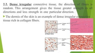
2.2. Dense irregularconnective tissue, the direction of fibers is
random. This arrangement gives the tissue greater strength in all
directions and less strength in one particular direction.
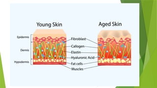
The dermis of the skin is an example of dense irregular connective
tissue rich in collagen fibers.
71.
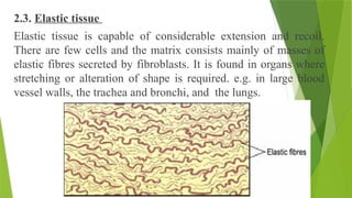
2.3. Elastic tissue
Elastictissue is capable of considerable extension and recoil.
There are few cells and the matrix consists mainly of masses of
elastic fibres secreted by fibroblasts. It is found in organs where
stretching or alteration of shape is required. e.g. in large blood
vessel walls, the trachea and bronchi, and the lungs.
72.
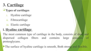
3. Cartilage
Typesof cartilages
i. Hyaline cartilage
ii. Fibrocartilage
iii. Elastic cartilage
i. Hyaline cartilage
The most common type of cartilage in the body, consists of short and
dispersed collagen fibers and contains large amounts of
proteoglycans.
The surface of hyaline cartilage is smooth, Both strong and flexible.
73.
It is foundin the rib cage and nose and covers bones where they
meet to form moveable joints.
It makes up a template of the embryonic skeleton before bone
formation.
A plate of hyaline cartilage at the ends of bone allows continued
growth until adulthood.
ii. Fibro cartilage
It is tough because it has thick bundles of collagen fibers dispersed
through its matrix.
The knee and jaw joints and the intervertebral discs are examples of
fibrocartilage.
74.
iii. Elastic cartilage
ItContains elastic fibers as well as collagen and proteoglycans.
This tissue gives rigid support as wellas elasticity.
Tug gently at your ear lobes, and notice that the lobes return to
their initial shape. The external ear contains elastic cartilage.
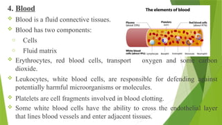
4. Blood
Bloodis a fluid connective tissues.
Blood has two components:
o Cells
o Fluid matrix
Erythrocytes, red blood cells, transport oxygen and some carbon
dioxide.
Leukocytes, white blood cells, are responsible for defending against
potentially harmful microorganisms or molecules.
Platelets are cell fragments involved in blood clotting.
Some white blood cells have the ability to cross the endothelial layer
that lines blood vessels and enter adjacent tissues.
77.
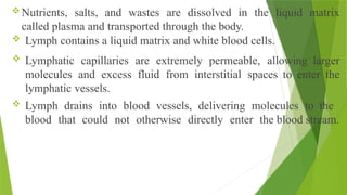
Nutrients, salts, andwastes are dissolved in the liquid matrix
called plasma and transported through the body.
Lymph contains a liquid matrix and white blood cells.
Lymphatic capillaries are extremely permeable, allowing larger
molecules and excess fluid from interstitial spaces to enter the
lymphatic vessels.
Lymph drains into blood vessels, delivering molecules to the
blood that could not otherwise directly enter the blood stream.
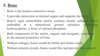
5. Bone
Boneis the hardest connective tissue.
It provides protection to internal organs and supports the body.
Bone’s rigid extracellular matrix contains mostly collagen fibers
embedded in a mineralized ground substance containing
hydroxyapatite, a form of calcium phosphate.
Both components of the matrix, organic and inorganic, contribute
to the unusual properties of bone.
Without collagen, bones would be brittle and shatter easily.
Without mineral crystals, bones would flex and provide little support.
81.
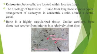
Osteocytes, bone cells,are located within lacunae (gap).
The histology of transverse tissue from long bone shows a typical
arrangement of osteocytes in concentric circles around a central
canal.
Bone is a highly vascularized tissue. Unlike cartilage, bone
tissue can recover from injuries in a relatively short time.
82.
Bone isa living tissue capable of changing its structure as the
result of the stresses to which it is subjected.
Like other connective tissues, bone consists of:
cells,
fibers, and
Matrix
It is hard because of the calcification of its extracellular matrix
and possesses a degree of elasticity because of the presence of
organic fibers.
Bone has a protective function.
83.
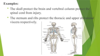
Examples:
The skullprotect the brain and vertebral column protect the
spinal cord from injury.
The sternum and ribs protect the thoracic and upper abdominal
viscera respectively.
84.
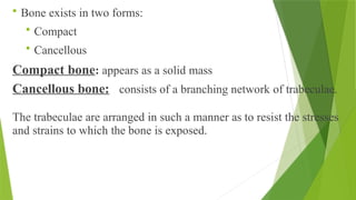
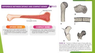
Bone existsin two forms:
Compact
Cancellous
Compact bone: appears as a solid mass
Cancellous bone: consists of a branching network of trabeculae.
The trabeculae are arranged in such a manner as to resist the stresses
and strains to which the bone is exposed.
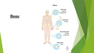
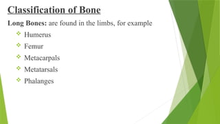
Classification of Bone
LongBones: are found in the limbs, for example
Humerus
Femur
Metacarpals
Metatarsals
Phalanges
88.
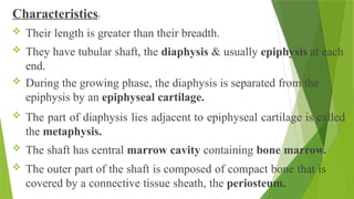
Characteristics:
Their lengthis greater than their breadth.
They have tubular shaft, the diaphysis & usually epiphysis at each
end.
During the growing phase, the diaphysis is separated from the
epiphysis by an epiphyseal cartilage.
The part of diaphysis lies adjacent to epiphyseal cartilage is called
the metaphysis.
The shaft has central marrow cavity containing bone marrow.
The outer part of the shaft is composed of compact bone that is
covered by a connective tissue sheath, the periosteum.
89.
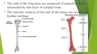
The endsof the long bone are composed of cancellous bone
surrounded by thin layer of compact bone.
The articular surfaces of the end of the bones are covered by
hyaline cartilage.
90.
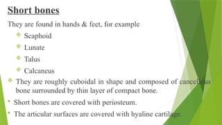
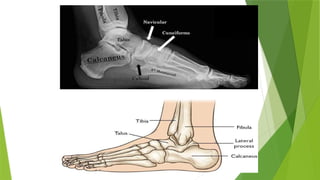
Short bones
They arefound in hands & feet, for example
Scaphoid
Lunate
Talus
Calcaneus
They are roughly cuboidal in shape and composed of cancellous
bone surrounded by thin layer of compact bone.
Short bones are covered with periosteum.
The articular surfaces are covered with hyaline cartilage.
93.
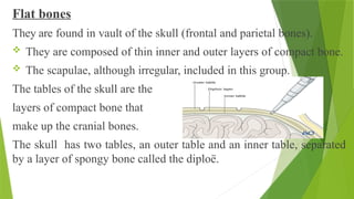
Flat bones
They arefound in vault of the skull (frontal and parietal bones).
They are composed of thin inner and outer layers of compact bone.
The scapulae, although irregular, included in this group.
The tables of the skull are the
layers of compact bone that
make up the cranial bones.
The skull has two tables, an outer table and an inner table, separated
by a layer of spongy bone called the diploë.
94.
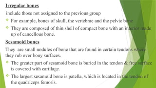
Irregular bones
include thosenot assigned to the previous group
For example, bones of skull, the vertebrae and the pelvic bone
They are composed of thin shell of compact bone with an interior made
up of cancellous bone.
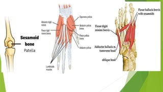
Sesamoid bones
They are small nodules of bone that are found in certain tendons where
they rub over bony surfaces.
The greater part of sesamoid bone is buried in the tendon & free surface
is covered with cartilage.
The largest sesamoid bone is patella, which is located in the tendon of
the quadriceps femoris.
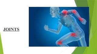
Definition: A sitewhere two or more bones come together, whether
or not movement occurs between them, is called a joint.
Types: joints are classified according to the tissues that lie between
the bones:
i. Fibrous joints
ii. Cartilaginous joints
iii. Synovial joints
98.
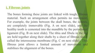
i. Fibrous joints
Thebones forming these joints are linked with tough, fibrous
material. Such an arrangement often permits no movement.
For example, the joints between the skull bones, the sutures,
are completely immovable (Fig. A on next slide), and the
healthy tooth is cemented into the mandible by the periodontal
ligament (Fig. B on next slide). The tibia and fibula in the leg
are held together along their shafts by a sheet of fibrous tissue
called the interosseous membrane (Fig. C on next slide) . This
fibrous joint allows a limited amount of movement and
stabilises the alignment of the bones.
99.
Fibrous joints: (A)Sutureof the skull (B) The periodontal ligament (C) The Interosseous membrane linking
the
tibia and fibula
100.
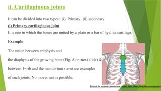
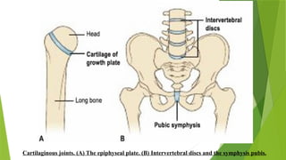
ii. Cartilaginous joints
Itcan be divided into two types: (i) Primary (ii) secondary
(i) Primary cartilaginous joint
It is one in which the bones are united by a plate or a bar of hyaline cartilage.
Example
The union between epiphysis and
the diaphysis of the growing bone (Fig. A on next slide) &
between 1st rib and the manubrium sterni are examples
of such joints. No movement is possible.
ii. Secondary cartilaginousjoint
It is one in which bones are united by a plate of fibrocartilage and
articular surface of the bones are covered by a thin layer of hyaline
cartilage.
Examples: Joints between vertebral bodies and the symphysis pubis
(Fig. B on previous slide).
A small amount of movement is possible. They are also shock
absorbent.
103.
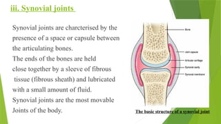
iii. Synovial joints
Synovialjoints are charcterised by the
presence of a space or capsule between
the articulating bones.
The ends of the bones are held
close together by a sleeve of fibrous
tissue (fibrous sheath) and lubricated
with a small amount of fluid.
Synovial joints are the most movable
Joints of the body. The basic structure of a synovial joint
104.
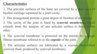
Characteristics
1. The articularsurfaces of the bone are covered by a thin layer of
hyaline cartilage separated by a joint cavity.
2. This arrangement permits a great degree of freedom of movement.
3. The cavity of the joint is lined by synovial membrane, which
extends from the margins of one articular surface to those of the
other.
4. The synovial membrane is protected on the outside by a tough
fibrous membrane referred to as the capsule of the joint.
5. The articular surfaces are lubricated by a viscous fluid called
synovial fluid, produced by synovial membrane.
105.
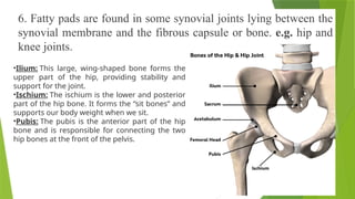
6. Fatty padsare found in some synovial joints lying between the
synovial membrane and the fibrous capsule or bone. e.g. hip and
knee joints.
•Ilium: This large, wing-shaped bone forms the
upper part of the hip, providing stability and
support for the joint.
•Ischium: The ischium is the lower and posterior
part of the hip bone. It forms the “sit bones” and
supports our body weight when we sit.
•Pubis: The pubis is the anterior part of the hip
bone and is responsible for connecting the two
hip bones at the front of the pelvis.
106.
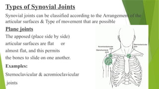
Types of SynovialJoints
Synovial joints can be classified according to the Arrangement of the
articular surfaces & Type of movement that are possible
Plane joints
The apposed (place side by side)
articular surfaces are flat or
almost flat, and this permits
the bones to slide on one another.
Examples:
Sternoclavicular & acromioclavicular
joints
107.
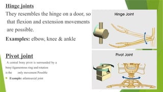
Hinge joints
They resemblesthe hinge on a door, so
that flexion and extension movements
are possible.
Examples: elbow, knee & ankle
Pivot joint
A central bony pivot is surrounded by a
bony-ligamentous ring and rotation
is the only movement Possible
Example: atlantoaxial joint
108.
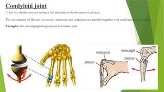
Condyloid joint
It hastwo distinct convex surfaces that articulate with two concave surfaces.
The movements of flexion, extension, abduction and adduction are possible together with small amount of rotation.
Examples: the metacarpophalangeal joint or knuckle joint
109.
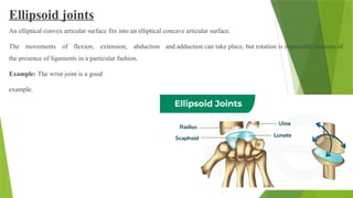
Ellipsoid joints
An ellipticalconvex articular surface fits into an elliptical concave articular surface.
The movements of flexion, extension, abduction and adduction can take place, but rotation is impossible because of
the presence of ligaments in a particular fashion.
Example: The wrist joint is a good
example.
110.
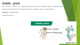
Saddle joints
The articularsurfaces are reciprocally concavoconvex and resemble a saddle on a horse’s back.
These joints permit flexion, extension, abduction, adduction, and very slight rotation.
Example: carpometacarpal
joint of the thumb
111.
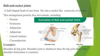
Ball-and-socket joints
A ball-shapedhead of one bone fits into a socket like concavity of another.
This arrangement permits free movements, including
• Flexion
• Extension
• Abduction
• Adduction
• Lateral rotation
• Circumduction
Examples:
Shoulder & hip joint. Shoulder joint is shallower than the hip joint that’s why
shoulder dislocation is common.
112.
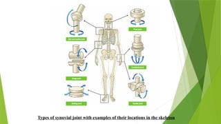
Types of synovialjoint with examples of their locations in the skeleton
Definition
A band orbundle of fibrous tissues that has the ability to
contract, producing movement in or maintaining the position of parts of
the body.
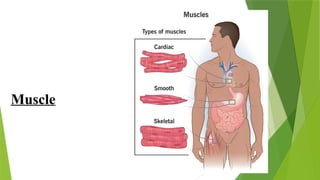
Skeletal Muscle
Skeletal muscles produce the movements of the skeleton.
They are sometimes called voluntary muscles and are made up of
striped muscle fibers.
A skeletal muscle has two or more attachments.
The attachment that moves the least is referred to as the origin, and the
one that moves the most is known as the insertion.
115.
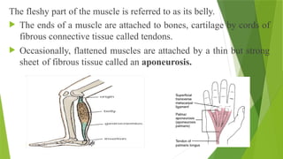
The fleshy partof the muscle is referred to as its belly.
The ends of a muscle are attached to bones, cartilage by cords of
fibrous connective tissue called tendons.
Occasionally, flattened muscles are attached by a thin but strong
sheet of fibrous tissue called an aponeurosis.
116.
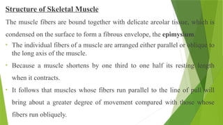
Structure of SkeletalMuscle
The muscle fibers are bound together with delicate areolar tissue, which is
condensed on the surface to form a fibrous envelope, the epimysium.
• The individual fibers of a muscle are arranged either parallel or oblique to
the long axis of the muscle.
• Because a muscle shortens by one third to one half its resting length
when it contracts.
• It follows that muscles whose fibers run parallel to the line of pull will
bring about a greater degree of movement compared with those whose
fibers run obliquely.
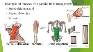
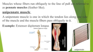
Muscles whose fibersrun obliquely to the line of pull are referred to
as pennate muscles (feather like).
unipennate muscle
A unipennate muscle is one in which the tendon lies along one side
of the muscle and the muscle fibers pass obliquely to it.
Example: Extensor digitorum longus
119.
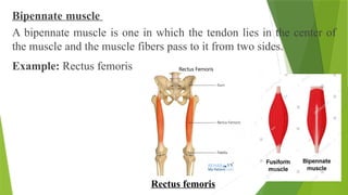
Bipennate muscle
A bipennatemuscle is one in which the tendon lies in the center of
the muscle and the muscle fibers pass to it from two sides.
Example: Rectus femoris
Rectus femoris
120.
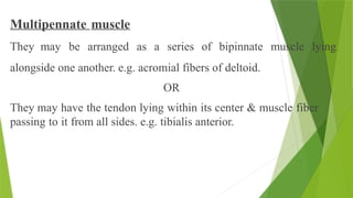
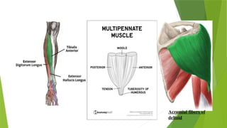
Multipennate muscle
They maybe arranged as a series of bipinnate muscle lying
alongside one another. e.g. acromial fibers of deltoid.
OR
They may have the tendon lying within its center & muscle fiber
passing to it from all sides. e.g. tibialis anterior.
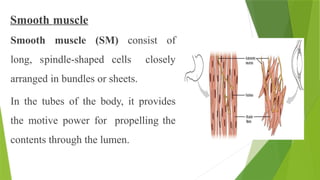
Smooth muscle
Smooth muscle(SM) consist of
long, spindle-shaped cells closely
arranged in bundles or sheets.
In the tubes of the body, it provides
the motive power for propelling the
contents through the lumen.
123.
• A waveof contraction of the circularly arranged fibers passes along
the tube, mixing the contents onward.
• In the storage organ such as urinary bladder & the uterus, the
fibers are irregularly arranged & intercalated with one another.
• In the walls of the blood vessels, the SM fibers are arranged
circularly & serve to modify the caliber of the lumen.
124.
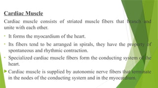
Cardiac Muscle
Cardiac muscleconsists of striated muscle fibers that branch and
unite with each other.
• It forms the myocardium of the heart.
• Its fibers tend to be arranged in spirals, they have the property of
spontaneous and rhythmic contraction.
• Specialized cardiac muscle fibers form the conducting system of the
heart.
Cardiac muscle is supplied by autonomic nerve fibers that terminate
in the nodes of the conducting system and in the myocardium.
125.
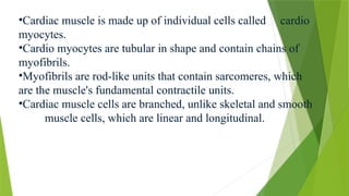
•Cardiac muscle ismade up of individual cells called cardio
myocytes.
•Cardio myocytes are tubular in shape and contain chains of
myofibrils.
•Myofibrils are rod-like units that contain sarcomeres, which
are the muscle's fundamental contractile units.
•Cardiac muscle cells are branched, unlike skeletal and smooth
muscle cells, which are linear and longitudinal.

![History of Bichat
Xavier Bichat, who lived a short life
(1771–1802), was prominent French anatomist
and physiologist during the time of revolution.
He played a key role in the creation of the science of histology. Indeed, he
was the first to see the organs of the body as being formed through the
specialization of simple, functional units (tissues). Bichat is also known as
one of the last of the major theorists of vitalism. [Vitalism is a scientific
theory that living organisms have a vital force, or life-force,
that is distinct from physical and chemical forces, and that this
force controls the development and activities of living
organisms].](https://image.slidesharecdn.com/bodytissues1-250917032454-2d23aff7/85/Body_Tissues-fully-detailed-notes-1-pptx-3-320.jpg)

![[Type of Hyaline
cartilage]](https://image.slidesharecdn.com/bodytissues1-250917032454-2d23aff7/85/Body_Tissues-fully-detailed-notes-1-pptx-75-320.jpg)
![Body_Tissues fully detailed notes [1].pptx](https://image.slidesharecdn.com/bodytissues1-250917032454-2d23aff7/85/Body_Tissues-fully-detailed-notes-1-pptx-126-320.jpg)


![ONFH[AVN HIP] -TRIPLE REGIME -A NOVAL SURGICAL CONCEPT .pptx](https://cdn.slidesharecdn.com/ss_thumbnails/onfhavnhip2026koaconcalicutdrgokuldevdrmashraf-260210064517-213ec005-thumbnail.jpg?width=640&height=640&fit=bounds)

