Downloaded 112 times


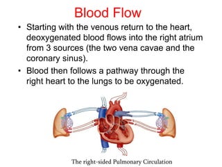


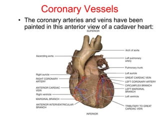
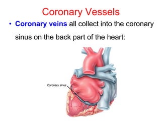


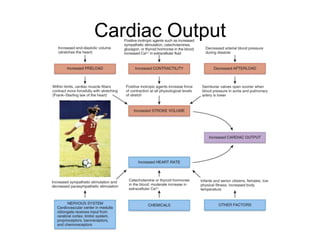

The document summarizes key aspects of heart anatomy and physiology. It describes the location of the heart in the thoracic cavity and its layers, including the epicardium, myocardium, and endocardium. It explains the heart's four chambers, valves that prevent backflow of blood, and the cardiac cycle of alternating atrial and ventricular contraction and relaxation. It also outlines the cardiac conduction system that coordinates heart contractions and generates the electrocardiogram.


![Human Reproduction [ Reproductive System ] Notes @irfanullah_mehar Irfanullah...](https://cdn.slidesharecdn.com/ss_thumbnails/humanreproductionreproductivesystemnotesirfanullahmeharirfanullahmeharjanantantra-260111172350-56e85778-thumbnail.jpg?width=640&height=640&fit=bounds)


