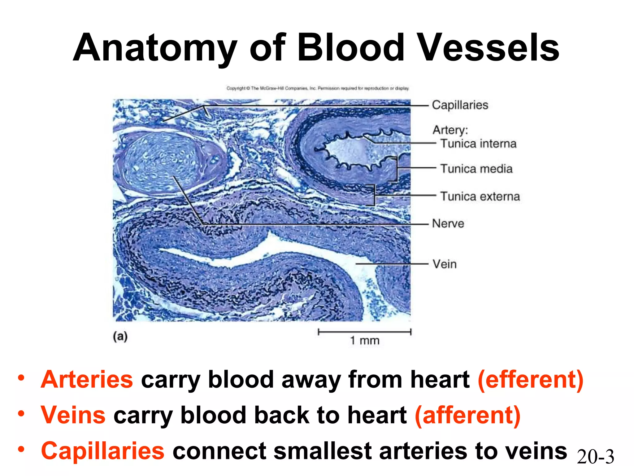
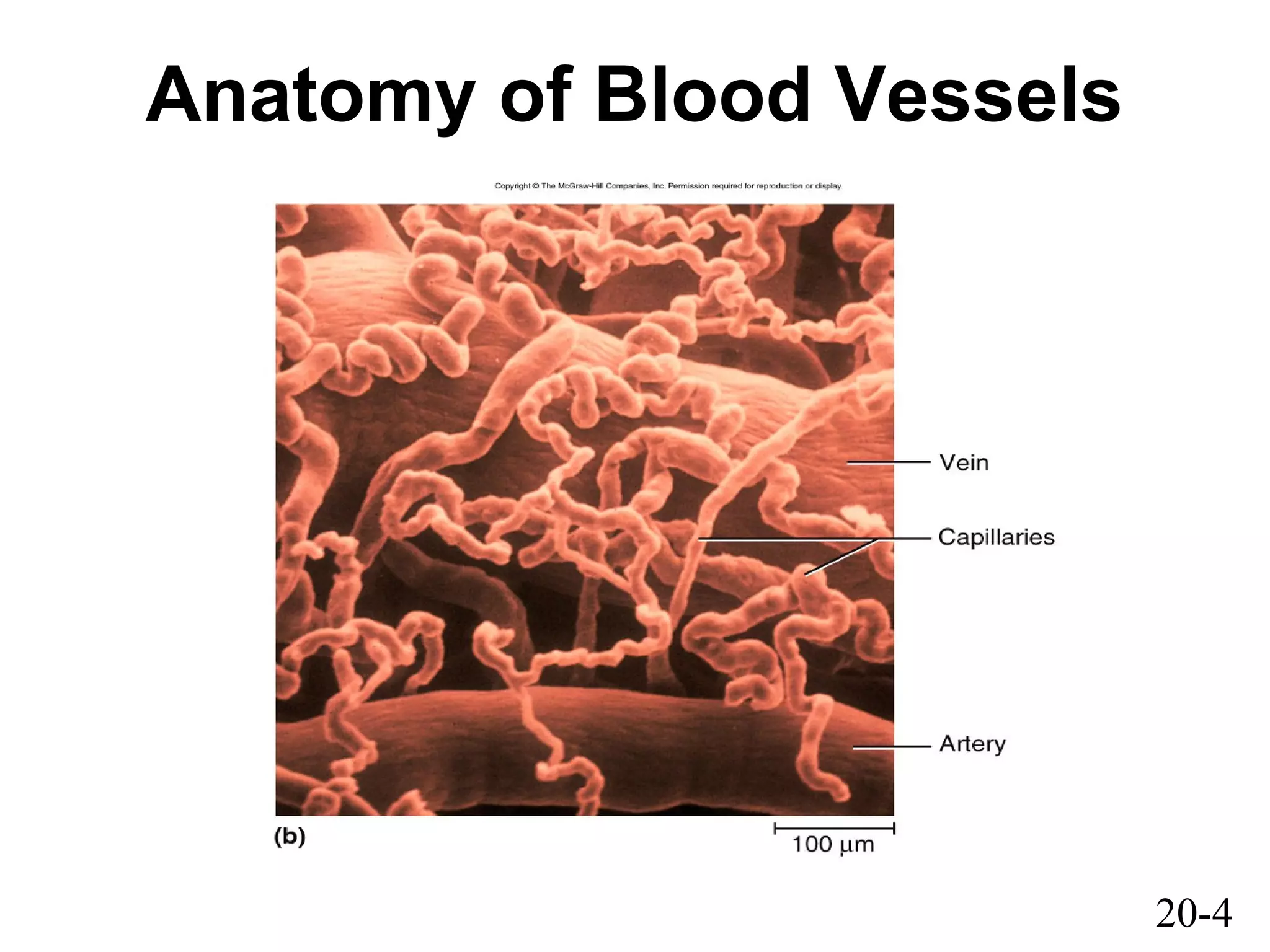
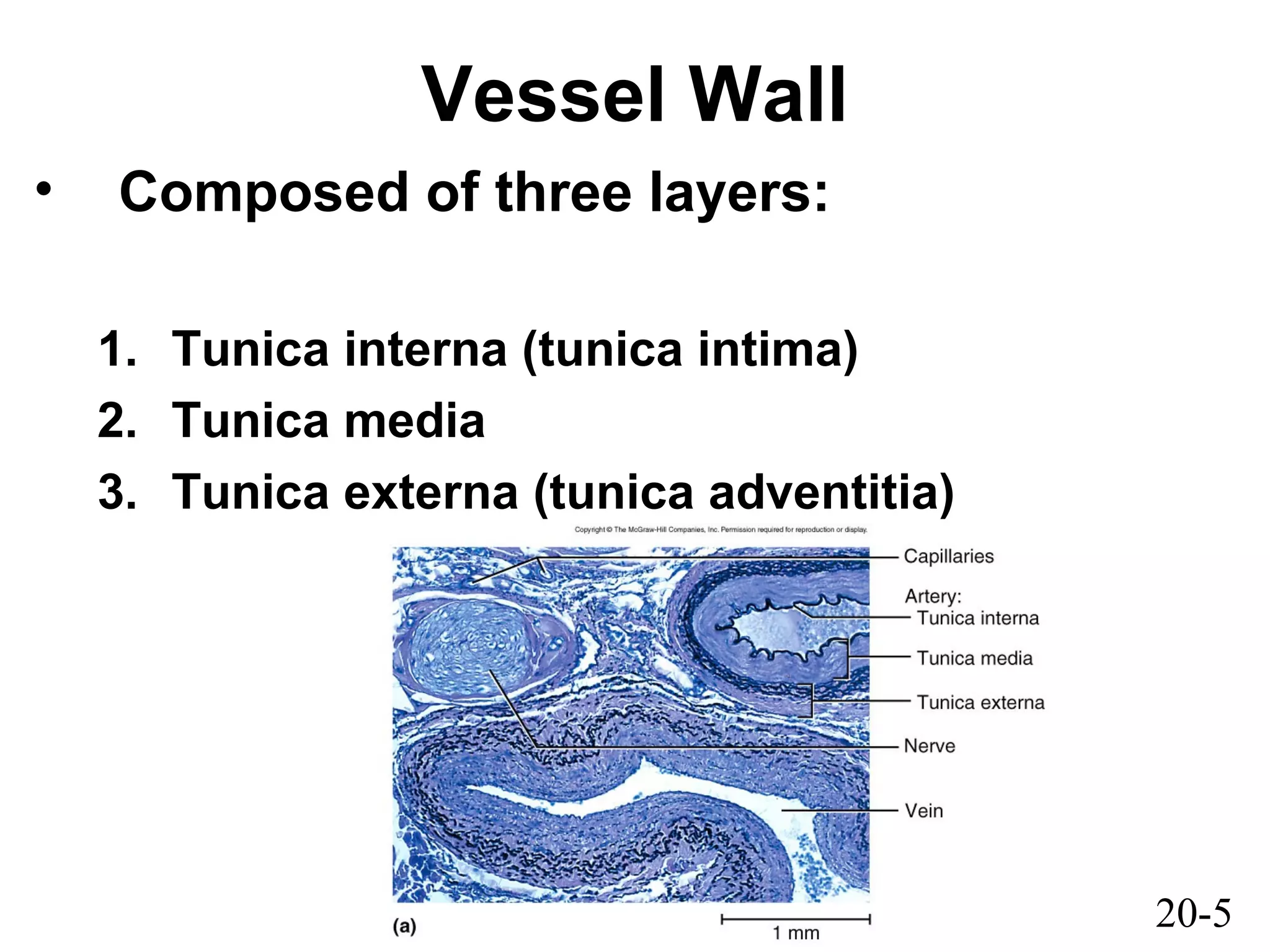
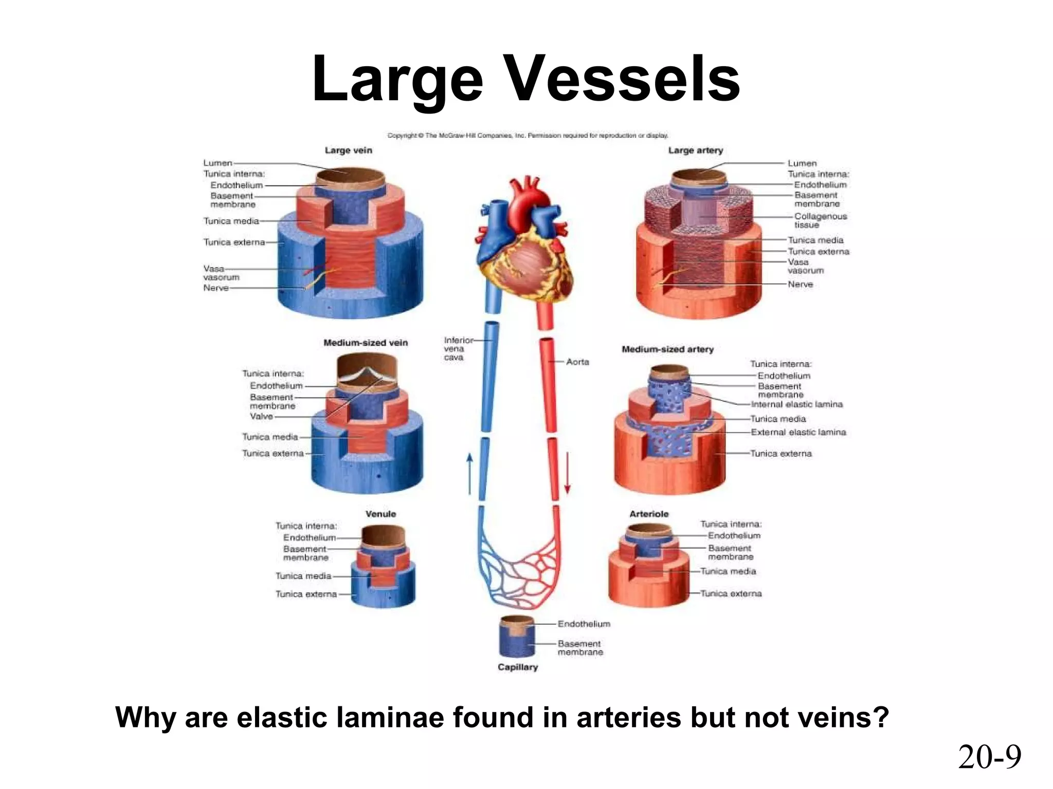
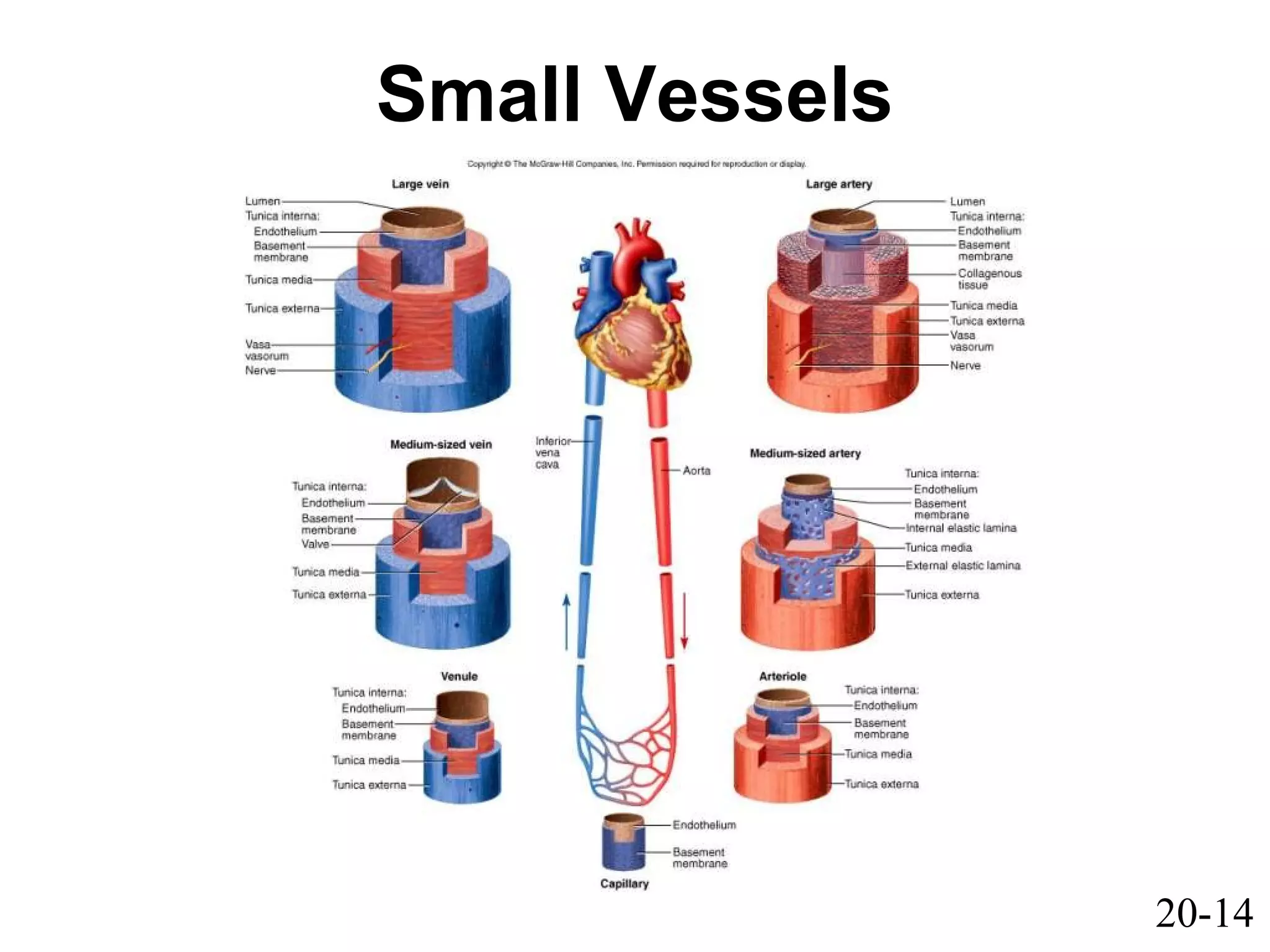
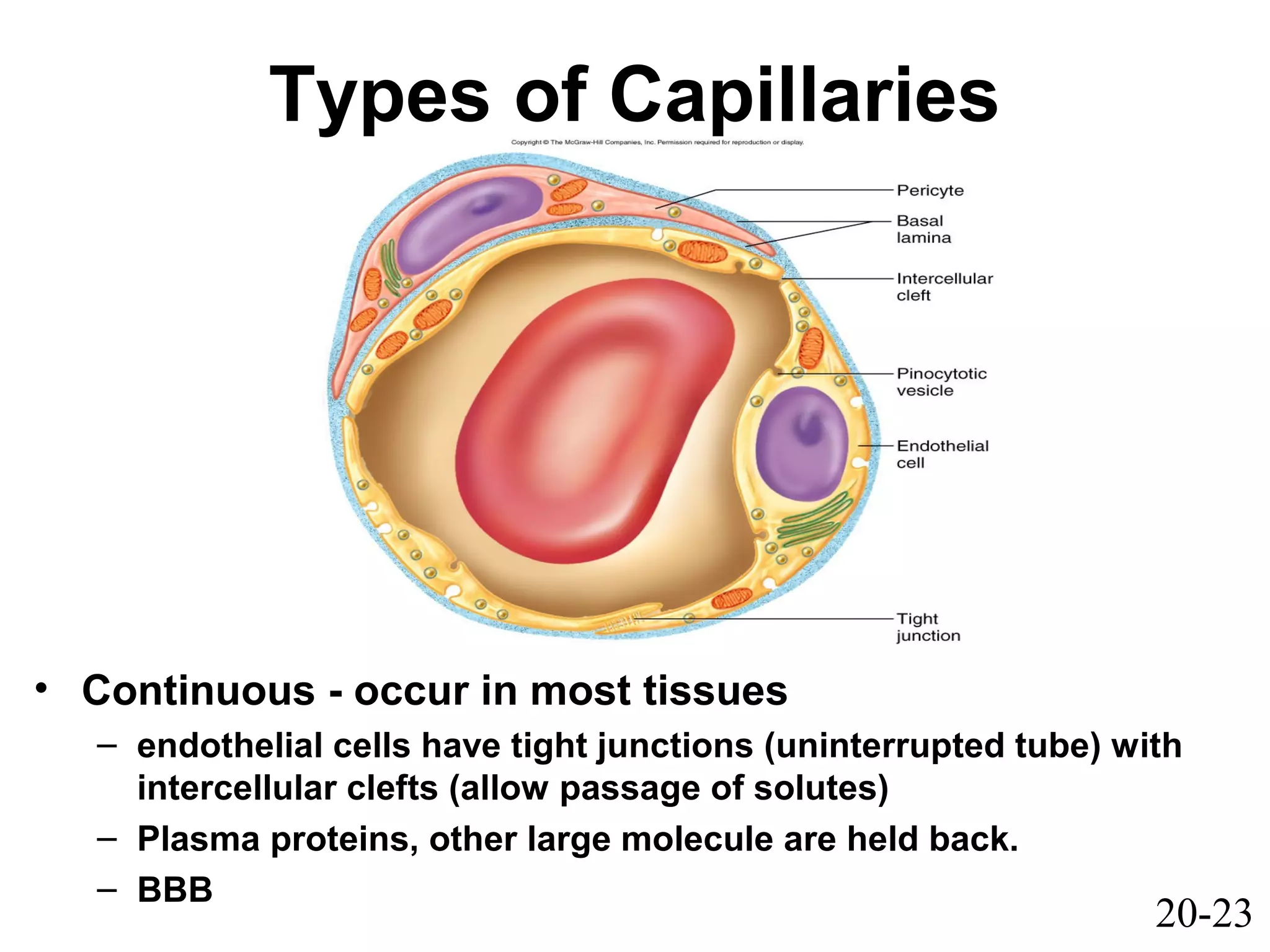
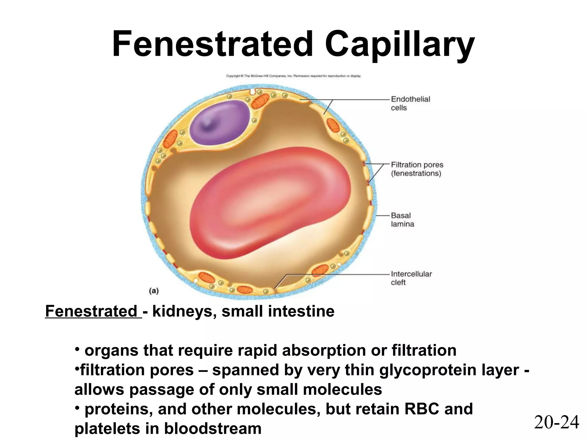
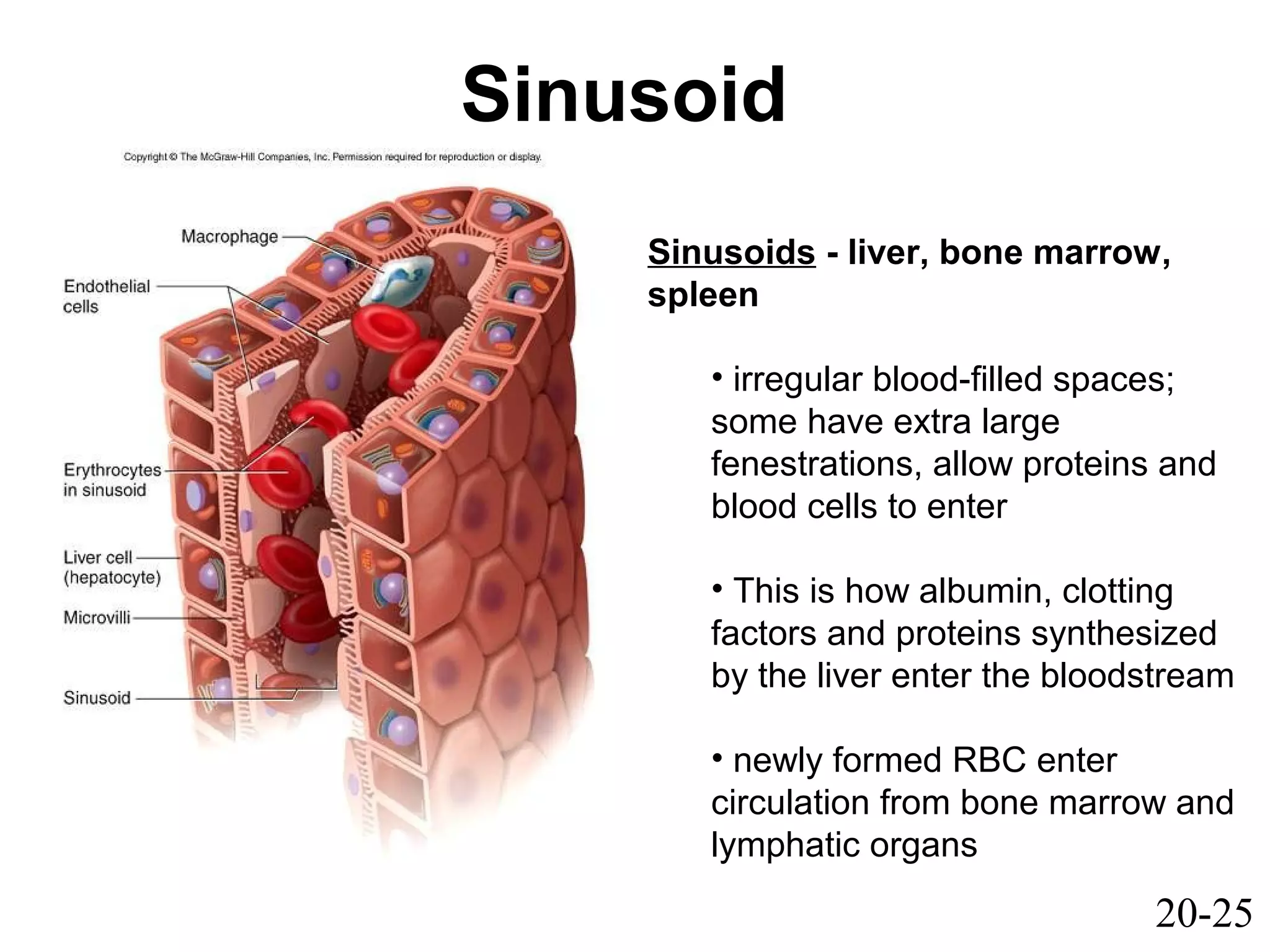
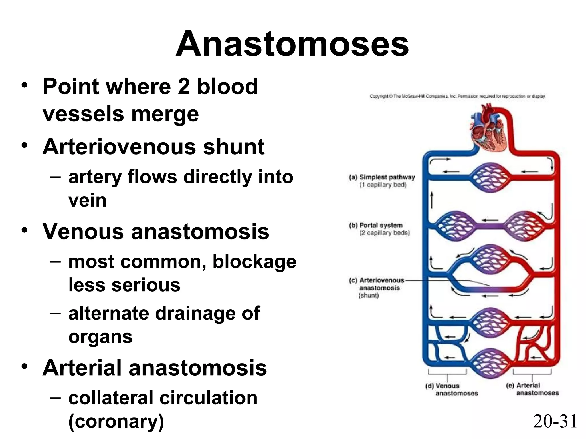
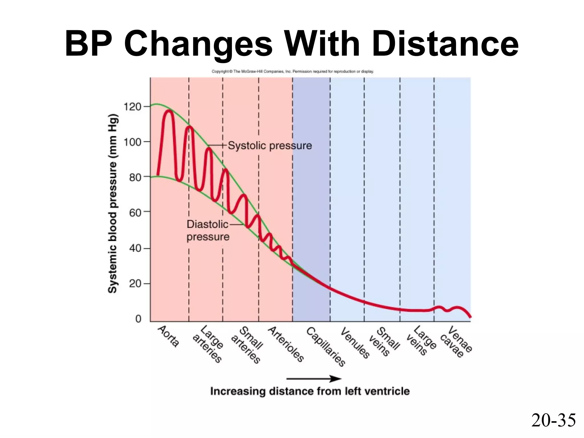
20-1
Chapter 20
Circulatory
System
Blood Vessels
and Circulation
20-2
Blood Vessels and Circulation
• General anatomy of blood vessels
• Blood pressure, resistance and flow
• Capillary exchange
• Venous return and circulatory shock
• Special circulatory routes
• Anatomy of
– pulmonary circuit
– systemic arteries and veins
20-3
Anatomy of Blood Vessels
• Arteries carry blood away from heart (efferent)
• Veins carry blood back to heart (afferent)
• Capillaries connect smallest arteries to veins
20-4
Anatomy of Blood Vessels
20-5
Vessel Wall
• Composed of three layers:
1. Tunica interna (tunica intima)
2. Tunica media
3. Tunica externa (tunica adventitia)
20-6
Vessel Wall
1. Tunica interna (intima)
– Acts as a selective permeable barrier to
materials entering or exiting the bloodstream.
– smooth inner layer that repels blood cells
and platelets
– simple squamous endothelium overlying a
basement membrane and layer of fibrous
tissue
– Secretes chemicals that stimulate dilation or
constriction of the vessel
20-7
Vessel Wall
2. Tunica media
– middle layer
– usually thickest; smooth muscle, collagen,
some elastic tissue
– 2 functions:
1. * strengthen the vessel and prevent blood
pressure from rupturing them
2. Provides vasomotion
20-8
Vessel Wall
3. Tunica externa (tunica adventitia)
– outermost layer
– loose connective tissue
– Function:
• Anchors vessel
• Provides passage for small nerves, lymphatic
vessels, and smaller blood vessels
• Vasa vasorum
20-9
Large Vessels
Why are elastic laminae found in arteries but not veins?
20-10
Arteries
Divided in to 3 catagories:
1. Conducting (elastic or large) artieries
2. Distributing (muscular or medium)
3. Resistance (small) arteries
20-11
Arteries
• Conducting (elastic) arteries - largest
– pulmonary, aorta and common carotid,
subclavian, common iliac
– tunica media consists of perforated sheets of
elastic tissue, alternating with thin layers of
smooth muscle, collagen and elastic fibers
– expand during systole, recoil during diastole;
lessens fluctuations in BP
– Recoil between heartbeats prevents the blood
pressure from dropping to low during the
quiescent period.
20-12
Medium Vessels
20-13
Arteries
• Distributing (muscular) arteries
– distributes blood to specific organs; femoral
splenic, renal, and brachial arteries
– smooth muscle layers constitute 3/4 of wall
thickness
20-14
Small Vessels
20-15
Arteries and Metarterioles
• Resistance (small) arteries
– arterioles control amount of blood to various
organs
• Metarterioles
– short vessels connect arterioles to capillaries
– muscle cells form a precapillary sphincter
about entrance to capillary
• Precapillary sphincters control which beds
are well perfused
– only 1/4 of the capillaries are open at a given
time
20-16
20-17
Arterial Sense Organs
• Major arteries above heart, monitor
pressure and chemistry
• Information is transmitted to the
brainstem to regulate heartbeat,
vasomotion, and respiration.
• 3 types of sensory receptors:
1. Carotid sinuses
2. Carotid bodies
3. Aortic bodies
20-18
Arterial Sense Organs
• Carotid sinuses
– These are baroreceptors
(pressure sensors)
– in walls of internal carotid
artery
– monitors BP – signaling
brainstem via CN IX
• HR ↓ and vessels dilate
thereby lowering BP
20-19
Arterial Sense Organs
• Carotid bodies
(chemoreceptors)
– oval bodies near carotids
– Innervated by CN IX, X
– monitor Δ blood chemistry
• adjust respiratory rate to
stabilize pH, CO2, and O2
• Aortic bodies
– in walls of aorta
– same function as carotid
bodies
20-20
Capillaries
• Allow passing of nutrient, waste, and hormones.
• AKA exchange vessels
• Composed of endothelium and basement
membrane.
• Scarce in tendons, ligaments, cornea, lens
• 3 types of cap. distinguished by the ease with
which they allow substances to pass.
– Continuous, Fenestrated, and Sinusoids
20-21
Control of Capillary Bed Perfusion
20-22
Control of Capillary Bed Perfusion
20-23
Types of Capillaries
• Continuous - occur in most tissues
– endothelial cells have tight junctions (uninterrupted tube) with
intercellular clefts (allow passage of solutes)
– Plasma proteins, other large molecule are held back.
– BBB
20-24
Fenestrated Capillary
Fenestrated - kidneys, small intestine
• organs that require rapid absorption or filtration
•filtration pores – spanned by very thin glycoprotein layer -
allows passage of only small molecules
• proteins, and other molecules, but retain RBC and
platelets in bloodstream
20-25
Sinusoid
Sinusoids - liver, bone marrow,
spleen
• irregular blood-filled spaces;
some have extra large
fenestrations, allow proteins and
blood cells to enter
• This is how albumin, clotting
factors and proteins synthesized
by the liver enter the bloodstream
• newly formed RBC enter
circulation from bone marrow and
lymphatic organs
20-26
Veins
• Veins
– thinner walls, flaccid
– less muscular and elastic tissue,
– expand easily, have high capacitance
– valves aid skeletal muscles in upward blood
flow
– Lower BP because further away from heart.
20-27
Veins
• Follow the direction of blood flow, smallest to
largest veins
• Postcapillary venules
– Receive blood directly from capillaries
– more porous than capillaries, thus exchange
fluid with surrounding tissues
• Muscular venules
– Receive blood from the postcapillary venules
20-28
Veins
• Medium veins
– Individual names; radial, ulnar, saphenous
– Infolding of tunica interna that meets in the
middle of lumen = venous valves
– Skeletal muscle pump (varicose veins)
• Venous sinuses
– veins with thin walls, large lumens, no smooth
muscle
– Coronary sinus, dural sinus of brain
20-29
Veins
• Large veins
– Venae cavae
– Pulmonary veins
– Jugular vein
– Renal veins
20-30
Circulatory Routes
• Most common route
– heart → arteries →
arterioles → capillaries →
venules → veins
• Portal system
– blood flows through
two consecutive
capillary networks
before returning to heart
• hypothalamus - anterior
pituitary
• found in kidneys
• between intestines - liver
20-31
Anastomoses
• Point where 2 blood
vessels merge
• Arteriovenous shunt
– artery flows directly into
vein
• Venous anastomosis
– most common, blockage
less serious
– alternate drainage of
organs
• Arterial anastomosis
– collateral circulation
(coronary)
20-32
Principles of Blood Flow
• Blood flow: amount of blood flowing
through a tissue in a given time (ml/min)
• Perfusion: rate of blood flow per given
mass of tissue (ml/min/g)
• Important for delivery of nutrients and
oxygen, and removal of metabolic
wastes
• Total flow = cardiac output (5.25 L/min)
20-33
Principles of Blood Flow
• Hemodynamics
– physical principles of blood flow based on
pressure and resistance
• F ∝ ∆P/R, (F = flow, ∆P = difference in pressure,
R = resistance to flow)
• The greater the pressure difference between two
points, the greater the flow;
• The greater the resistance (R) the less the flow.
20-34
Blood Pressure
• Force that blood exerts against a vessel
wall
• Measured at brachial artery of arm
• Systolic pressure: BP during ventricular
systole
• Diastolic pressure: BP during ventricular
diastole
• Normal value, young adult: 120/75 mm Hg
• Pulse pressure: systolic - diastolic
– important measure of stress exerted on small
arteries
20-35
BP Changes With Distance
20-36
Blood Pressure
• Importance of arterial elasticity
– expansion and recoil maintains steady flow of
blood throughout cardiac cycle, smoothes out
pressure fluctuations and ↓ stress on small
arteries
• BP rises with age: arteries less distensible
• BP determined by cardiac output, blood
volume and peripheral resistance
» Three variables: blood viscosity, vessel length, and
vessel radius.
20-37
Abnormalities of Blood Pressure
• Hypertension
– chronic resting BP > 140/90
– consequences
• can weaken small arteries and cause aneurysms
• Hypotension
– chronic low resting BP
– caused by blood loss, dehydration, anemia
20-38
Peripheral Resistance
• The opposition to flow that blood
encounters in vessels away from the
heart.
• Blood viscosity - by RBC’s and albumin
↓ viscosity with anemia, hypoproteinemia
↑ viscosity with polycythemia , dehydration
• Vessel length
– pressure and flow ↓ with distance (friction)
• Vessel radius - very powerful influence
over flow
– most adjustable variable, controls resistance
quickly
– vasomotion: change in vessel radius
20-39
20-40
Peripheral Resistance
• Vessel radius (cont.)
– laminar flow - flows in layers, faster in center
– blood flow (F) proportional to the fourth power
of radius (r), F ∝ r4
• arterioles can constrict to 1/3 of fully
relaxed radius (see fig. 20.12)
20-41
Peripheral Resistance
• Example:
– Lets say a blood vessel has a 1-mm radius when fully constricted
and a 3-mm radius when fully relaxed. (F ∂ r 4
)
r = 1mm r 4
= 1 4
= 1 F = 1 mm/sec
r = 2mm r 4
= 2 4
= 16 F = 16 mm/sec
r = 3mm r 4
= 3 4
= 81 F = 81 mm/sec
– 3X ↑ in radius results in 81X ↑ in flow
– Demonstrates that vessel radius exerts a very powerful influence
over flow.
20-42
20-43
Flow at Different Points
• From aorta to capillaries, flow ↓ for 3
reasons
– greater distance, more friction to ↓ flow
– smaller radii of arterioles and capillaries
– farther from heart, greater total cross
sectional area
• From capillaries to vena cava, flow ↑ again
– large amount of blood forced into smaller
channels
– never regains velocity of large arteries
20-44
Regulation of BP and Flow
• Local control
• Neural control
• Hormonal control
20-45
Local Control of BP and Flow
• Metabolic theory of autoregulation
– tissue inadequately perfused, wastes
accumulate = vasodilation
• Vasoactive chemicals
– substances that stimulate vasomotion;
histamine, bradykinin (trauma, exercise)
• Reactive hyperemia
– blood supply cut off then restored
• Angiogenesis - growth of new vessels
– regrowth of uterine lining, around
obstructions, exercise, malignant tumors
– controlled by growth factors and inhibitors
20-46
Neural Control of BP and Flow
• Vasomotor center of medulla oblongata:
– sympathetic control stimulates most vessels
to constrict, but dilates vessels in skeletal and
cardiac muscle
– integrates three autonomic reflexes
• baroreflexes
• chemoreflexes
• medullary ischemic reflex
20-47
Neural Control: Baroreflex
• Changes in BP detected by stretch receptors
(baroreceptors), in large arteries above heart
– aortic arch
– aortic sinuses (behind aortic valve cusps)
– carotid sinus (base of each internal carotid artery)
• Autonomic negative feedback response
– baroreceptors send constant signals to brainstem
↑ BP causes rate of signals to rise, inhibits vasomotor
center, ↓ sympathetic tone, vasodilation causes BP ↓
↓ BP causes rate of signals to drop, excites vasomotor
center, ↑ sympathetic tone, vasoconstriction and BP ↑
20-48
Baroreflex
Negative Feedback Response
20-49
Neural Control: Chemoreflex
• Chemoreceptors in aortic bodies and
carotid bodies
– located in aortic arch, subclavian arteries,
external carotid arteries
• Autonomic response to changes in blood
chemistry
– pH, O2, CO2
– primary role: adjust respiration
– secondary role: vasomotion
• hypoxemia, chemoreceptors, instruct vasomotor
center to cause vasoconstriction, ↑ BP, ↑ lung
perfusion and gas exchange
20-50
Other Inputs to Vasomotor Center
• Medullary ischemic reflex
– inadequate perfusion of brainstem
• cardiac and vasomotor centers send sympathetic
signals to heart and blood vessels
∀↑ cardiac output and causes widespread
vasoconstriction
∀↑ BP
• Other brain centers
– stress, anger, arousal can also ↑ BP
20-51
• Angiotensinogen (prohormone produced by liver)
↓ Renin (kidney enzyme released by low BP)
• Angiotensin I
↓ ACE (angiotensin-converting enzyme in lungs)
ACE inhibitors block this enzyme lowering BP
• Angiotensin II
– very potent vasoconstrictor
Hormonal Control of BP and Flow
20-52
Hormonal Control of BP and Flow
• Aldosterone
– promotes Na+
and water retention by kidneys
– increases blood volume and pressure
• Atrial natriuretic factor (↑ urinary sodium excretion)
– generalized vasodilation
• ADH (water retention)
– pathologically high concentrations, vasoconstriction
• Epinephrine and norepinephrine effects
– most blood vessels
• binds to α-adrenergic receptors, vasoconstriction
– skeletal and cardiac muscle blood vessels
• binds to β-adrenergic receptors, vasodilation
20-53
Routing of Blood Flow
• Localized vasoconstriction
– pressure downstream drops, pressure
upstream rises
– enables routing blood to different organs as
needed
• Arterioles - most control over peripheral
resistance
– located on proximal side of capillary beds
– most numerous
– more muscular by diameter
20-54
Blood Flow in Response to Needs
• Arterioles shift blood flow with changing priorities
20-55
Blood Flow Comparison
• During exercise
↑ perfusion of lungs, myocardium and skeletal
muscles ↓ perfusion of kidneys and digestive tract
20-56
Capillary Exchange
• Only occurs across capillary walls
between blood and surrounding tissues
• 3 routes across endothelial cells
– intercellular clefts
– fenestrations
– through cytoplasm
• Mechanisms involved
– diffusion, transcytosis, filtration and
reabsorption
20-57
Capillary Exchange - Diffusion
• Most important mechanism
• Lipid soluble substances
– steroid hormones, O2 and CO2 diffuse easily
• Insoluble substances
– glucose and electrolytes must pass through
channels, fenestrations or intercellular clefts
• Large particles - proteins, held back
20-58
Capillary Exchange - Transcytosis
• Pinocytosis - transport vesicles across cell -
exocytosis
• Important for fatty acids, albumin and some
hormones (insulin)
20-59
Capillary Exchange -
Filtration and Reabsorption
• Opposing forces
– blood (hydrostatic) pressure drives fluid out of
capillary
• high on arterial end of capillary, low on venous end
– colloid osmotic pressure (COP) draws fluid
into capillary
• results from plasma proteins (albumin)- more in
blood
• oncotic pressure = net COP (blood COP - tissue COP)
• Hydrostatic pressure
– physical force exerted against a surface by a
liquid, (BP is an example)
20-60
Capillary Filtration and Reabsorption
• Capillary filtration at arterial end
• Capillary reabsorption at venous end
• Variations
– location
(glomeruli- devoted to filtration
alveolar cap.- devoted to absorption)
– activity or trauma
(↑ filtration)
20-61
Causes of Edema
∀ ↑ Capillary filtration (↑ capillary BP or permeability)
– poor venous return
• congestive heart failure - pulmonary edema
• insufficient muscular activity
– kidney failure (water retention, hypertension)
– histamine makes capillaries more permeable
∀ ↓ Capillary reabsorption
– hypoproteinemia (oncotic pressure ∝ blood albumin)
cirrhosis, famine, burns, kidney disease
• Obstructed lymphatic drainage
20-62
Consequences of Edema
• Tissue necrosis
– oxygen delivery and waste removal impaired
• Pulmonary edema
– suffocation
• Cerebral edema
– headaches, nausea, seizures and coma
• Circulatory shock
– excess fluid in tissue spaces causes low
blood volume and low BP
20-63
Mechanisms of Venous Return
• Pressure gradient
– 7-13 mm Hg venous pressure towards heart
• venules (12-18 mm Hg) to central venous pressure
(~5 mm Hg)
• Gravity drains blood from head and neck
• Skeletal muscle pump in the limbs
• Thoracic pump
– inhalation - thoracic cavity expands (pressure
↓) abdominal pressure ↑, forcing blood
upward
• Cardiac suction of expanding atrial space
20-64
Skeletal Muscle Pump
20-65
Venous Return and Physical Activity
• Exercise ↑ venous return in many ways
– heart beats faster, harder - ↑ CO and BP
– vessels of skeletal muscles, lungs and heart dilate ↑
flow
↑ respiratory rate ↑ action of thoracic pump
↑ skeletal muscle pump
• Venous pooling occurs with inactivity
– venous pressure not enough force blood upward
– with prolonged standing, CO may be low enough to
cause dizziness or syncope
• prevented by tensing leg muscles, activate skeletal m. pump
– jet pilots wear pressure suits
20-66
Circulatory Shock
• Any state where cardiac output
insufficient to meet metabolic needs
– cardiogenic shock - inadequate pumping of
heart (MI)
– low venous return (LVR) shock - 3 principle
forms
1. hypovolemic shock - most common
– loss of blood volume: trauma, burns, dehydration
2. obstructed venous return shock
– tumor or aneurysm
3. venous pooling (vascular) shock
– next slide
20-67
LVR Shock
• Venous pooling (vascular) shock
– long periods of standing, sitting or widespread
vasodilation
– neurogenic shock - loss of vasomotor tone,
vasodilation
• causes from emotional shock to brainstem injury
• Septic shock
– bacterial toxins trigger vasodilation and ↑ capillary
permeability
• Anaphylactic shock
– severe immune reaction to antigen, histamine release,
generalized vasodilation, ↑ capillary permeability
20-68
Responses to Circulatory Shock
• Compensated shock
• Decompensated shock
20-69
Compensated shock
• Homeostatic mechanisms bring about
recovery
∀↓ BP triggers baroreflex and production of
angiotensin II, both stimulate
vasoconstriction
• If person faints and falls to horizontal
position, gravity restores blood flow to
brain; quicker if feet are raised
20-70
Decompensated shock
• Life threatening positive feedback loops occur
↓ CO → myocardial ischemia and infarction →
↓ CO
– slow circulation → disseminated intravascular
coagulation → slow circulation
– ischemia and acidosis of brainstem → ↓
vasomotor tone, vasodilation → ↓ CO →
ischemia and acidosis of brainstem
20-71
Special Circulatory Routes- Brain
• Total perfusion kept constant
– seconds of deprivation causes loss of consciousness
– 4-5 minutes causes irreversible brain damage
– flow can be shifted from one active region to another
• Responds to changes in BP and chemistry
– cerebral arteries: dilate as BP ↓, constrict as BP rises
– main chemical stimulus: pH
• CO2 + H2O → H2 CO3 → H+
+ (HCO3)-
• hypercapnia (CO2 ↑) in brain, pH ↓, triggers vasodilation
• hypocapnia, ↑ pH, vasoconstriction
– occurs with hyperventilation, may lead to ischemia,
dizziness and sometimes syncope
20-72
TIA’s and CVA’s
• TIA’s - transient ischemic attacks
– dizziness, loss of vision, weakness, paralysis,
headache or aphasia; lasts from a moment to a few
hours, often early warning of impending stroke
• CVA - cerebral vascular accident (stroke)
– brain infarction caused by ischemia
• atherosclerosis, thrombosis, ruptured aneurysm
– effects range from unnoticeable to fatal
• blindness, paralysis, loss of sensation, loss of speech
common
– recovery depends on surrounding neurons, collateral
circulation
20-73
Special Circulatory Routes -
Skeletal Muscle
• Highly variable flow
• At rest
– arterioles constrict, total flow about 1L/min
• During exercise
– arterioles dilate in response to epinephrine and
sympathetic nerves
– precapillary sphincters dilate due to lactic acid, CO2
– blood flow can increase 20 fold
• Muscular contraction impedes flow
– isometric contraction causes fatigue faster than
isotonic
20-74
Special Circulatory Routes -
Lungs
• Low pulmonary blood pressure
– flow slower, more time for gas exchange
– capillary fluid absorption
• oncotic pressure overrides hydrostatic pressure
• Unique response to hypoxia
– pulmonary arteries constrict, redirects flow to
better ventilated region
20-75
Pulmonary Circulation
• Pulmonary trunk to pulmonary arteries to lungs
– lobar branches for each lobe (3 right, 2 left)
• Pulmonary veins return to left atrium
– increased O2 and reduced CO2 levels
20-76
Pulmonary Capillaries Near Alveoli
• Basketlike
capillary beds
surround
alveoli
• Exchange of
gases with air
at alveoli
20-77
Major Systemic Arteries
• Supplies oxygen and nutrients to all organs
20-78
Major Branches of Aorta
• Ascending aorta
– right and left coronary arteries supply heart
• Aortic arch
– brachiocephalic
• right common carotid supplying right side of head
• right subclavian supplying right shoulder and upper limb
– left common carotid supplying left side of head
– left subclavian supplying shoulder and upper limb
• Descending aorta
– thoracic aorta above diaphragm
– abdominal aorta below diaphragm
20-79
Major Branches of the Aorta
20-80
Arteries of the Head and Neck
• Common carotid to internal and external carotids
– external carotid supplies most external head structures
20-81
Arterial Supply of Brain
• Paired vertebral aa. combine to form basilar artery on pons
• Circle of Willis on base of brain formed from anastomosis of
basilar and internal carotid aa
• Supplies brain, internal ear and orbital structures
– anterior, middle and posterior cerebral
– superior, anterior and posterior cerebellar
20-82
Arteries of the Upper Limb
• Subclavian
passes between
clavicle and 1st
rib
• Vessel changes
names as passes
to different
regions
– subclavian to
axillary to brachial
to radial and ulnar
– brachial used for
BP and radial
artery for pulse
20-83
Arteries of the Thorax
• Thoracic aorta supplies viscera and body wall
– bronchial, esophageal and mediastinal branches
– posterior intercostal and phrenic arteries
• Internal thoracic, anterior intercostal and
pericardiophrenic arise from subclavian artery
20-84
Major Branches of Abdominal Aorta
20-85
Celiac Trunk Branches
• Branches of celiac trunk supply upper
abdominal viscera -- stomach, spleen, liver and
pancreas
20-86
Mesenteric Arteries
20-87
Arteries of the Lower Limb
• Branches to the lower limb arise from external
iliac branch of the common iliac artery
20-88
Arterial Pressure Points
• Some major arteries close to surface -- allows
palpation for pulse and serve as pressure
points to reduce arterial bleeding
20-89
Major Systemic Veins
• Deep veins run parallel to arteries while
superficial veins have many anastomoses
20-90
Deep Veins of Head and Neck
• Large, thin-walled dural sinuses form in between
layers of dura mater (drain brain to internal
jugular vein)
20-91
Superficial Veins of Head and Neck
• Branches of internal and external jugular veins
drain the external structures of the head
• Upper limb is drained by subclavian vein
20-92
Superficial and Deep Veins of Upper
Limb
20-93
Inferior Vena Cava and Branches
• Notice absence of veins draining the viscera ---
stomach, spleen, pancreas and intestines
20-94
Veins of Hepatic Portal System
• Drains blood from viscera (stomach, spleen and
intestines) to liver so that nutrients are absorbed
20-95
Superficial and Deep Veins of Lower Limb

Chap20 powerpoint l

  • 1.
  • 2.
    20-2 Blood Vessels andCirculation • General anatomy of blood vessels • Blood pressure, resistance and flow • Capillary exchange • Venous return and circulatory shock • Special circulatory routes • Anatomy of – pulmonary circuit – systemic arteries and veins
  • 3.
    20-3 Anatomy of BloodVessels • Arteries carry blood away from heart (efferent) • Veins carry blood back to heart (afferent) • Capillaries connect smallest arteries to veins
  • 4.
  • 5.
    20-5 Vessel Wall • Composedof three layers: 1. Tunica interna (tunica intima) 2. Tunica media 3. Tunica externa (tunica adventitia)
  • 6.
    20-6 Vessel Wall 1. Tunicainterna (intima) – Acts as a selective permeable barrier to materials entering or exiting the bloodstream. – smooth inner layer that repels blood cells and platelets – simple squamous endothelium overlying a basement membrane and layer of fibrous tissue – Secretes chemicals that stimulate dilation or constriction of the vessel
  • 7.
    20-7 Vessel Wall 2. Tunicamedia – middle layer – usually thickest; smooth muscle, collagen, some elastic tissue – 2 functions: 1. * strengthen the vessel and prevent blood pressure from rupturing them 2. Provides vasomotion
  • 8.
    20-8 Vessel Wall 3. Tunicaexterna (tunica adventitia) – outermost layer – loose connective tissue – Function: • Anchors vessel • Provides passage for small nerves, lymphatic vessels, and smaller blood vessels • Vasa vasorum
  • 9.
    20-9 Large Vessels Why areelastic laminae found in arteries but not veins?
  • 10.
    20-10 Arteries Divided in to3 catagories: 1. Conducting (elastic or large) artieries 2. Distributing (muscular or medium) 3. Resistance (small) arteries
  • 11.
    20-11 Arteries • Conducting (elastic)arteries - largest – pulmonary, aorta and common carotid, subclavian, common iliac – tunica media consists of perforated sheets of elastic tissue, alternating with thin layers of smooth muscle, collagen and elastic fibers – expand during systole, recoil during diastole; lessens fluctuations in BP – Recoil between heartbeats prevents the blood pressure from dropping to low during the quiescent period.
  • 12.
  • 13.
    20-13 Arteries • Distributing (muscular)arteries – distributes blood to specific organs; femoral splenic, renal, and brachial arteries – smooth muscle layers constitute 3/4 of wall thickness
  • 14.
  • 15.
    20-15 Arteries and Metarterioles •Resistance (small) arteries – arterioles control amount of blood to various organs • Metarterioles – short vessels connect arterioles to capillaries – muscle cells form a precapillary sphincter about entrance to capillary • Precapillary sphincters control which beds are well perfused – only 1/4 of the capillaries are open at a given time
  • 16.
  • 17.
    20-17 Arterial Sense Organs •Major arteries above heart, monitor pressure and chemistry • Information is transmitted to the brainstem to regulate heartbeat, vasomotion, and respiration. • 3 types of sensory receptors: 1. Carotid sinuses 2. Carotid bodies 3. Aortic bodies
  • 18.
    20-18 Arterial Sense Organs •Carotid sinuses – These are baroreceptors (pressure sensors) – in walls of internal carotid artery – monitors BP – signaling brainstem via CN IX • HR ↓ and vessels dilate thereby lowering BP
  • 19.
    20-19 Arterial Sense Organs •Carotid bodies (chemoreceptors) – oval bodies near carotids – Innervated by CN IX, X – monitor Δ blood chemistry • adjust respiratory rate to stabilize pH, CO2, and O2 • Aortic bodies – in walls of aorta – same function as carotid bodies
  • 20.
    20-20 Capillaries • Allow passingof nutrient, waste, and hormones. • AKA exchange vessels • Composed of endothelium and basement membrane. • Scarce in tendons, ligaments, cornea, lens • 3 types of cap. distinguished by the ease with which they allow substances to pass. – Continuous, Fenestrated, and Sinusoids
  • 21.
  • 22.
  • 23.
    20-23 Types of Capillaries •Continuous - occur in most tissues – endothelial cells have tight junctions (uninterrupted tube) with intercellular clefts (allow passage of solutes) – Plasma proteins, other large molecule are held back. – BBB
  • 24.
    20-24 Fenestrated Capillary Fenestrated -kidneys, small intestine • organs that require rapid absorption or filtration •filtration pores – spanned by very thin glycoprotein layer - allows passage of only small molecules • proteins, and other molecules, but retain RBC and platelets in bloodstream
  • 25.
    20-25 Sinusoid Sinusoids - liver,bone marrow, spleen • irregular blood-filled spaces; some have extra large fenestrations, allow proteins and blood cells to enter • This is how albumin, clotting factors and proteins synthesized by the liver enter the bloodstream • newly formed RBC enter circulation from bone marrow and lymphatic organs
  • 26.
    20-26 Veins • Veins – thinnerwalls, flaccid – less muscular and elastic tissue, – expand easily, have high capacitance – valves aid skeletal muscles in upward blood flow – Lower BP because further away from heart.
  • 27.
    20-27 Veins • Follow thedirection of blood flow, smallest to largest veins • Postcapillary venules – Receive blood directly from capillaries – more porous than capillaries, thus exchange fluid with surrounding tissues • Muscular venules – Receive blood from the postcapillary venules
  • 28.
    20-28 Veins • Medium veins –Individual names; radial, ulnar, saphenous – Infolding of tunica interna that meets in the middle of lumen = venous valves – Skeletal muscle pump (varicose veins) • Venous sinuses – veins with thin walls, large lumens, no smooth muscle – Coronary sinus, dural sinus of brain
  • 29.
    20-29 Veins • Large veins –Venae cavae – Pulmonary veins – Jugular vein – Renal veins
  • 30.
    20-30 Circulatory Routes • Mostcommon route – heart → arteries → arterioles → capillaries → venules → veins • Portal system – blood flows through two consecutive capillary networks before returning to heart • hypothalamus - anterior pituitary • found in kidneys • between intestines - liver
  • 31.
    20-31 Anastomoses • Point where2 blood vessels merge • Arteriovenous shunt – artery flows directly into vein • Venous anastomosis – most common, blockage less serious – alternate drainage of organs • Arterial anastomosis – collateral circulation (coronary)
  • 32.
    20-32 Principles of BloodFlow • Blood flow: amount of blood flowing through a tissue in a given time (ml/min) • Perfusion: rate of blood flow per given mass of tissue (ml/min/g) • Important for delivery of nutrients and oxygen, and removal of metabolic wastes • Total flow = cardiac output (5.25 L/min)
  • 33.
    20-33 Principles of BloodFlow • Hemodynamics – physical principles of blood flow based on pressure and resistance • F ∝ ∆P/R, (F = flow, ∆P = difference in pressure, R = resistance to flow) • The greater the pressure difference between two points, the greater the flow; • The greater the resistance (R) the less the flow.
  • 34.
    20-34 Blood Pressure • Forcethat blood exerts against a vessel wall • Measured at brachial artery of arm • Systolic pressure: BP during ventricular systole • Diastolic pressure: BP during ventricular diastole • Normal value, young adult: 120/75 mm Hg • Pulse pressure: systolic - diastolic – important measure of stress exerted on small arteries
  • 35.
  • 36.
    20-36 Blood Pressure • Importanceof arterial elasticity – expansion and recoil maintains steady flow of blood throughout cardiac cycle, smoothes out pressure fluctuations and ↓ stress on small arteries • BP rises with age: arteries less distensible • BP determined by cardiac output, blood volume and peripheral resistance » Three variables: blood viscosity, vessel length, and vessel radius.
  • 37.
    20-37 Abnormalities of BloodPressure • Hypertension – chronic resting BP > 140/90 – consequences • can weaken small arteries and cause aneurysms • Hypotension – chronic low resting BP – caused by blood loss, dehydration, anemia
  • 38.
    20-38 Peripheral Resistance • Theopposition to flow that blood encounters in vessels away from the heart. • Blood viscosity - by RBC’s and albumin ↓ viscosity with anemia, hypoproteinemia ↑ viscosity with polycythemia , dehydration • Vessel length – pressure and flow ↓ with distance (friction) • Vessel radius - very powerful influence over flow – most adjustable variable, controls resistance quickly – vasomotion: change in vessel radius
  • 39.
  • 40.
    20-40 Peripheral Resistance • Vesselradius (cont.) – laminar flow - flows in layers, faster in center – blood flow (F) proportional to the fourth power of radius (r), F ∝ r4 • arterioles can constrict to 1/3 of fully relaxed radius (see fig. 20.12)
  • 41.
    20-41 Peripheral Resistance • Example: –Lets say a blood vessel has a 1-mm radius when fully constricted and a 3-mm radius when fully relaxed. (F ∂ r 4 ) r = 1mm r 4 = 1 4 = 1 F = 1 mm/sec r = 2mm r 4 = 2 4 = 16 F = 16 mm/sec r = 3mm r 4 = 3 4 = 81 F = 81 mm/sec – 3X ↑ in radius results in 81X ↑ in flow – Demonstrates that vessel radius exerts a very powerful influence over flow.
  • 42.
  • 43.
    20-43 Flow at DifferentPoints • From aorta to capillaries, flow ↓ for 3 reasons – greater distance, more friction to ↓ flow – smaller radii of arterioles and capillaries – farther from heart, greater total cross sectional area • From capillaries to vena cava, flow ↑ again – large amount of blood forced into smaller channels – never regains velocity of large arteries
  • 44.
    20-44 Regulation of BPand Flow • Local control • Neural control • Hormonal control
  • 45.
    20-45 Local Control ofBP and Flow • Metabolic theory of autoregulation – tissue inadequately perfused, wastes accumulate = vasodilation • Vasoactive chemicals – substances that stimulate vasomotion; histamine, bradykinin (trauma, exercise) • Reactive hyperemia – blood supply cut off then restored • Angiogenesis - growth of new vessels – regrowth of uterine lining, around obstructions, exercise, malignant tumors – controlled by growth factors and inhibitors
  • 46.
    20-46 Neural Control ofBP and Flow • Vasomotor center of medulla oblongata: – sympathetic control stimulates most vessels to constrict, but dilates vessels in skeletal and cardiac muscle – integrates three autonomic reflexes • baroreflexes • chemoreflexes • medullary ischemic reflex
  • 47.
    20-47 Neural Control: Baroreflex •Changes in BP detected by stretch receptors (baroreceptors), in large arteries above heart – aortic arch – aortic sinuses (behind aortic valve cusps) – carotid sinus (base of each internal carotid artery) • Autonomic negative feedback response – baroreceptors send constant signals to brainstem ↑ BP causes rate of signals to rise, inhibits vasomotor center, ↓ sympathetic tone, vasodilation causes BP ↓ ↓ BP causes rate of signals to drop, excites vasomotor center, ↑ sympathetic tone, vasoconstriction and BP ↑
  • 48.
  • 49.
    20-49 Neural Control: Chemoreflex •Chemoreceptors in aortic bodies and carotid bodies – located in aortic arch, subclavian arteries, external carotid arteries • Autonomic response to changes in blood chemistry – pH, O2, CO2 – primary role: adjust respiration – secondary role: vasomotion • hypoxemia, chemoreceptors, instruct vasomotor center to cause vasoconstriction, ↑ BP, ↑ lung perfusion and gas exchange
  • 50.
    20-50 Other Inputs toVasomotor Center • Medullary ischemic reflex – inadequate perfusion of brainstem • cardiac and vasomotor centers send sympathetic signals to heart and blood vessels ∀↑ cardiac output and causes widespread vasoconstriction ∀↑ BP • Other brain centers – stress, anger, arousal can also ↑ BP
  • 51.
    20-51 • Angiotensinogen (prohormoneproduced by liver) ↓ Renin (kidney enzyme released by low BP) • Angiotensin I ↓ ACE (angiotensin-converting enzyme in lungs) ACE inhibitors block this enzyme lowering BP • Angiotensin II – very potent vasoconstrictor Hormonal Control of BP and Flow
  • 52.
    20-52 Hormonal Control ofBP and Flow • Aldosterone – promotes Na+ and water retention by kidneys – increases blood volume and pressure • Atrial natriuretic factor (↑ urinary sodium excretion) – generalized vasodilation • ADH (water retention) – pathologically high concentrations, vasoconstriction • Epinephrine and norepinephrine effects – most blood vessels • binds to α-adrenergic receptors, vasoconstriction – skeletal and cardiac muscle blood vessels • binds to β-adrenergic receptors, vasodilation
  • 53.
    20-53 Routing of BloodFlow • Localized vasoconstriction – pressure downstream drops, pressure upstream rises – enables routing blood to different organs as needed • Arterioles - most control over peripheral resistance – located on proximal side of capillary beds – most numerous – more muscular by diameter
  • 54.
    20-54 Blood Flow inResponse to Needs • Arterioles shift blood flow with changing priorities
  • 55.
    20-55 Blood Flow Comparison •During exercise ↑ perfusion of lungs, myocardium and skeletal muscles ↓ perfusion of kidneys and digestive tract
  • 56.
    20-56 Capillary Exchange • Onlyoccurs across capillary walls between blood and surrounding tissues • 3 routes across endothelial cells – intercellular clefts – fenestrations – through cytoplasm • Mechanisms involved – diffusion, transcytosis, filtration and reabsorption
  • 57.
    20-57 Capillary Exchange -Diffusion • Most important mechanism • Lipid soluble substances – steroid hormones, O2 and CO2 diffuse easily • Insoluble substances – glucose and electrolytes must pass through channels, fenestrations or intercellular clefts • Large particles - proteins, held back
  • 58.
    20-58 Capillary Exchange -Transcytosis • Pinocytosis - transport vesicles across cell - exocytosis • Important for fatty acids, albumin and some hormones (insulin)
  • 59.
    20-59 Capillary Exchange - Filtrationand Reabsorption • Opposing forces – blood (hydrostatic) pressure drives fluid out of capillary • high on arterial end of capillary, low on venous end – colloid osmotic pressure (COP) draws fluid into capillary • results from plasma proteins (albumin)- more in blood • oncotic pressure = net COP (blood COP - tissue COP) • Hydrostatic pressure – physical force exerted against a surface by a liquid, (BP is an example)
  • 60.
    20-60 Capillary Filtration andReabsorption • Capillary filtration at arterial end • Capillary reabsorption at venous end • Variations – location (glomeruli- devoted to filtration alveolar cap.- devoted to absorption) – activity or trauma (↑ filtration)
  • 61.
    20-61 Causes of Edema ∀↑ Capillary filtration (↑ capillary BP or permeability) – poor venous return • congestive heart failure - pulmonary edema • insufficient muscular activity – kidney failure (water retention, hypertension) – histamine makes capillaries more permeable ∀ ↓ Capillary reabsorption – hypoproteinemia (oncotic pressure ∝ blood albumin) cirrhosis, famine, burns, kidney disease • Obstructed lymphatic drainage
  • 62.
    20-62 Consequences of Edema •Tissue necrosis – oxygen delivery and waste removal impaired • Pulmonary edema – suffocation • Cerebral edema – headaches, nausea, seizures and coma • Circulatory shock – excess fluid in tissue spaces causes low blood volume and low BP
  • 63.
    20-63 Mechanisms of VenousReturn • Pressure gradient – 7-13 mm Hg venous pressure towards heart • venules (12-18 mm Hg) to central venous pressure (~5 mm Hg) • Gravity drains blood from head and neck • Skeletal muscle pump in the limbs • Thoracic pump – inhalation - thoracic cavity expands (pressure ↓) abdominal pressure ↑, forcing blood upward • Cardiac suction of expanding atrial space
  • 64.
  • 65.
    20-65 Venous Return andPhysical Activity • Exercise ↑ venous return in many ways – heart beats faster, harder - ↑ CO and BP – vessels of skeletal muscles, lungs and heart dilate ↑ flow ↑ respiratory rate ↑ action of thoracic pump ↑ skeletal muscle pump • Venous pooling occurs with inactivity – venous pressure not enough force blood upward – with prolonged standing, CO may be low enough to cause dizziness or syncope • prevented by tensing leg muscles, activate skeletal m. pump – jet pilots wear pressure suits
  • 66.
    20-66 Circulatory Shock • Anystate where cardiac output insufficient to meet metabolic needs – cardiogenic shock - inadequate pumping of heart (MI) – low venous return (LVR) shock - 3 principle forms 1. hypovolemic shock - most common – loss of blood volume: trauma, burns, dehydration 2. obstructed venous return shock – tumor or aneurysm 3. venous pooling (vascular) shock – next slide
  • 67.
    20-67 LVR Shock • Venouspooling (vascular) shock – long periods of standing, sitting or widespread vasodilation – neurogenic shock - loss of vasomotor tone, vasodilation • causes from emotional shock to brainstem injury • Septic shock – bacterial toxins trigger vasodilation and ↑ capillary permeability • Anaphylactic shock – severe immune reaction to antigen, histamine release, generalized vasodilation, ↑ capillary permeability
  • 68.
    20-68 Responses to CirculatoryShock • Compensated shock • Decompensated shock
  • 69.
    20-69 Compensated shock • Homeostaticmechanisms bring about recovery ∀↓ BP triggers baroreflex and production of angiotensin II, both stimulate vasoconstriction • If person faints and falls to horizontal position, gravity restores blood flow to brain; quicker if feet are raised
  • 70.
    20-70 Decompensated shock • Lifethreatening positive feedback loops occur ↓ CO → myocardial ischemia and infarction → ↓ CO – slow circulation → disseminated intravascular coagulation → slow circulation – ischemia and acidosis of brainstem → ↓ vasomotor tone, vasodilation → ↓ CO → ischemia and acidosis of brainstem
  • 71.
    20-71 Special Circulatory Routes-Brain • Total perfusion kept constant – seconds of deprivation causes loss of consciousness – 4-5 minutes causes irreversible brain damage – flow can be shifted from one active region to another • Responds to changes in BP and chemistry – cerebral arteries: dilate as BP ↓, constrict as BP rises – main chemical stimulus: pH • CO2 + H2O → H2 CO3 → H+ + (HCO3)- • hypercapnia (CO2 ↑) in brain, pH ↓, triggers vasodilation • hypocapnia, ↑ pH, vasoconstriction – occurs with hyperventilation, may lead to ischemia, dizziness and sometimes syncope
  • 72.
    20-72 TIA’s and CVA’s •TIA’s - transient ischemic attacks – dizziness, loss of vision, weakness, paralysis, headache or aphasia; lasts from a moment to a few hours, often early warning of impending stroke • CVA - cerebral vascular accident (stroke) – brain infarction caused by ischemia • atherosclerosis, thrombosis, ruptured aneurysm – effects range from unnoticeable to fatal • blindness, paralysis, loss of sensation, loss of speech common – recovery depends on surrounding neurons, collateral circulation
  • 73.
    20-73 Special Circulatory Routes- Skeletal Muscle • Highly variable flow • At rest – arterioles constrict, total flow about 1L/min • During exercise – arterioles dilate in response to epinephrine and sympathetic nerves – precapillary sphincters dilate due to lactic acid, CO2 – blood flow can increase 20 fold • Muscular contraction impedes flow – isometric contraction causes fatigue faster than isotonic
  • 74.
    20-74 Special Circulatory Routes- Lungs • Low pulmonary blood pressure – flow slower, more time for gas exchange – capillary fluid absorption • oncotic pressure overrides hydrostatic pressure • Unique response to hypoxia – pulmonary arteries constrict, redirects flow to better ventilated region
  • 75.
    20-75 Pulmonary Circulation • Pulmonarytrunk to pulmonary arteries to lungs – lobar branches for each lobe (3 right, 2 left) • Pulmonary veins return to left atrium – increased O2 and reduced CO2 levels
  • 76.
    20-76 Pulmonary Capillaries NearAlveoli • Basketlike capillary beds surround alveoli • Exchange of gases with air at alveoli
  • 77.
    20-77 Major Systemic Arteries •Supplies oxygen and nutrients to all organs
  • 78.
    20-78 Major Branches ofAorta • Ascending aorta – right and left coronary arteries supply heart • Aortic arch – brachiocephalic • right common carotid supplying right side of head • right subclavian supplying right shoulder and upper limb – left common carotid supplying left side of head – left subclavian supplying shoulder and upper limb • Descending aorta – thoracic aorta above diaphragm – abdominal aorta below diaphragm
  • 79.
  • 80.
    20-80 Arteries of theHead and Neck • Common carotid to internal and external carotids – external carotid supplies most external head structures
  • 81.
    20-81 Arterial Supply ofBrain • Paired vertebral aa. combine to form basilar artery on pons • Circle of Willis on base of brain formed from anastomosis of basilar and internal carotid aa • Supplies brain, internal ear and orbital structures – anterior, middle and posterior cerebral – superior, anterior and posterior cerebellar
  • 82.
    20-82 Arteries of theUpper Limb • Subclavian passes between clavicle and 1st rib • Vessel changes names as passes to different regions – subclavian to axillary to brachial to radial and ulnar – brachial used for BP and radial artery for pulse
  • 83.
    20-83 Arteries of theThorax • Thoracic aorta supplies viscera and body wall – bronchial, esophageal and mediastinal branches – posterior intercostal and phrenic arteries • Internal thoracic, anterior intercostal and pericardiophrenic arise from subclavian artery
  • 84.
    20-84 Major Branches ofAbdominal Aorta
  • 85.
    20-85 Celiac Trunk Branches •Branches of celiac trunk supply upper abdominal viscera -- stomach, spleen, liver and pancreas
  • 86.
  • 87.
    20-87 Arteries of theLower Limb • Branches to the lower limb arise from external iliac branch of the common iliac artery
  • 88.
    20-88 Arterial Pressure Points •Some major arteries close to surface -- allows palpation for pulse and serve as pressure points to reduce arterial bleeding
  • 89.
    20-89 Major Systemic Veins •Deep veins run parallel to arteries while superficial veins have many anastomoses
  • 90.
    20-90 Deep Veins ofHead and Neck • Large, thin-walled dural sinuses form in between layers of dura mater (drain brain to internal jugular vein)
  • 91.
    20-91 Superficial Veins ofHead and Neck • Branches of internal and external jugular veins drain the external structures of the head • Upper limb is drained by subclavian vein
  • 92.
    20-92 Superficial and DeepVeins of Upper Limb
  • 93.
    20-93 Inferior Vena Cavaand Branches • Notice absence of veins draining the viscera --- stomach, spleen, pancreas and intestines
  • 94.
    20-94 Veins of HepaticPortal System • Drains blood from viscera (stomach, spleen and intestines) to liver so that nutrients are absorbed
  • 95.
    20-95 Superficial and DeepVeins of Lower Limb

Editor's Notes

  • #2 <number>