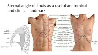
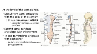
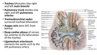
ANATOMICAL AND CLINICAL
EVENTS AT OR NEAR THE
STERNAL ANGLE OF LOUIS
OLUWOLE AKINOLA
Sternal angle of Louis as a useful anatomical
and clinical landmark
At the level of the sternal angle,
• Manubrium sterni articulates
with the body of the sternum
• to form manubriosternal joint
• a secondary cartilaginous joint or
symphysis
• Second costal cartilage
articulates with the sternum
• T4 and T5 vertebrae articulate
with each other
• an intervertebral disc intervening
between them
• Superior and
inferior
mediastina are
continuous
• at the imaginary
transverse sternal
plane
• Trachea bifurcates into right
and left main bronchi
• Pulmonary trunk divides into
right and left pulmonary
arteries
• Tracheobronchial nodes
surround tracheal bifurcation
• Azygos vein joins SVC from
behind
• Deep cardiac plexus of nerves
lies anterior to the bifurcation
of the trachea
• Ligamentum arteriosum
connects the aortic arch to the
left pulmonary artery
• Ascending aorta is continuous
with the aortic arch anteriorly,
• while the aortic arch is
continuous with the descending
thoracic aorta posteriorly
• Thoracic duct
crosses from the
right to the left of
midline
• as it ascends on the
vertebral column
•Auscultation for the
pulmonary and aortic
valves
• At the 2nd intercostal
space
• Close to sternal margin
• Pulmonary valve
auscultated near the
left sternal margin
• and aortic valve
auscultated near the
right sternal margin

12_dEvents at Sternal Angle of Louis.pdf

  • 1.
    ANATOMICAL AND CLINICAL EVENTSAT OR NEAR THE STERNAL ANGLE OF LOUIS OLUWOLE AKINOLA
  • 2.
    Sternal angle ofLouis as a useful anatomical and clinical landmark
  • 3.
    At the levelof the sternal angle, • Manubrium sterni articulates with the body of the sternum • to form manubriosternal joint • a secondary cartilaginous joint or symphysis • Second costal cartilage articulates with the sternum • T4 and T5 vertebrae articulate with each other • an intervertebral disc intervening between them
  • 4.
    • Superior and inferior mediastinaare continuous • at the imaginary transverse sternal plane
  • 5.
    • Trachea bifurcatesinto right and left main bronchi • Pulmonary trunk divides into right and left pulmonary arteries • Tracheobronchial nodes surround tracheal bifurcation • Azygos vein joins SVC from behind • Deep cardiac plexus of nerves lies anterior to the bifurcation of the trachea • Ligamentum arteriosum connects the aortic arch to the left pulmonary artery
  • 6.
    • Ascending aortais continuous with the aortic arch anteriorly, • while the aortic arch is continuous with the descending thoracic aorta posteriorly
  • 7.
    • Thoracic duct crossesfrom the right to the left of midline • as it ascends on the vertebral column
  • 8.
    •Auscultation for the pulmonaryand aortic valves • At the 2nd intercostal space • Close to sternal margin • Pulmonary valve auscultated near the left sternal margin • and aortic valve auscultated near the right sternal margin