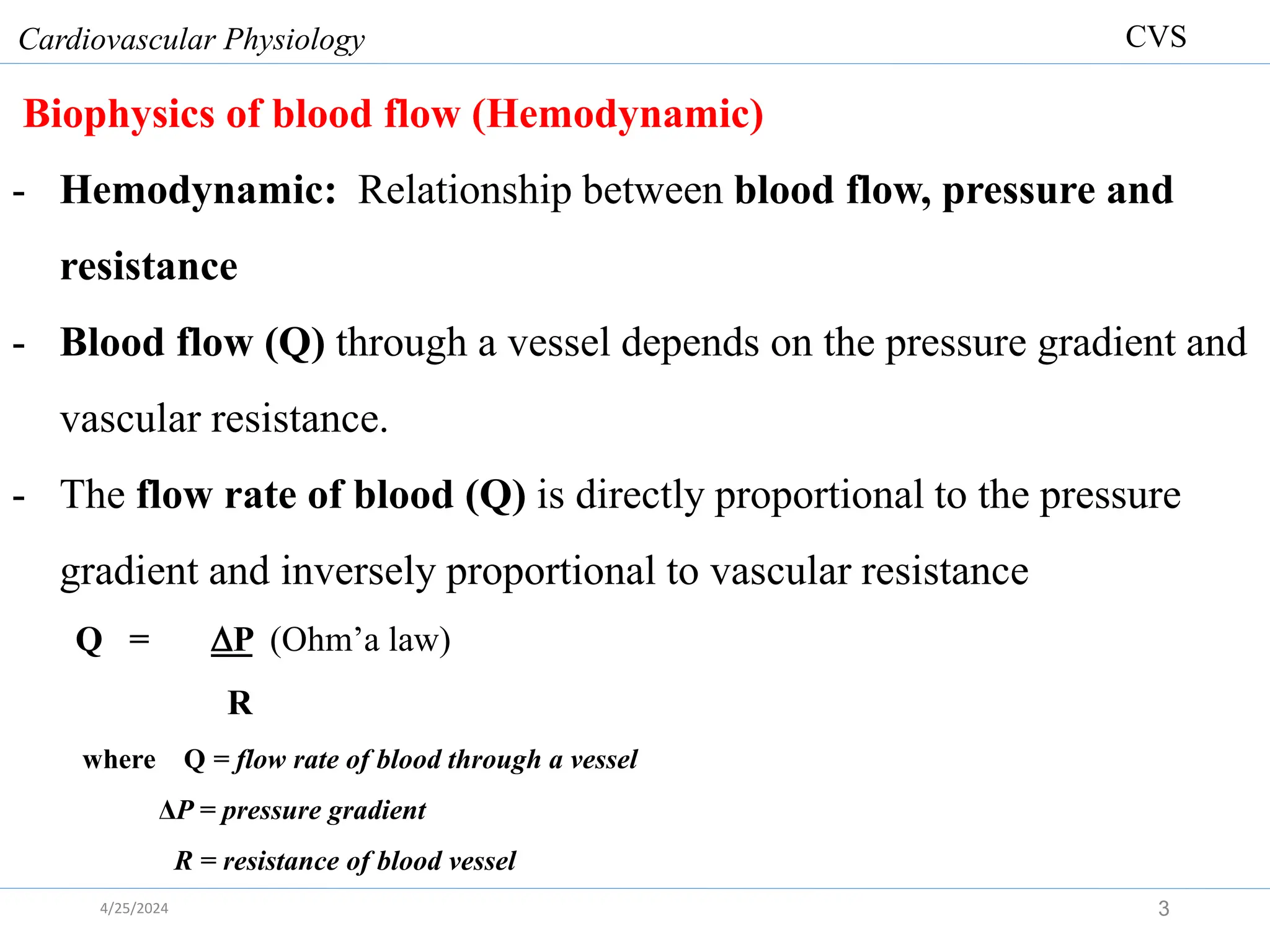
2. Outlines to be covered in this module
Blood flow
Peripheral circulation
Arterial system
o Arterial blood pressure
o Arterioles & its control mechanism
Microcirculation
o Capillaries
Venous system
Regulation of flow through blood vessels
Hypertension
Circulatory shock.
Cardiovascular Physiology
2
CVS
4/25/2024
3. Biophysics of blood flow (Hemodynamic)
- Hemodynamic: Relationship between blood flow, pressure and
resistance
- Blood flow (Q) through a vessel depends on the pressure gradient and
vascular resistance.
- The flow rate of blood (Q) is directly proportional to the pressure
gradient and inversely proportional to vascular resistance
Q = P (Ohm’a law)
R
where Q = flow rate of blood through a vessel
ΔP = pressure gradient
R = resistance of blood vessel
Cardiovascular Physiology
3
CVS
4/25/2024
4. Pressure Gradient-
- The pressure gradient is the difference in pressure between the
beginning and the end of a vessel.
- Blood flows from an area of higher pressure to an area of lower
pressure down a pressure gradient.
- Contraction of the heart imparts pressure to the blood, which is the
main driving force for flow through a vessel.
- Because of frictional losses (resistance), the pressure drops as blood
flows throughout the vessel’s length.
- Note that the difference in pressure between the two ends of a vessel,
not the absolute pressures within the vessel, determines flow rate
Cardiovascular Physiology
4
CVS
4/25/2024
7. Resistance to flow (R)
- Is a measure of the hindrance or opposition to blood flow through the
vessel
R= measure of friction between
Blood vessel & moving fluid.
Fluid molecules within themselves.
- As resistance to flow increases, it is more difficult for blood to pass
through the vessel, so flow rate decreases
- Viscosity - This is friction of molecules in the moving stream of fluid
Cardiovascular Physiology
7
CVS
4/25/2024
8. Vascular resistance to BF is
- directly proportional to viscosity of the blood(η)
- directly proportional to vessel length (L)
- inversely proportional to the 4th power of radius of the vessel(r4 )
R = 8.η. L
π r4
- The lengths of the tubes/blood vessels/ are approximately the same,
and the viscosity of the fluid is constant
- Therefore, differences in resistance offered by the tubes are due
solely to differences in their radii.
- Obviously, the widest tubes have the greatest flows.
Cardiovascular Physiology
8
CVS
4/25/2024
11. Types of blood flow
1.Laminar flow
Normal and noiseless.
All elements of the fluid move in a stream line,
that are parallel to the axis of the tube.
No fluid move in radial or circumferential direction.
Layer of fluid in contact with the wall is motionless (thin layer,
adherent to wall, hence motionless).
Fluid that move along the axis of the tube has max.Velocity.
Cardiovascular Physiology
11
CVS
4/25/2024
13. 2.Turbulent blood flow
Various elements of fluid move irregularly in axial radial and
circumferential direction
More pressure required to drive blood than laminar
(Energy wasted in propelling blood radially & axially)
Often accompanied by noise (murmurs) because of more driving
pressure.
Occur at valves and aorta (normal) & at site of blood clot
(pathological)
Cardiovascular Physiology
13
CVS
4/25/2024
16. Components of the vascular tree
- The vascular tree consists of arteries, arterioles, capillaries, venules,
and veins.
- Arteries- carry blood from the heart to the organs
- When a small artery reaches the organ it is supplying, it branches into
numerous arterioles.
- Arterioles branch further within the organs into capillaries
- Capillaries rejoin to form small venules, which further merge to form
small veins that leave the organs.
- The small veins progressively unite to form larger veins that
eventually empty into the heart.
Cardiovascular Physiology
16
CVS
4/25/2024
18. The Vessel Wall
The walls of the arteries and veins have three layers called tunics:
1. Tunica interna (intima)
- Inner lining in direct contact with blood
- Endothelium continuous with endocardial lining of heart
- Active role in vessel-related activities such as secretion of
vasoconstrictors and vasodilators .
2. Tunica media
- the middle layer, is usually the thickest.
- the thickness varies among vessel types
- It consists of smooth muscle, collagen, and sometimes elastic tissue.
- The smooth muscle regulates diameter of the lumen
3. Tunica externa
- is the outermost layer.
- Helps anchor vessel to surrounding tissue
Cardiovascular Physiology
18
CVS
4/25/2024
21. Arterial system:
1. Arteries
- Vessels that transport blood away from the heart
- Structurally it consists of
Larger arteries, Elastic (conducting) arteries
Medium arteries, Muscular (distributing) arteries
Arterioles, tiny arteries
Metarteriole
Cardiovascular Physiology
21
CVS
4/25/2024
22. 1. Conducting (elastic) arteries
- Found in largest arteries, aorta, pulmonary arteries, and common
carotid arteries.
- Their tunica media consists of numerous sheets of elastic tissue, this
makes the arteries to have elastic properties
- Functions.
- To conduct blood to medium-sized arteries
- To act as a pressure reservoir to provide the driving force for
blood when the heart is relaxing.
Cardiovascular Physiology
22
CVS
4/25/2024
24. Function as pressure reservoir
- As the heart pumps blood into the arteries during ventricular systole, a
greater volume of blood enters the arteries
- The highly elastic arteries expand to temporarily hold this excess
volume of ejected blood
- When the heart relaxes and temporarily stops pumping blood into the
arteries, the stretched arterial walls passively recoil.
- This recoil exerts pressure on the blood in the arteries during diastole.
- The pressure pushes the excess blood contained in the arteries into the
vessels downstream.
- This ensuring continued blood flow to the organs when the heart is
relaxing and not pumping blood into the system
Cardiovascular Physiology
24
CVS
4/25/2024
27. 2. Muscular arteries/ distributing arteries
- Tunica media contains more smooth muscle and fewer elastic fibers
than elastic arteries
- Walls relatively thick
- Capable of great vasoconstriction/ vasodilatation to adjust rate of
blood flow
- The brachial, femoral, and splenic arteries are examples of
distributing arteries.
Cardiovascular Physiology
27
CVS
4/25/2024
29. 3. Arterioles
- Each nutrient artery entering an organ branches six to eight times to be
called arterioles.
- The arterioles themselves branch two to five times to be capillaries.
- Internal diameters of arterioles is only 10 to 15 micrometers.
- Capillaries are originate from the terminal arterioles called metarterioles
- The metarterioles do not have a continuous muscular coat, but smooth
muscle fibers encircle the vessel at intermittent points.
- The junction of metarterioles and capillaries is encircled by smooth
muscle fiber called precapillary sphincter.
- This sphincter can open and close the entrance to the capillary.
Cardiovascular Physiology
29
CVS
4/25/2024
31. Arterioles are major resistance vessels and their diameters can
change many fold.
1. Increased degree of resistance help to regulate arterial blood
pressure
- Arterioles converts the pulsatile systolic-to-diastolic pressure swings
in the arteries into the non-fluctuating pressure present in the
capillaries by
- Dropping mean arterial pressure from 93 mm Hg to capillary
pressure of 35 mm Hg.
2. To distribute cardiac output among systemic organs, depending on
body’s momentary needs
Cardiovascular Physiology
31
CVS
4/25/2024
32. Mechanisms involved in adjusting arteriolar resistance
- The MAP is identical throughout the body
- Differences in flows between organs depend entirely on the relative
resistances offered by the arterioles of each organ.
- Arterioles contain smooth muscle, which can
contract and decrease the vessel radius (vasoconstriction) or
relax and cause the vessel radius to increase (vasodilation)
- Thus the pattern of blood-flow distribution depends upon the degree
of arteriolar smooth-muscle contraction within each organ and tissue.
Cardiovascular Physiology
32
CVS
4/25/2024
33. How can the arterioles resistance be changed?
Arteriolar smooth muscle
Possesses a large degree of spontaneous activity
Can contract independent of any neural, hormonal, or paracrine input).
This spontaneous contractile activity is called intrinsic tone.
The intrinsic tone/ level of contraction can be increased or decreased
by external signals, such as neurotransmitters.
An increase in contractile force above the vessel’s intrinsic tone
causes vasoconstriction, whereas a decrease in contractile force causes
vasodilation.
Cardiovascular Physiology
33
CVS
4/25/2024
35. -The mechanisms controlling vasoconstriction and vasodilation in
arterioles fall into two general categories:
(1) local controls
(2) extrinsic controls.
Cardiovascular Physiology
35
CVS
4/25/2024
37. Local Controls
- The term local controls denotes mechanisms independent of nerves or
hormones by which organs and tissues alter their own arteriolar
resistances, thereby self-regulating their blood flows.
- It does include changes caused by autocrine/paracrine agents.
- This self-regulation includes the phenomena of
- active hyperemia
- reactive hyperemia
- flow autoregulation, and
- local response to injury.
Cardiovascular Physiology
37
CVS
4/25/2024
38. Active Hyperemia
- Most organs and tissues manifest an increased blood flow (hyperemia)
when their metabolic activity is increased this is termed active
hyperemia.
- For example, the blood flow to exercising skeletal muscle increases in
direct proportion to the increased activity of the muscle.
- Active hyperemia is the direct result of arteriolar dilation in the more
active organ or tissue.
- Local chemical changes in the extracellular fluid surrounding the
arterioles causes arteriolar smooth muscle dilation
- Some of these local chemical changes that occur in the extracellular fluid
are PO2, PCO2, H+ , bradykinine & other
Cardiovascular Physiology
38
CVS
4/25/2024
39. Reactive Hyperemia
- When blood flow to a tissue is blocked for few seconds and then is
unblocked, the flow through tissue increases almost 4-7 times
normal.
- During the period of no blood flow, the arterioles in the affected organ
or tissue dilate, owing to the local factors described above.
- The excess blood flow lasts long enough to repay the tissue oxygen
deficit that has occurred during occlusion.
Cardiovascular Physiology
39
CVS
4/25/2024
42. Flow Autoregulation
- Autoregulation – maintenance of constant blood flow to an organ in
spite of fluctuations in blood pressure.
- For example, when arterial pressure in an organ is reduced, local
controls cause - arteriolar vasodilation, which tends to maintain
flow relatively constant.
- The opposite events occur when, for various reasons, arterial pressure
increases
Cardiovascular Physiology
42
CVS
4/25/2024
43. 43
Arterial
pressure
in organ
Blood flow
to organ
PO2 ,
metabolites,
vessel-wall
stretch
in organ
Arteriolar
dilation
in organ
Restoration
of blood
flow toward
normal
in organ
Flow Autoregulation
4/25/2024
45. Two basic mechanisms that explain local control of blood flow
1. Myogenic theory
Increase in blood flow
Stretches the vessel
Contraction of vascular smooth muscle
Decrease blood flow back to normal
Cardiovascular Physiology
45
CVS
4/25/2024
46. 2. Metabolic theory
Increase in rate of metabolism
Accumulation of vasodilator substances in active tissues
Blood vessels dilate
Increase blood flow
Vasodilator metabolites
O2 tension, H, CO2 tension, Temperature, K+, lactate,
Adenosine, Histamine.
Cardiovascular Physiology
46
CVS
4/25/2024
47. Response to Injury
- Tissue injury causes a variety of substances to be released locally
from cells or generated from plasma precursors.
- These substances make arteriolar smooth muscle relax and cause
vasodilation in an injured area.
- This phenomenon, a part of the general process known as
inflammation
Cardiovascular Physiology
47
CVS
4/25/2024
48. Extrinsic Controls
Sympathetic Nerves
- Most arterioles receive a rich supply of sympathetic postganglionic
nerve fibers.
- These neurons release mainly norepinephrine, which binds to alpha-
adrenergic receptors on the vascular smooth muscle to cause
vasoconstriction.
- Increase sympathetic stimulation causes arteriolar vasoconstriction.
- Decrease sympathetic stimulation causes arteriolar vasodilation.
Parasympathetic Nerves
- There is no parasympathetic innervations to arterioles.
Cardiovascular Physiology
48
CVS
4/25/2024
51. Hormones
Epinephrine
- Bind to alphaadrenergic receptors on arteriolar smooth muscle and
cause vasoconstriction.
- The arterioles in skeletal muscle have a large number of beta adrenergic
receptors, circulating epinephrine usually causes vasodilation.
Angiotensin II- constricts most arterioles.
Vasopressin - released from the posterior pituitary gland causes arteriolar
constriction
Atrial natriuretic factor-is the hormone secreted by the cardiac atria is a
potent vasodilator.
Cardiovascular Physiology
51
CVS
4/25/2024
53. 4. Microcirculatory unit
The arterioles, capillaries, and venules are collectively referred to as
the microcirculation, because they are only visible through a
microscope.
- The microcirculatory vessels are all located within the organs.
The microcirculation refers to the smallest blood vessels in the body:
- the smallest arterioles
- the metarterioles
- the precapillary sphincters
- the capillaries
- the small venules
Cardiovascular Physiology
53
CVS
4/25/2024
55. Capillary Beds
- A microcirculation moving from arterioles to venules
- Consist of two types of vessels:
Vascular shunts – metarteriole–thoroughfare channel connecting an
arteriole directly with a postcapillary venule
True capillaries – the actual exchange vessels
Cardiovascular Physiology
55
CVS
4/25/2024
58. True Capillaries
- The capillary wall is thin & consists of a single layer of endothelial
cells surrounded by a very thin basement membrane.
- The total thickness of the capillary wall is only about 0.5 micrometer.
- The internal diameter of the capillary is 4 to 9 micrometers.
- Lumen narrow, thus RBC change shape to pass through
- The peripheral circulation of the whole body has about 10 billion
capillaries with a total surface area estimated to be 500 to 700 square
meters
- Velocity of blood flow=1mm/s
- Therefore ideal for exchange (nutritional flow)
Cardiovascular Physiology
58
CVS
4/25/2024
60. Types of Capillaries
According to the size of the pores capillaries are classified into three:
- Continuous capillaries
- Fenestrated capillaries
- Sinusoids capillaries
Cardiovascular Physiology
60
CVS
4/25/2024
62. Continuous Capillaries
- Continuous capillaries are abundant in the skin and muscles
- Endothelial cells provide an uninterrupted lining
- Adjacent cells are connected with tight junctions
- Intercellular clefts about 4 nm wide between them ,allow the passage
of fluids & small solutes, such as glucose.
- But plasma proteins, other large molecules, and formed elements are
held back.
- Brain capillaries do not have clefts (blood-brain barrier).
Cardiovascular Physiology
62
CVS
4/25/2024
63. Fenestrated Capillaries
- Found wherever active capillary absorption or filtrate formation
occurs (e.g., glomerular capillaries of kidney, pancreas and salivary
glands and small intestines)
Characterized by:
- Fenestrations are about 20 to 100 nm in diameter
- They allow for the rapid passage of small molecules
- Fenestrate keep plasma proteins back
- Greater permeability than continuous capillaries
Cardiovascular Physiology
63
CVS
4/25/2024
64. Sinusoids Capillaries
- The clefts between the capillary endothelial cells are wide open
- Found in the liver, bone marrow and spleen
- Allow large molecules (proteins and blood cells) to pass between the
blood and surrounding tissues
Cardiovascular Physiology
64
CVS
4/25/2024
65. Capillary Exchange
- Capillary exchange refers to the two-way movement of fluid between
the blood and surrounding tissues.
- Substances pass between the blood and tissue fluid by diffusion,
transcytosis, filtration, and reabsorption
Movement across capillary influenced by:
- Nature of substance (size, shape, lipid solubility)
- Balance between hydrostatic and colloid osmotic pressure force
across membrane (Starling’s law)
- Capillary surface area available for exchange
- Physical characteristics of capillary membrane (continuous,
fenestrated, discontinuous)
Cardiovascular Physiology
65
CVS
4/25/2024
66. Diffusion
- The most important mechanism of exchange is diffusion.
- Glucose and oxygen, being more concentrated in the systemic blood
than in the tissue fluid, diffuse out of the blood.
- Carbon dioxide and other wastes, being more concentrated in the
tissue fluid, diffuse into the blood.
- Lipid-soluble substances as steroid hormones, O2, CO2 diffuse easily
through the plasma membranes.
- Substances insoluble in lipids, such as glucose and electrolytes, must
pass through membrane channels.
Cardiovascular Physiology
66
CVS
4/25/2024
67. Transcytosis
- Transcytosis is a process in which endothelial cells pick up droplets
of fluid on one side of the plasma membrane by pinocytosis,
transport the vesicles across the cell, and discharge the fluid on the
other side by exocytosis.
- Fatty acids, albumin, and some hormones such as insulin move
across the endothelium by this mechanism.
Cardiovascular Physiology
67
CVS
4/25/2024
71. Filtration and Reabsorption
- Fluid filters out of the arterial end of a capillary and osmotically
reenters it at the venous end.
- The rate of filtration and reabsorption at any point along the
capillary depends on a balance of forces.
- The rate of filtration and reabsorption at any point along the capillary
depends on a balance of forces – STARLINGS FORCES.
- These forces are hydrostatic and osmotic pressure on each side of
membrane
Cardiovascular Physiology
71
CVS
4/25/2024
73. 1. Capillary blood pressure (PC)
- Is the hydrostatic pressure exerted on the inside of the capillary walls
by blood.
- This pressure tends to force fluid out of the capillaries into the
interstitial fluid.
- On average, the hydrostatic pressure is 35 mm Hg at the arteriolar
end and 16 mm Hg at the capillary’s venular end.
Cardiovascular Physiology
73
CVS
4/25/2024
74. 2. Plasma-colloid osmotic pressure (𝛑P)
- Is a force caused by colloidal dispersion of plasma proteins.
- Plasma has a higher protein concentration and a lower water
concentration than interstitial fluid does.
- It encourages fluid movement into the capillaries
- Plasma-colloid osmotic pressure averages 26 mm Hg.
3. Interstitial fluid hydrostatic pressure (PIF)
- Is the fluid pressure exerted on the outside of the capillary wall by
interstitial fluid.
- This pressure tends to force fluid into the capillaries.
- The value of this pressure varies among tissues, (1 mm Hg )
Cardiovascular Physiology
74
CVS
4/25/2024
75. 4. Interstitial fluid–colloid osmotic pressure (𝛑IF)
- It is a pressure exerted by the small fraction of plasma proteins that
leak across the capillary walls into the interstitial spaces
- The leaked proteins exert an osmotic effect that tends to promote
movement of fluid out of the capillaries into the interstitial fluid.
- The interstitial fluid–colloid osmotic pressure is essentially zero mm
Hg
Cardiovascular Physiology
75
CVS
4/25/2024
76. Net filtration pressure in Arterial end (NFP)
Ultrafiltration pressure= Outward pressure - Inward pressure
NFP = (Pc + π IF ) – (πp + IF)
=(35 mm Hg +1) – (26 mm Hg + 0 mm Hg )
= 10 mm Hg
- This 10 mm Hg cause filtration to occur.
Cardiovascular Physiology
76
CVS
4/25/2024
77. Net reabsorption pressure in Venous end
Ultrafiltration pressure= Outward pressure - Inward pressure
NFP = (Pc + π IF ) – (πp + PIF)
= (16 mm Hg +1) – (26 mm Hg + 0 mm Hg )
= - 9 mm Hg
Net inward pressure of 9 mm Hg = Reabsorption pressure
- Reabsorption of fluid takes place as this inward pressure gradient
forces fluid back into the capillary at its venular end.
Cardiovascular Physiology
77
CVS
4/25/2024
80. Venous part of the systemic circulation
Structure of veins :
Thin walled
Small amount of elastic tissues and smooth muscle
Veins of dependent parts of body have valves
Valves prevent backflow of venous blood
Function of veins:
Store large quantity(>60%) of blood
(making blood available when required)
Propel blood forward by means of “venous pump”
Regulate cardiac output
Cardiovascular Physiology
80
CVS
4/25/2024
81. Venous Return.
Def:- Volume of blood returning from systemic circulation to heart.
Any factor/condition increases venous return increases End diastolic
volume.
Cardiovascular Physiology
81
CVS
4/25/2024
82. Factors regulating venous return
1. Pressure gradient
- Pressure generated by the heart is the most important force in venous
flow.
- Pressure in the venules ranges from 12 to 18 mmHg, and pressure at
the point where the venae cavae enter the heart, called central venous
pressure, averages 4.6 mmHg.
- Thus, there is a venous pressure gradient (P) of about 7 to 13
mmHg favoring the flow of blood toward the heart
- Increased pressure gradient causes increased venous return
Cardiovascular Physiology
82
CVS
4/25/2024
83. 2. Effect of gravity on Venous Return Veins
When a person is in upright position
- 500-700ml blood rapidly dislocated from thorax to lower extremities
- hydrostatic pressure superimposed on pressure generated by heart
-This adds hydrostatic pressure of 90mmHg
This increased hydrostatic pressure below the heart causes
1. Increase the distensiblity of veins, so that their capacity is increased
which in turn decreases venous return.
2. Marked increase in capillary blood pressure causes excessive fluid to
filter out of capillary beds in the lower extremities, producing localized
edema decreases venous return
NB:- Vessels above heart has negative hydrostatic pressure
Cardiovascular Physiology
83
CVS
4/25/2024
85. Countering the Effects of Gravity on the Venous System
The gravitational effects can be counteract by
A. Sympathetically induced venous vasoconstriction
- the resultant fall in mean arterial pressure that occurs when a person
moves from a lying-down to an upright position triggers
sympathetically induced venous vasoconstriction, and promotes the
return of blood to the heart
B. The skeletal muscle pump
Cardiovascular Physiology
85
CVS
4/25/2024
86. 3. Effect of Skeletal Muscle Activity on Venous Return
- Many large veins in the extremities lie between skeletal muscles.
- Muscle contraction compresses the veins.
- This external venous compression decreases venous capacity and
increases venous pressure.
- Increased muscular activity pushes more blood out of the veins and
into the heart.
- This pumping action, known as the skeletal muscle pump
Cardiovascular Physiology
86
CVS
4/25/2024
88. 4. Effect of Respiratory Activity on Venous Return
The respiratory pump.
- The respiratory pump moves blood up toward the heart as pressure
changes in the ventral body cavity during breathing.
- As we inhale, abdominal pressure increases, squeezing local veins
and forcing blood toward the heart.
- At the same time, the pressure in the chest decreases, allowing
thoracic veins to expand and speeding blood entry into the right
atrium.
- A decreased in intr-thoracic pressure lower right atrial pressure and
thus facilitating venous return.
Cardiovascular Physiology
88
CVS
4/25/2024
91. Blood pressure (BP)
Def/n: ~ Pressure exerted by circulating blood upon the
walls of blood vessels.
During each beat: BP varies between maximum (systole) &
minimum (diastole)….120/80mmHg
Average blood pressure (mmHg) increases with age.
95/65 → for 1 year
100/65 → for 6-9 years
110/65-140/90 → for Adults
BP is measured in millimeters of mercury (mmHg).
Cardiovascular Physiology
91
CVS
4/25/2024
92. Average blood pressure (mmHg) increases with age.
Cardiovascular Physiology
92
CVS
4/25/2024
93. Arterial pressure indices
■ Systolic blood pressure (SBP)
- is the highest arterial pressure during a cardiac cycle.
- is measured after the heart contracts (systole) and blood is ejected
into the arterial system.
- averages 120 mm Hg
■ Diastolic blood pressure (DBP)
- is the lowest arterial pressure during a cardiac cycle.
- is measured when the heart is relaxed (diastole)
- averages 80 mm Hg.
■ Pulse pressure (PP)
- Is the difference between systolic and diastolic blood pressure.
- Pulse pressure is palpated in the peripheral arteries.
- It is detected during systole not in diastole.
- When blood pressure is 120/80, pulse pressure is 40 mm Hg (120
minus 80 mm Hg)
PP=SBP-DBP
Cardiovascular Physiology
93
CVS
4/25/2024
94. ■ Mean arterial pressure (MAP)
- Is the average pressure driving blood forward into the tissues
throughout the cardiac cycle.
- The value of arterial pressure remains closer to diastolic than to
systolic pressure for a longer portion of each cardiac cycle.
- At resting heart rate, about two thirds of the cardiac cycle is spent in
diastole and only one third in systole.
MAP= DBP +1/3 PP
MAP = 80 + (1/3) 40 = 93 mm Hg
MAP = Mean arterial blood pressure
DBP = Diastolic blood pressure
PP = Pulse pressure
Cardiovascular Physiology
94
CVS
4/25/2024
97. Blood Pressure Measurement
-Blood pressure can be measured
1. Directly by connecting a pressure-measuring device to a needle inserted
in an artery.
2. Indirectly by means of a sphygmomanometer, an externally applied
inflatable cuff attached to a pressure gauge.
- During the determination of blood pressure, a stethoscope is placed
over the brachial artery at the inside bend of the elbow just below the
cuff.
- No sound can be detected either when blood is not flowing through the
vessel or when blood is flowing in the normal, smooth laminar flow
- Turbulent blood flow, in contrast, creates vibrations that can be heard.
Cardiovascular Physiology
97
CVS
4/25/2024
99. Cardiovascular Physiology
99
CVS
When cuff pressure is greater than 120 mm Hg
- The brachial artery is occluded
- No blood flows through the vessel.
- No sound is heard because no blood is flowing.
When cuff pressure is between 120 and 80 mm Hg:
- Blood flow through the vessel is turbulent
-The first sound (Korotkoff sounds) is heard at peak systolic
pressure.
-Intermittent sounds are produced by turbulent spurts of flow as
blood pressure cyclically exceeds cuff pressure.
When cuff pressure is less than 80 mm Hg
- No sound is heard throughout the cardiac cycle:
- Blood flows through the vessel in smooth, laminar fashion.
- The last sound is heard at minimum diastolic pressure.
- Thereafter because of uninterrupted, smooth, laminar flow.
4/25/2024
104. Mean arterial pressure (MAP) REGULATION
- Mean arterial pressure is the main driving force for propelling blood
to the tissues.
- Cardiovascular system is designed to monitor the MAP closely and
held at approximately 93 mm Hg.
- Blood pressure is regulated by controlling cardiac output and total
peripheral resistance.
- Determined by CO and Total Peripheral vascular resistance (TPVR)
Cardiovascular Physiology
104
CVS
MAP= CO .TPVR
4/25/2024
105. Classification of regulatory process of MAP
Regulatory processes classified into 3 groups according to time of action
1. Short term control mechanisms
2. Intermediate-term control mechanisms
3. Long-term control mechanisms
Cardiovascular Physiology
105
CVS
4/25/2024
108. A. Short-term control mechanisms
Predominantly vasomotor adjustment and neuronal control
Include:
Baroreceptor (Stretch) reflexes:
Chemoreceptor reflexes
Ischemic reflexes of CNS
Common characteristics:
Rapid onset of action (within few sec.)
Response vigorous, but if activated continuously, within a few
days, it either dies out completely- (baroreceptors)
or attenuated -(chemoreceptors, CNS Ischemic response)
Cardiovascular Physiology
108
CVS
4/25/2024
109. 1. Baroreceptor reflexes
Baroreceptors convey information about:
mean arterial pressure (MAP)
Amplitude of pressure fluctuation
Steepness of pressure rise rate of pressure change
More sensitive to than pressure (P/t)
More sensitive to sudden change
More sensitive to decrease than increase pressure
Stimulation of baroreceptors: stretch
Cardiovascular Physiology
109
CVS
4/25/2024
110. The baroreceptor reflex includes:-
1. A receptor
- found in carotid sinus and aortic arch baroreceptors
- sensitive to changes in mean arterial pressure.
- When MAP , the baroreceptors increases the rate of firing in the
corresponding afferent neurons.
- Conversely, a in MAP slows the rate of firing generated in the afferent
neurons by the baroreceptors.
2. The afferent neurons
- contains vagus & glossopharyngial nerves
- convoy signal to the medula oblongata
3. The integrating center
- receives the afferent impulses about the state of MAP
- is located at the cardiovascular control center, in the medulla
4. The efferent pathway
- is the autonomic nervous system.
5. The effectors- Heart and blood vessels
Cardiovascular Physiology
110
CVS
4/25/2024
111. N.B.
- The carotid baroreceptors provide the vasomotor center with
information regarding arterial pressure within the range of 50–200
mm Hg,
- whereas the aortic baroreceptors can only provide pressure
information within the range of 100–200 mm Hg.
Cardiovascular Physiology
111
CVS
4/25/2024
112. Baroreceptor reflexes for increased blood pressure
If an arterial blood pressure increase for any reason:-
- Baroreceptors increase their firing rate to the medulla and this induces
(1) Decreased sympathetic activity, which resulted in
- decreased heart rate
- decreased ventricular contractility
- decreased stroke volume
- decreased arteriolar constriction
- decreased venous constriction
(2) Increased parasympathetic activities, which resulted in
- decreased heart rate
The net result is
- decreased cardiac output
- decreased total peripheral resistance (arteriolar constriction), and
- return of blood pressure toward normal.
Cardiovascular Physiology
112
CVS
4/25/2024
113. Baroreceptor reflexes for decreased blood pressure
If arterial pressure decreases as during a hemorrhage,
- Baroreceptors decrease their firing rate to the medulla and this induces
(1) increased sympathetic activity, which resulted in
- increased heart rate
- increased ventricular contractility
- Increased stroke volume
- increased arteriolar constriction
- increased venous constriction
(2) decreased parasympathetic activities, which resulted in
- increased heart rate
The net result is
- an increased cardiac output
- increased total peripheral resistance (arteriolar constriction), and
- return of blood pressure toward normal.
Cardiovascular Physiology
113
CVS
4/25/2024
122. 2. Arterial chemoreceptors
- A chemoreflex is an autonomic response to changes in blood
chemistry such as decreased PO2, increased PCO2, & increased H+
- It is initiated by chemoreceptors called carotid and aortic bodies
(located near aortic & carotid sinus).
- The primary role of chemoreflexes is to adjust respiration to changes
in blood chemistry by doing hyperventilation (increased minute
volume).
- Stimulation of chemoreceptors in the medulla oblongata act through
the vasomotor center to cause widespread vasoconstriction. This
increases overall Blood Pressure.
Cardiovascular Physiology
122
CVS
4/25/2024
126. 3. CNS ischemic response
- Raises arterial pressure in response to diminished blood flow in
vasomotor center of the brain.
- CNS ischemic response is stimulated by
BP (<60 mm Hg) brain ischemia
Local hypoxia, hypercapnia & acidosis in medulla
- The cardiac and vasomotor centers of the medulla oblongata send
sympathetic signals to the heart and blood vessels that induce
(1) an increase in heart rate and contraction force and
(2) widespread vasoconstriction.
- These actions raise the blood pressure and, ideally, restore normal
perfusion of the brain.
This control system is “last ditch” mechanism for blood pressure
control.
Cardiovascular Physiology
126
CVS
4/25/2024
127. B. Intermediate term control
1. Stress-relaxation or reverse stress relaxation:
Pressure stress on veins veins relax &pressure
(stress relaxation)
intravascular volume opposite effect.
Severe bleeding → reverse stress-relaxation mechanism,
i.e blood vessels tighten up around the blood that is left→
re-establish normal circulation. This mechanism cannot correct
changes that are about 30% above normal or 15% below normal.
Cardiovascular Physiology
127
CVS
4/25/2024
128. 2. Capillary fluid shifts:- in response to changes in
capillary pressure
Fluid moves into interstitial fluid space
when BP rises→ normal BP.
3. Beginning of arteriolar vasoconstriction by Ang. II
Cardiovascular Physiology
128
CVS
4/25/2024
129. 4. Cardiac stretch receptor reflex
Atrial natriuretic peptide (ANP)= stretch receptors
- Released from atria in response to increase atrial pressure
- During increases in venous return ----- RA stretch
Elicits 2 reflexes to increase CO
1.Stretch of SA node ---->increase HR, which helps to
pump extra blood that is returning to the heart.
Cardiovascular Physiology
129
CVS
4/25/2024
130. 2. Bain-bridge-reflex
- When the right sided filling pressure increased, the wall of the atrium
is stretched. The stretch receptor present in the right atrial wall sends
signal to Vasomotor center of medulla(VMC) via afferent vagal nerve.
- The VMC in turn inhibit parasympathetic activity to increase heart
rate and increase sympathetic NS to increase contractility.
- This reflex causes an increase in HR, which also helps to pump out
the excess venous return
Cardiovascular Physiology
130
CVS
4/25/2024
134. C. Long term control
-Renin-Angiotension-Aldosterone system (long-term control (RASS)
-Long term control mechanism of control of ABP is shown below
Cardiovascular Physiology
134
CVS
4/25/2024
135. Angiotensin II.
- This is a potent vasoconstrictor that raises the blood pressure.
Aldosterone.
- This “salt-retaining hormone” primarily promotes Na retention by the
kidneys. Since water follows sodium osmotically, Na retention
promotes water retention, thus promoting a higher blood volume and
pressure.
Antidiuretic hormone.
- ADH primarily promotes water retention, but at pathologically high
concentrations it is also a vasoconstrictor—hence its alternate name,
vasopressin. Both of these effects raise blood pressure.
Cardiovascular Physiology
135
CVS
4/25/2024
138. Circulatory shock
Exists when there is a generalized, severe reduction in blood
supply and the metabolic needs of the tissues are not meet.
Occurrence of low arterial pressure even with all
cardiovascular compensatory mechanisms activated.
Primary Disturbances
Severely depressed myocardial functional ability or
Inadequate cardiac filling due to low mean circulatory
filling pressure.
Cardiovascular Physiology
138
CVS
4/25/2024
140. 1. Hypovolemic shock
Accompanies significant hemorrhage (usually greater than 20% of
blood volume), severe burns, chronic diarrhea, or prolonged
vomiting.
Induce shock by depleting body fluids and thus circulating blood
volume.
Pulmonary embolus:- evoke a shock that resembles
hypovolemic shock in that left ventricular filling may be
compromised.
large emboli not only reduce cardiac output but interfere with
gas exchange in the lungs.
Cardiovascular Physiology
140
CVS
4/25/2024
141. 2. Anaphylactic shock
Occurs as a result of a severe allergic reaction to an antigen .
(e.g. insect bites, antibiotics, certain foods).
It is also called an "immediate hypersensitivity reaction" which
is mediated by several substances (such as histamine, prostaglandins,
leukotrienes, bradykinin).
It results in peripheral vasodilatation and increases microvascular
permeability.
Cardiovascular Physiology
141
CVS
4/25/2024
142. 3. Septic shock
It is caused by vasodilator effects of substances released into the
circulating blood by infective agents.
One of the most common is endotoxin, a lipopolysaccharide
released from bacteria.
It induces the formation of a NO synthase (called inducible nitric
oxide synthase to distinguish it from the normally present
constitutive nitric oxide synthase) in endothelial cells, vascular
smooth muscle, and macrophages.
Cardiovascular Physiology
142
CVS
4/25/2024
143. 4. Neurogenic shock
It is produced by loss of vascular tone due to inhibition of the
normal tonic activity of the sympathetic vasoconstrictor nerves &
often occurs with deep general anesthesia or in reflex response to
deep pain associated with traumatic injuries.
The transient vasovagal syncope evoked by strong emotions is
a mild form of neurogenic shock.
Cardiovascular Physiology
143
CVS
4/25/2024
145. Stages of Shock
Because the characteristics of circulatory shock change
with different degrees of severity, shock is divided into
the following three major stages:
1) A non progressive stage (Compensated stage):
Normal circulatory compensatory mechanisms cause full recovery
without help from outside therapy.
2) A progressive stage:
- Without therapy, the shock becomes steadily worse until death.
3) An irreversible stage:
- All forms of known therapy are inadequate to save the person’s
life, even though, for the moment, the person is still alive.
Cardiovascular Physiology
145
CVS
4/25/2024