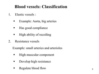
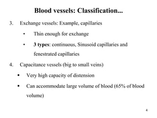
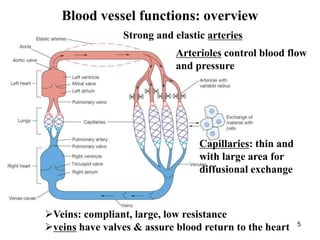
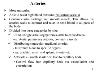
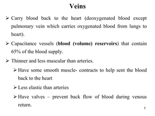
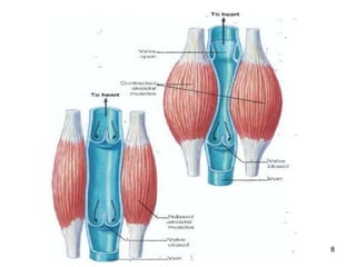
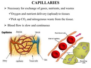
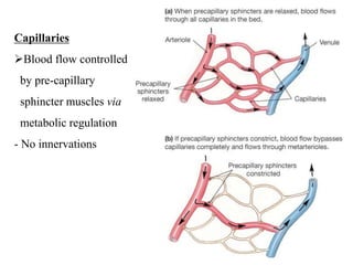
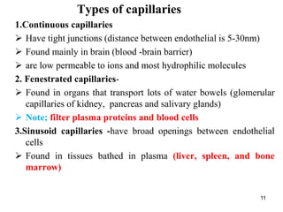
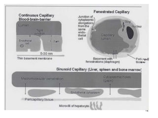
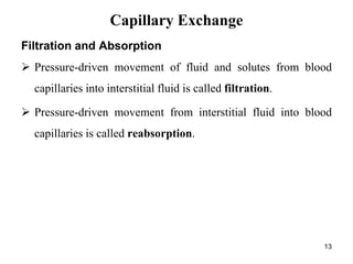
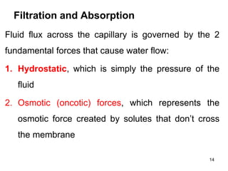
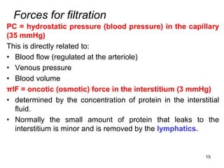
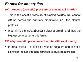
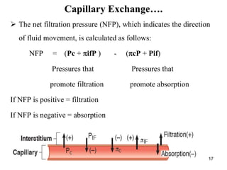
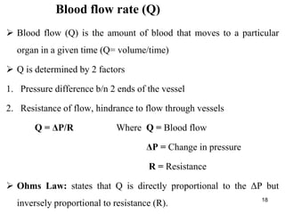
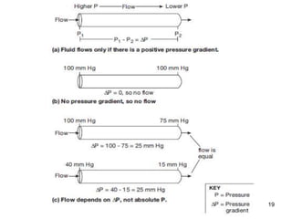
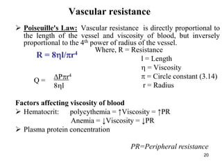
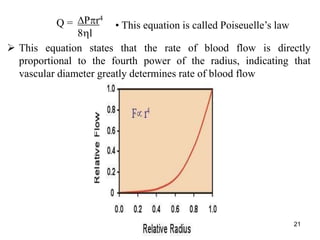
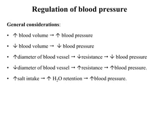
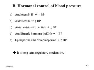
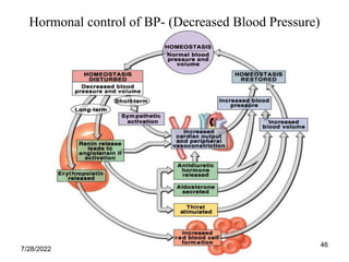
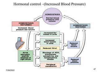
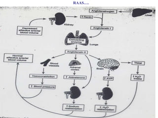
1
HEMODYNAMICS
2
HEMODYNAMICS
 Hemodynamics is the study of the relationship between flow,
pressure and resistance and other physical principles of blood
circulation
 Blood vessels: types, histology
 Blood flow: types
 Blood pressure & regulation
 Microcirculation
3
Blood vessels: Classification
1. Elastic vessels :
 Example: Aorta, big arteries
 Has good compliance
 High ability of recoiling
2. Resistance vessels
Example: small arteries and arterioles
 High muscular component
 Develop high resistance
 Regulate blood flow
3. Exchange vessels: Example, capillaries
• Thin enough for exchange
• 3 types: continuous, Sinusoid capillaries and
fenestrated capillaries
4. Capacitance vessels (big to small veins)
 Very high capacity of distension
 Can accommodate large volume of blood (65% of blood
volume)
4
Blood vessels: Classification...
5
Blood vessel functions: overview
Strong and elastic arteries
Arterioles control blood flow
and pressure
Capillaries: thin and
with large area for
diffusional exchange
Veins: compliant, large, low resistance
veins have valves & assure blood return to the heart
6
Arteries
 More muscular.
 Able to resist high blood pressure (resistance vessels)
 Contain elastic cartilage and smooth muscle, This allows the
arteries walls to contract and relax to send blood to all parts of
the body.
 Divided into three categories by size.
 Conducting(elastic/large)arteries-Able to expand/recoil.
-eg. Aorta, pulmonary arteries, common carotids.
 Distributing (muscular, medium) arteries
- Distribute blood to specific organs.
- eg. brachial, renal, and splenic arteries etc
 Arterioles – smallest arteries; lead to capillary beds.
• Control flow into capillary beds via vasodilation and
constriction
Veins
7
 Carry blood back to the heart (deoxygenated blood except
pulmonary vein which carries oxygenated blood from lungs to
heart).
 Capacitance vessels (blood (volume) reservoirs) that contain
65% of the blood supply.
 Thinner and less muscular than arteries.
Have some smooth muscle- contracts to help sent the blood
back to the heart
Less elastic than arteries
Have valves – prevent back flow of blood during venous
return.
8
9
 Necessary for exchange of gases, nutrients, and wastes
Oxygen and nutrient delivery (upload) to tissues
Pick up CO2 and nitrogenous waste from the tissue.
 Blood flow is slow and continuous
CAPILLARIES
10
10
Capillaries
Blood flow controlled
by pre-capillary
sphincter muscles via
metabolic regulation
- No innervations
Types of capillaries
11
1.Continuous capillaries
 Have tight junctions (distance between endothelial is 5-30nm)
 Found mainly in brain (blood -brain barrier)
 are low permeable to ions and most hydrophilic molecules
2. Fenestrated capillaries-
 Found in organs that transport lots of water bowels (glomerular
capillaries of kidney, pancreas and salivary glands)
 Note; filter plasma proteins and blood cells
3.Sinusoid capillaries -have broad openings between endothelial
cells
 Found in tissues bathed in plasma (liver, spleen, and bone
marrow)
12
12
Filtration and Absorption
 Pressure-driven movement of fluid and solutes from blood
capillaries into interstitial fluid is called filtration.
 Pressure-driven movement from interstitial fluid into blood
capillaries is called reabsorption.
13
Capillary Exchange
Filtration and Absorption
Fluid flux across the capillary is governed by the 2
fundamental forces that cause water flow:
1. Hydrostatic, which is simply the pressure of the
fluid
2. Osmotic (oncotic) forces, which represents the
osmotic force created by solutes that don’t cross
the membrane
14
Forces for filtration
PC = hydrostatic pressure (blood pressure) in the capillary
(35 mmHg)
This is directly related to:
• Blood flow (regulated at the arteriole)
• Venous pressure
• Blood volume
πIF = oncotic (osmotic) force in the interstitium (3 mmHg)
• determined by the concentration of protein in the interstitial
fluid.
• Normally the small amount of protein that leaks to the
interstitium is minor and is removed by the lymphatics.
15
Forces for absorption
πC = oncotic (osmotic) pressure of plasma (28 mmHg)
• This is the oncotic pressure of plasma solutes that cannot
diffuse across the capillary membrane, i.e., the plasma
proteins.
• Albumin is the most abundant plasma protein and thus the
biggest contributor to this force.
PIF = hydrostatic pressure in the interstitium (0 mmHg)
• In most cases it is close to zero or negative and is not a
significant factor affecting filtration versus reabsorption.
16
17
 The net filtration pressure (NFP), which indicates the direction
of fluid movement, is calculated as follows:
NFP = (Pc + ifP ) - (cP + Pif)
Pressures that Pressures that
promote filtration promote absorption
If NFP is positive = filtration
If NFP is negative = absorption
Capillary Exchange….
18
Blood flow rate (Q)
 Blood flow (Q) is the amount of blood that moves to a particular
organ in a given time (Q= volume/time)
 Q is determined by 2 factors
1. Pressure difference b/n 2 ends of the vessel
2. Resistance of flow, hindrance to flow through vessels
Q = ΔP/R Where Q = Blood flow
ΔP = Change in pressure
R = Resistance
 Ohms Law: states that Q is directly proportional to the ΔP but
inversely proportional to resistance (R).
19
20
Vascular resistance
 Poiseuille's Law: Vascular resistance is directly proportional to
the length of the vessel and viscosity of blood, but inversely
proportional to the 4th power of radius of the vessel.
Where, R = Resistance
l = Length
 = Viscosity
 = Circle constant (3.14)
r = Radius
Factors affecting viscosity of blood
 Hematocrit: polycythemia = ↑Viscosity = ↑PR
Anemia = ↓Viscosity = ↓PR
 Plasma protein concentration
R = 8l/r4
Pr4
8l
Q =
PR=Peripheral resistance
 This equation states that the rate of blood flow is directly
proportional to the fourth power of the radius, indicating that
vascular diameter greatly determines rate of blood flow
21
Pr4
8l
Q = • This equation is called Poiseuelle’s law
22
Velocity of blood flow (V)
 Velocity is a measure of how fast blood flows past a point.
• Velocity of blood flow through a tube equals the flow rate divided
by the tubes cross-sectional area.
V = Q/A
A= r2 Q=
 Velocity (v) is directly proportional to the pressure difference (P)
and diameter of the vessels, but inversely related to the viscosity of
blood and length of the blood vessel
v = Pr2 /8l
Pr4
8l
23
 Lowest velocity
 Largest total
cross sectional
area
 Hydrostatic
pressure drops
slightly
Capillary Blood Flow-Velocity
Figure. The velocity of flow depends on the total cross-sectional area
24
Change in velocity of BF, volume, P, and R along blood vessels
25
Types of Blood Flow
 There are two types of blood flow inside the vessels
1. Laminar flow
2. Turbulent flow
1. Laminar blood flow also called streamline flow
Blood flows in a steady rate through long smooth vessels
Blood cells move in a straight line
Blood cells at the centre of the vessel move faster
Laminar flow is silent
2. Turbulent flow
Blood flows in all directions in a vessel, continuously mixed
Turbulent blood flow produces sound
Occurs at blood vessels with high elastic content and during
high velocity
26
27
Laminar vs turbulent flow the reynold’s number
28
Regulation of blood flow
 Each organ can control the rate of BF based on the degree of
requirement of O2, nutrients and removal of wastes.
 Two types of regulation
A. Intrinsic Regulation of Blood Flow (Autoregulation)
• It is localized regulation of vascular resistance and blood flow.
• Includes - Myogenic
- Metabolic
B. Extrinsic Regulation of Blood Flow
• Extrinsic regulation refers to control by the autonomic nervous
system and endocrine system (hormonal)
29
A. Intrinsic Regulation of Blood Flow
i. Myogenic Control Mechanisms
• Stretching of small blood vessels at high pressure causes the smooth
muscle of the vessel wall to contract.
• Conversely, at low pressures, the muscles relax.
 Blood flow will maintain as required.
ii. Metabolic Control Mechanisms
Local vasodilation within an organ can occur as a result of
metabolism
Chemicals that promote vasodilation include:
• O2 , CO2 concentrations, pH (due to CO2, lactic acid, etc.),
release of adenosine the tissue cells.
7/28/2022
30
B. Extrinsic Regulation of Blood Flow
 Includes
Autonomic nervous system (Sympathetic and
parasympathetic)
Endocrine system (hormonal)
Norepinephrine and epinephrine.
Angiotensin II.
Vasopressin (ADH)
7/28/2022
31
Arterial blood pressure-ABP
 BP is a force exerted by the blood on the wall of the blood
vessels
 Has 2 components: systolic and diastolic pressure
 Expressed as : BP=systolic/diastolic
 Normal Ps: 90 – 130 mm Hg (120 mm Hg)
Pd: 60 – 90 mm Hg (80 mm Hg)
Normal adult BP=120/80
32
Pulse pressure- up and down fluctuation
of arterial pressure.
Ppulse = Ps-Pd
where;
Ps = systolic pressure
Pd= diastolic pressure
. it is about ~ 40mmHg
7/28/2022 32
33
Mean arterial pressure(MAP)
• MAP is average BP over a cardiac cycle.
• Mainly determined by CO, and total peripheral vascular resistance
(TPVR)
MAP= CO X TPR
MAP also ~ Pd + 1/3 (Ps-Pd.)
 it is about 93-100 mmHg
where;
Ps = systolic pressure
Pd= diastolic pressure
7/28/2022 33
General considerations:
•  blood volume   blood pressure
•  blood volume   blood pressure
• diameter of blood vessel  resistance   blood pressure
• diameter of blood vessel  resistance  blood pressure.
• salt intake   H2O retention  blood pressure.
Regulation of blood pressure
35
35
 Two types
A. Short-term controlling mechanisms
 Baroreceptor reflex
 Chemoreceptor reflex
 CNS ischemic reflex
B. Long-term controlling mechanisms
 Hormonal mechanism
 The Renin-angiotensin-aldosterone system
Regulation of blood pressure
 Baroreceptors are stretch-sensitive mechanoreceptors which are
located on:
• walls of the carotid sinus
• Aortic arch
 Regulate arterial pressure by increasing firing when stretched
(high pressure) and conversely, slowing firing when relaxed (low
pressure
36
The Baroreceptor Reflex
37
38
39
Regulation of Blood Pressure
Figure The baroreceptor reflex: the response to increased blood pressure
40
41
Chemoreceptor reflex
 At low O2 or high CO2 or H+ (as occurs during low
pressure because of blood flow), chemoreceptors are
stimulated.
 Chemoreceptors excite the vasomotor center, which
elevates the arterial pressure.
42
43
43
Chemoreceptor
44
44
CNS ischemic reflexes
 If blood flow is decreased to the vasomotor center in the lower
brainstem and CO2 accumulates, which stimulates sympathetic
area of medulla of brain.
 Very strong sympathetic stimulator causing major
vasoconstriction and cardiac acceleration.
 Sometimes called the “last ditch stand”.
7/28/2022
45
B. Hormonal control of blood pressure
a) Angiotensin II  ↑ BP
b) Aldosterone  ↑ BP
c) Atrial natriuretic peptide  ↓ BP
d) Antidiruetic hormone (ADH)  ↑ BP
e) Epinephrine and Norepinephrine  ↑ BP
 it is long term regulatory mechanism.
7/28/2022
46
Hormonal control of BP- (Decreased Blood Pressure)
7/28/2022
47
Hormonal control –(Increased Blood Pressure)
7/28/2022
48
Renin-Angiotension-Aldosterone system (RAAS)
 The kidneys control the level of H2O and NaCl in the body, thus
controlling the volume of the extracellular fluid and blood.
 By controlling blood volume, the kidneys control arterial
pressure.
 Increased arterial pressure results in increased renal output of
H2O (pressure diuresis) and salt (pressure natiuresis).
 It is long-term control system
7/28/2022
49
RAAS….

A CVS-Physiology2.ppt

  • 1.
  • 2.
    2 HEMODYNAMICS  Hemodynamics isthe study of the relationship between flow, pressure and resistance and other physical principles of blood circulation  Blood vessels: types, histology  Blood flow: types  Blood pressure & regulation  Microcirculation
  • 3.
    3 Blood vessels: Classification 1.Elastic vessels :  Example: Aorta, big arteries  Has good compliance  High ability of recoiling 2. Resistance vessels Example: small arteries and arterioles  High muscular component  Develop high resistance  Regulate blood flow
  • 4.
    3. Exchange vessels:Example, capillaries • Thin enough for exchange • 3 types: continuous, Sinusoid capillaries and fenestrated capillaries 4. Capacitance vessels (big to small veins)  Very high capacity of distension  Can accommodate large volume of blood (65% of blood volume) 4 Blood vessels: Classification...
  • 5.
    5 Blood vessel functions:overview Strong and elastic arteries Arterioles control blood flow and pressure Capillaries: thin and with large area for diffusional exchange Veins: compliant, large, low resistance veins have valves & assure blood return to the heart
  • 6.
    6 Arteries  More muscular. Able to resist high blood pressure (resistance vessels)  Contain elastic cartilage and smooth muscle, This allows the arteries walls to contract and relax to send blood to all parts of the body.  Divided into three categories by size.  Conducting(elastic/large)arteries-Able to expand/recoil. -eg. Aorta, pulmonary arteries, common carotids.  Distributing (muscular, medium) arteries - Distribute blood to specific organs. - eg. brachial, renal, and splenic arteries etc  Arterioles – smallest arteries; lead to capillary beds. • Control flow into capillary beds via vasodilation and constriction
  • 7.
    Veins 7  Carry bloodback to the heart (deoxygenated blood except pulmonary vein which carries oxygenated blood from lungs to heart).  Capacitance vessels (blood (volume) reservoirs) that contain 65% of the blood supply.  Thinner and less muscular than arteries. Have some smooth muscle- contracts to help sent the blood back to the heart Less elastic than arteries Have valves – prevent back flow of blood during venous return.
  • 8.
  • 9.
    9  Necessary forexchange of gases, nutrients, and wastes Oxygen and nutrient delivery (upload) to tissues Pick up CO2 and nitrogenous waste from the tissue.  Blood flow is slow and continuous CAPILLARIES
  • 10.
    10 10 Capillaries Blood flow controlled bypre-capillary sphincter muscles via metabolic regulation - No innervations
  • 11.
    Types of capillaries 11 1.Continuouscapillaries  Have tight junctions (distance between endothelial is 5-30nm)  Found mainly in brain (blood -brain barrier)  are low permeable to ions and most hydrophilic molecules 2. Fenestrated capillaries-  Found in organs that transport lots of water bowels (glomerular capillaries of kidney, pancreas and salivary glands)  Note; filter plasma proteins and blood cells 3.Sinusoid capillaries -have broad openings between endothelial cells  Found in tissues bathed in plasma (liver, spleen, and bone marrow)
  • 12.
  • 13.
    Filtration and Absorption Pressure-driven movement of fluid and solutes from blood capillaries into interstitial fluid is called filtration.  Pressure-driven movement from interstitial fluid into blood capillaries is called reabsorption. 13 Capillary Exchange
  • 14.
    Filtration and Absorption Fluidflux across the capillary is governed by the 2 fundamental forces that cause water flow: 1. Hydrostatic, which is simply the pressure of the fluid 2. Osmotic (oncotic) forces, which represents the osmotic force created by solutes that don’t cross the membrane 14
  • 15.
    Forces for filtration PC= hydrostatic pressure (blood pressure) in the capillary (35 mmHg) This is directly related to: • Blood flow (regulated at the arteriole) • Venous pressure • Blood volume πIF = oncotic (osmotic) force in the interstitium (3 mmHg) • determined by the concentration of protein in the interstitial fluid. • Normally the small amount of protein that leaks to the interstitium is minor and is removed by the lymphatics. 15
  • 16.
    Forces for absorption πC= oncotic (osmotic) pressure of plasma (28 mmHg) • This is the oncotic pressure of plasma solutes that cannot diffuse across the capillary membrane, i.e., the plasma proteins. • Albumin is the most abundant plasma protein and thus the biggest contributor to this force. PIF = hydrostatic pressure in the interstitium (0 mmHg) • In most cases it is close to zero or negative and is not a significant factor affecting filtration versus reabsorption. 16
  • 17.
    17  The netfiltration pressure (NFP), which indicates the direction of fluid movement, is calculated as follows: NFP = (Pc + ifP ) - (cP + Pif) Pressures that Pressures that promote filtration promote absorption If NFP is positive = filtration If NFP is negative = absorption Capillary Exchange….
  • 18.
    18 Blood flow rate(Q)  Blood flow (Q) is the amount of blood that moves to a particular organ in a given time (Q= volume/time)  Q is determined by 2 factors 1. Pressure difference b/n 2 ends of the vessel 2. Resistance of flow, hindrance to flow through vessels Q = ΔP/R Where Q = Blood flow ΔP = Change in pressure R = Resistance  Ohms Law: states that Q is directly proportional to the ΔP but inversely proportional to resistance (R).
  • 19.
  • 20.
    20 Vascular resistance  Poiseuille'sLaw: Vascular resistance is directly proportional to the length of the vessel and viscosity of blood, but inversely proportional to the 4th power of radius of the vessel. Where, R = Resistance l = Length  = Viscosity  = Circle constant (3.14) r = Radius Factors affecting viscosity of blood  Hematocrit: polycythemia = ↑Viscosity = ↑PR Anemia = ↓Viscosity = ↓PR  Plasma protein concentration R = 8l/r4 Pr4 8l Q = PR=Peripheral resistance
  • 21.
     This equationstates that the rate of blood flow is directly proportional to the fourth power of the radius, indicating that vascular diameter greatly determines rate of blood flow 21 Pr4 8l Q = • This equation is called Poiseuelle’s law
  • 22.
    22 Velocity of bloodflow (V)  Velocity is a measure of how fast blood flows past a point. • Velocity of blood flow through a tube equals the flow rate divided by the tubes cross-sectional area. V = Q/A A= r2 Q=  Velocity (v) is directly proportional to the pressure difference (P) and diameter of the vessels, but inversely related to the viscosity of blood and length of the blood vessel v = Pr2 /8l Pr4 8l
  • 23.
    23  Lowest velocity Largest total cross sectional area  Hydrostatic pressure drops slightly Capillary Blood Flow-Velocity Figure. The velocity of flow depends on the total cross-sectional area
  • 24.
    24 Change in velocityof BF, volume, P, and R along blood vessels
  • 25.
    25 Types of BloodFlow  There are two types of blood flow inside the vessels 1. Laminar flow 2. Turbulent flow 1. Laminar blood flow also called streamline flow Blood flows in a steady rate through long smooth vessels Blood cells move in a straight line Blood cells at the centre of the vessel move faster Laminar flow is silent 2. Turbulent flow Blood flows in all directions in a vessel, continuously mixed Turbulent blood flow produces sound Occurs at blood vessels with high elastic content and during high velocity
  • 26.
  • 27.
    27 Laminar vs turbulentflow the reynold’s number
  • 28.
    28 Regulation of bloodflow  Each organ can control the rate of BF based on the degree of requirement of O2, nutrients and removal of wastes.  Two types of regulation A. Intrinsic Regulation of Blood Flow (Autoregulation) • It is localized regulation of vascular resistance and blood flow. • Includes - Myogenic - Metabolic B. Extrinsic Regulation of Blood Flow • Extrinsic regulation refers to control by the autonomic nervous system and endocrine system (hormonal)
  • 29.
    29 A. Intrinsic Regulationof Blood Flow i. Myogenic Control Mechanisms • Stretching of small blood vessels at high pressure causes the smooth muscle of the vessel wall to contract. • Conversely, at low pressures, the muscles relax.  Blood flow will maintain as required. ii. Metabolic Control Mechanisms Local vasodilation within an organ can occur as a result of metabolism Chemicals that promote vasodilation include: • O2 , CO2 concentrations, pH (due to CO2, lactic acid, etc.), release of adenosine the tissue cells. 7/28/2022
  • 30.
    30 B. Extrinsic Regulationof Blood Flow  Includes Autonomic nervous system (Sympathetic and parasympathetic) Endocrine system (hormonal) Norepinephrine and epinephrine. Angiotensin II. Vasopressin (ADH) 7/28/2022
  • 31.
    31 Arterial blood pressure-ABP BP is a force exerted by the blood on the wall of the blood vessels  Has 2 components: systolic and diastolic pressure  Expressed as : BP=systolic/diastolic  Normal Ps: 90 – 130 mm Hg (120 mm Hg) Pd: 60 – 90 mm Hg (80 mm Hg) Normal adult BP=120/80
  • 32.
    32 Pulse pressure- upand down fluctuation of arterial pressure. Ppulse = Ps-Pd where; Ps = systolic pressure Pd= diastolic pressure . it is about ~ 40mmHg 7/28/2022 32
  • 33.
    33 Mean arterial pressure(MAP) •MAP is average BP over a cardiac cycle. • Mainly determined by CO, and total peripheral vascular resistance (TPVR) MAP= CO X TPR MAP also ~ Pd + 1/3 (Ps-Pd.)  it is about 93-100 mmHg where; Ps = systolic pressure Pd= diastolic pressure 7/28/2022 33
  • 34.
    General considerations: • blood volume   blood pressure •  blood volume   blood pressure • diameter of blood vessel  resistance   blood pressure • diameter of blood vessel  resistance  blood pressure. • salt intake   H2O retention  blood pressure. Regulation of blood pressure
  • 35.
    35 35  Two types A.Short-term controlling mechanisms  Baroreceptor reflex  Chemoreceptor reflex  CNS ischemic reflex B. Long-term controlling mechanisms  Hormonal mechanism  The Renin-angiotensin-aldosterone system Regulation of blood pressure
  • 36.
     Baroreceptors arestretch-sensitive mechanoreceptors which are located on: • walls of the carotid sinus • Aortic arch  Regulate arterial pressure by increasing firing when stretched (high pressure) and conversely, slowing firing when relaxed (low pressure 36 The Baroreceptor Reflex
  • 37.
  • 38.
  • 39.
    39 Regulation of BloodPressure Figure The baroreceptor reflex: the response to increased blood pressure
  • 40.
  • 41.
    41 Chemoreceptor reflex  Atlow O2 or high CO2 or H+ (as occurs during low pressure because of blood flow), chemoreceptors are stimulated.  Chemoreceptors excite the vasomotor center, which elevates the arterial pressure.
  • 42.
  • 43.
  • 44.
    44 44 CNS ischemic reflexes If blood flow is decreased to the vasomotor center in the lower brainstem and CO2 accumulates, which stimulates sympathetic area of medulla of brain.  Very strong sympathetic stimulator causing major vasoconstriction and cardiac acceleration.  Sometimes called the “last ditch stand”. 7/28/2022
  • 45.
    45 B. Hormonal controlof blood pressure a) Angiotensin II  ↑ BP b) Aldosterone  ↑ BP c) Atrial natriuretic peptide  ↓ BP d) Antidiruetic hormone (ADH)  ↑ BP e) Epinephrine and Norepinephrine  ↑ BP  it is long term regulatory mechanism. 7/28/2022
  • 46.
    46 Hormonal control ofBP- (Decreased Blood Pressure) 7/28/2022
  • 47.
    47 Hormonal control –(IncreasedBlood Pressure) 7/28/2022
  • 48.
    48 Renin-Angiotension-Aldosterone system (RAAS) The kidneys control the level of H2O and NaCl in the body, thus controlling the volume of the extracellular fluid and blood.  By controlling blood volume, the kidneys control arterial pressure.  Increased arterial pressure results in increased renal output of H2O (pressure diuresis) and salt (pressure natiuresis).  It is long-term control system 7/28/2022
  • 49.