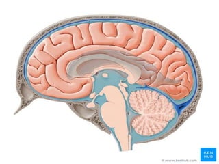
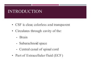
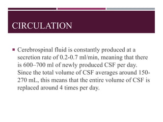
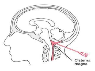
CEREBROSPINAL
FLUID
INTRODUCTION
• CSF is clear, colorless and transparent
• Circulates through cavity of the:
- Brain
- Subarachnoid space
- Central canal of spinal cord
• Part of Extracellular fluid (ECF)
ANATOMY
 CSF is predominantly, secreted by the choroid
plexuses. Brain interstitial fluid, ependyma and
capillaries may also play a poorly defined role in
CSF secretion.
 Most CSF is secreted by the specialized tissue called
the choroid plexus, which is located within the
lateral, third and fourth ventricles. The secretion of
CSF equals its removal, so there is around 150-270
milliliters of cerebrospinal fluid within the CNS at
all times.
 The main functions of CSF are to cushion the brain
and spinal cord when they’re struck with
mechanical force, to provide basic immunological
protection to the CNS, to remove metabolic waste,
as well as to transport neuromodulators and
neurotransmitters. CSF is also very useful for
clinical diagnosis, and its samples are usually
obtained from the subarachnoid space (SAS) by
lumbar puncture.
PRESSURE EXCERTED BY CSF
 Lateral recumbent position : 10 to 18 cm ofwater
 Lying position : 13 cm of water
 Sitting position : 30 cm of water
 Certain events like coughing and crying
increase the pressure by decreasing
adsorption.
CIRCULATION
 Cerebrospinal fluid is constantly produced at a
secretion rate of 0.2-0.7 ml/min, meaning that there
is 600–700 ml of newly produced CSF per day.
Since the total volume of CSF averages around 150-
270 mL, this means that the entire volume of CSF is
replaced around 4 times per day.
The CSF passes from the lateral ventricles to the third
ventricle through the interventricular foramen (of
Monro).
From the third ventricle, the CSF flows through the
cerebral aqueduct (of Sylvius) to the fourth ventricle.
From the fourth ventricle, some CSF flows through a
narrow passage called the obex and enters the central canal
of the spinal cord. However, the majority of CSF passes
through the apertures of the fourth ventricle; the median
aperture (of Magendie) and two lateral apertures (of
Luschka). Via these openings, the CSF enters the cisterna
magma and cerebellopontine cisterns, respectively.
From there, the CSF flows through the subarachnoid space of the brain and
spinal cord.
It is finally reabsorbed into the dural venous sinuses through arachnoid
granulations.
ABSORPTION
 CSF is mostly by the arachnoid villi into
dural sinuses and spinal veins.
 Small amount is absorbed along the
perineural spaces into cervical lymphatics
and into perivascular spaces.
 Normally , about 500 mL of CSF is
formed everyday and an equal amount
is absorbed.
FUNCTION
 Protection: the CSF protects the brain from
damage by "buffering" the brain. In other
words, the CSF acts to cushion a blow to
the head and lessen the impact.
 Buoyancy: because the brain is immersed
in fluid, the net weight of the brain is
reduced from about 1,400 gm to about 50
gm. Therefore, pressure at the base of the
brain is reduced.
 Excretion of waste products: the one-way flow
from the CSF to the blood takes potentially
harmful metabolites, drugs and other substances
away from the brain.
 Regulation of cranial content volume: Regulation
of this volume is essential, because brain may be
affected if volume is increases . It is prevented by
greater absorption of CSF to give space for the
cranial contents.
COLLECTION
CSF is collected by two methods :
BY LUMBAR PUNCTURE
BY CISTERNAL PUNCTURE
LUMBAR PUNCTURE
A lumbar puncture (spinal tap) is performed in your
lower back, in the lumbar region. During a lumbar
puncture, a needle is inserted between two lumbar
bones (vertebrae) to remove a sample of
cerebrospinal fluid.
lumbar puncture can help diagnose serious
infections, such as meningitis; other disorders of the
central nervous system, such as Guillain-Barre
syndrome and multiple sclerosis; or cancers of the
brain or spinal cord.
CISTERNAL PUNCTURE
A sub occipital puncture or cisternal puncture is a
diagnostic procedure that can be performed in order
to collect a sample of cerebrospinal fluid (CSF) for
biochemical, microbiological, and cytological
analysis, or rarely to relieve increased intracranial
pressure.
Cisternal puncture puncture of the cisterna cerebello-
medullaris with a hollow needle inserted just
between the occipital bone, to obtain a specimen of
cerebrospinal fluid.
BLOOD-CSF BARRIER
 It represents a barrier between the circulating blood and the CSF space.
 It control permeation of circulating solutes, including drugs needed in the
treatment of CNS diseases such as epilepsy or encephalitis.
 Transfer mechanisms of the BCB include active transport as well as passive
transport of hydrophilic compounds.
BLOOD BRAIN BARRIER AND CSF BARRIER
The BBB is sealed by tight junctions and does not show any permanent
fenestration. the BCB has several fenestrations (gap junctions) and
pinocytosis vesicles, which form a macrofilter for proteins
CSF DISORDER
 Hydrocephalus
 -Abnrmal accumulation of csf in the skul with enlarge
head
 2 Types of hydrocephalus
 Communicating
 Non -communicating
CONTINUED
 Non communicating
 Due to any obstruction of foramen from which csf escape result in
dilation of ventricular cavity
 Communicating
 Due to blockage of arachnoid villi
MULTIPLE SCLEROSIS
 MS is a chronic condition in which your immune system destroys the
protective covering of your nerves, which is called myelin.
 People with MS may have a variety of symptoms that are constant or
come and go. They include numbness or pain in their arms and legs,
vision problems, and trouble walking.
 CSF analysis may be done. The fluid may also show signs that your
immune system isn’t functioning normally. This can include high levels of
IgG
SPINA BIFIDA
 Spina bifida is a birth defect that occurs when the
spine and spinal cord don't form properly.
 It's a type of neural tube defect.
 The neural tube is the structure in a developing
embryo that eventually becomes the baby's brain,
spinal cord and the tissues that enclose them.
DIAGNOSIS
 prenatal screening tests to check for spina bifida and
other birth defects.
 Spina bifida can be screened with maternal blood
tests, but typically the diagnosis is made with
ultrasound.
REFERANCE
 https://fpnotebook.com/mobile/neuro/csf/index.htm
 https://www.healthline.com/health/csf-analysis#in-ms
 https://www.mayoclinic.org/diseases-
conditions/hydrocephalus/symptoms-causes/syc-20373604
 https://www.ncbi.nlm.nih.gov/books/NBK27998/
 https://www.researchgate.net/figure/Comparison-between-the-structure-of-the-
Blood-Brain-Barrier-BBB-and-the-blood-CSF_fig1_321985691
Cerebro Spinal Fluid

Cerebro Spinal Fluid

  • 1.
  • 3.
    INTRODUCTION • CSF isclear, colorless and transparent • Circulates through cavity of the: - Brain - Subarachnoid space - Central canal of spinal cord • Part of Extracellular fluid (ECF)
  • 4.
    ANATOMY  CSF ispredominantly, secreted by the choroid plexuses. Brain interstitial fluid, ependyma and capillaries may also play a poorly defined role in CSF secretion.  Most CSF is secreted by the specialized tissue called the choroid plexus, which is located within the lateral, third and fourth ventricles. The secretion of CSF equals its removal, so there is around 150-270 milliliters of cerebrospinal fluid within the CNS at all times.
  • 5.
     The mainfunctions of CSF are to cushion the brain and spinal cord when they’re struck with mechanical force, to provide basic immunological protection to the CNS, to remove metabolic waste, as well as to transport neuromodulators and neurotransmitters. CSF is also very useful for clinical diagnosis, and its samples are usually obtained from the subarachnoid space (SAS) by lumbar puncture.
  • 7.
    PRESSURE EXCERTED BYCSF  Lateral recumbent position : 10 to 18 cm ofwater  Lying position : 13 cm of water  Sitting position : 30 cm of water  Certain events like coughing and crying increase the pressure by decreasing adsorption.
  • 9.
    CIRCULATION  Cerebrospinal fluidis constantly produced at a secretion rate of 0.2-0.7 ml/min, meaning that there is 600–700 ml of newly produced CSF per day. Since the total volume of CSF averages around 150- 270 mL, this means that the entire volume of CSF is replaced around 4 times per day.
  • 10.
    The CSF passesfrom the lateral ventricles to the third ventricle through the interventricular foramen (of Monro). From the third ventricle, the CSF flows through the cerebral aqueduct (of Sylvius) to the fourth ventricle. From the fourth ventricle, some CSF flows through a narrow passage called the obex and enters the central canal of the spinal cord. However, the majority of CSF passes through the apertures of the fourth ventricle; the median aperture (of Magendie) and two lateral apertures (of Luschka). Via these openings, the CSF enters the cisterna magma and cerebellopontine cisterns, respectively.
  • 11.
    From there, theCSF flows through the subarachnoid space of the brain and spinal cord. It is finally reabsorbed into the dural venous sinuses through arachnoid granulations.
  • 13.
    ABSORPTION  CSF ismostly by the arachnoid villi into dural sinuses and spinal veins.  Small amount is absorbed along the perineural spaces into cervical lymphatics and into perivascular spaces.  Normally , about 500 mL of CSF is formed everyday and an equal amount is absorbed.
  • 14.
    FUNCTION  Protection: theCSF protects the brain from damage by "buffering" the brain. In other words, the CSF acts to cushion a blow to the head and lessen the impact.  Buoyancy: because the brain is immersed in fluid, the net weight of the brain is reduced from about 1,400 gm to about 50 gm. Therefore, pressure at the base of the brain is reduced.
  • 15.
     Excretion ofwaste products: the one-way flow from the CSF to the blood takes potentially harmful metabolites, drugs and other substances away from the brain.  Regulation of cranial content volume: Regulation of this volume is essential, because brain may be affected if volume is increases . It is prevented by greater absorption of CSF to give space for the cranial contents.
  • 16.
    COLLECTION CSF is collectedby two methods : BY LUMBAR PUNCTURE BY CISTERNAL PUNCTURE
  • 17.
    LUMBAR PUNCTURE A lumbarpuncture (spinal tap) is performed in your lower back, in the lumbar region. During a lumbar puncture, a needle is inserted between two lumbar bones (vertebrae) to remove a sample of cerebrospinal fluid. lumbar puncture can help diagnose serious infections, such as meningitis; other disorders of the central nervous system, such as Guillain-Barre syndrome and multiple sclerosis; or cancers of the brain or spinal cord.
  • 19.
    CISTERNAL PUNCTURE A suboccipital puncture or cisternal puncture is a diagnostic procedure that can be performed in order to collect a sample of cerebrospinal fluid (CSF) for biochemical, microbiological, and cytological analysis, or rarely to relieve increased intracranial pressure. Cisternal puncture puncture of the cisterna cerebello- medullaris with a hollow needle inserted just between the occipital bone, to obtain a specimen of cerebrospinal fluid.
  • 21.
    BLOOD-CSF BARRIER  Itrepresents a barrier between the circulating blood and the CSF space.  It control permeation of circulating solutes, including drugs needed in the treatment of CNS diseases such as epilepsy or encephalitis.  Transfer mechanisms of the BCB include active transport as well as passive transport of hydrophilic compounds.
  • 23.
    BLOOD BRAIN BARRIERAND CSF BARRIER The BBB is sealed by tight junctions and does not show any permanent fenestration. the BCB has several fenestrations (gap junctions) and pinocytosis vesicles, which form a macrofilter for proteins
  • 24.
    CSF DISORDER  Hydrocephalus -Abnrmal accumulation of csf in the skul with enlarge head  2 Types of hydrocephalus  Communicating  Non -communicating
  • 26.
    CONTINUED  Non communicating Due to any obstruction of foramen from which csf escape result in dilation of ventricular cavity  Communicating  Due to blockage of arachnoid villi
  • 27.
    MULTIPLE SCLEROSIS  MSis a chronic condition in which your immune system destroys the protective covering of your nerves, which is called myelin.  People with MS may have a variety of symptoms that are constant or come and go. They include numbness or pain in their arms and legs, vision problems, and trouble walking.  CSF analysis may be done. The fluid may also show signs that your immune system isn’t functioning normally. This can include high levels of IgG
  • 29.
    SPINA BIFIDA  Spinabifida is a birth defect that occurs when the spine and spinal cord don't form properly.  It's a type of neural tube defect.  The neural tube is the structure in a developing embryo that eventually becomes the baby's brain, spinal cord and the tissues that enclose them.
  • 31.
    DIAGNOSIS  prenatal screeningtests to check for spina bifida and other birth defects.  Spina bifida can be screened with maternal blood tests, but typically the diagnosis is made with ultrasound.
  • 32.
    REFERANCE  https://fpnotebook.com/mobile/neuro/csf/index.htm  https://www.healthline.com/health/csf-analysis#in-ms https://www.mayoclinic.org/diseases- conditions/hydrocephalus/symptoms-causes/syc-20373604  https://www.ncbi.nlm.nih.gov/books/NBK27998/  https://www.researchgate.net/figure/Comparison-between-the-structure-of-the- Blood-Brain-Barrier-BBB-and-the-blood-CSF_fig1_321985691