This document summarizes key information about cerebrospinal fluid (CSF):
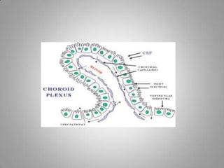
- CSF is clear fluid that circulates in the brain and spinal cord cavities. It is produced by the choroid plexus and absorbed into veins.
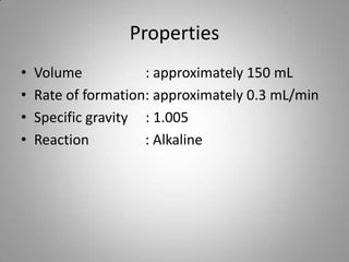
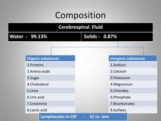
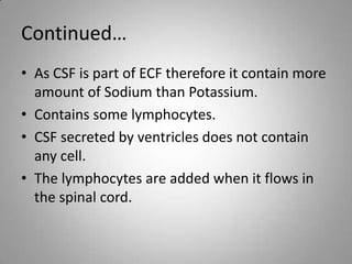
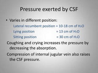
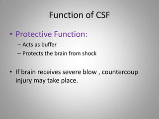
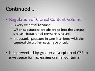
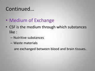
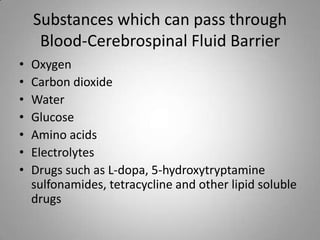
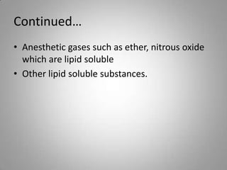
- CSF composition is 99% water with small amounts of proteins, sugars, and electrolytes. It acts as a buffer and regulates cranial pressure.
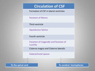
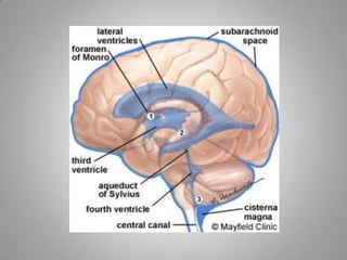
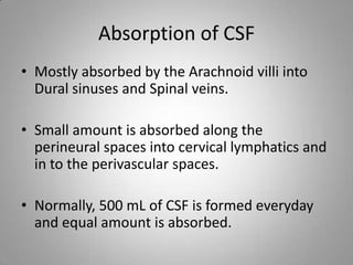
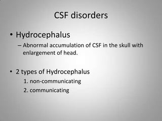
- CSF flows from the ventricles through the brain and spinal cord, and is ultimately absorbed into veins by the arachnoid villi or spinal veins. Hydrocephalus occurs when CSF circulation is blocked.