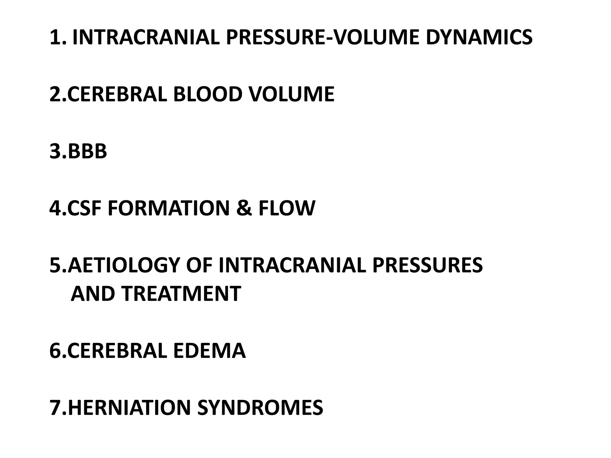
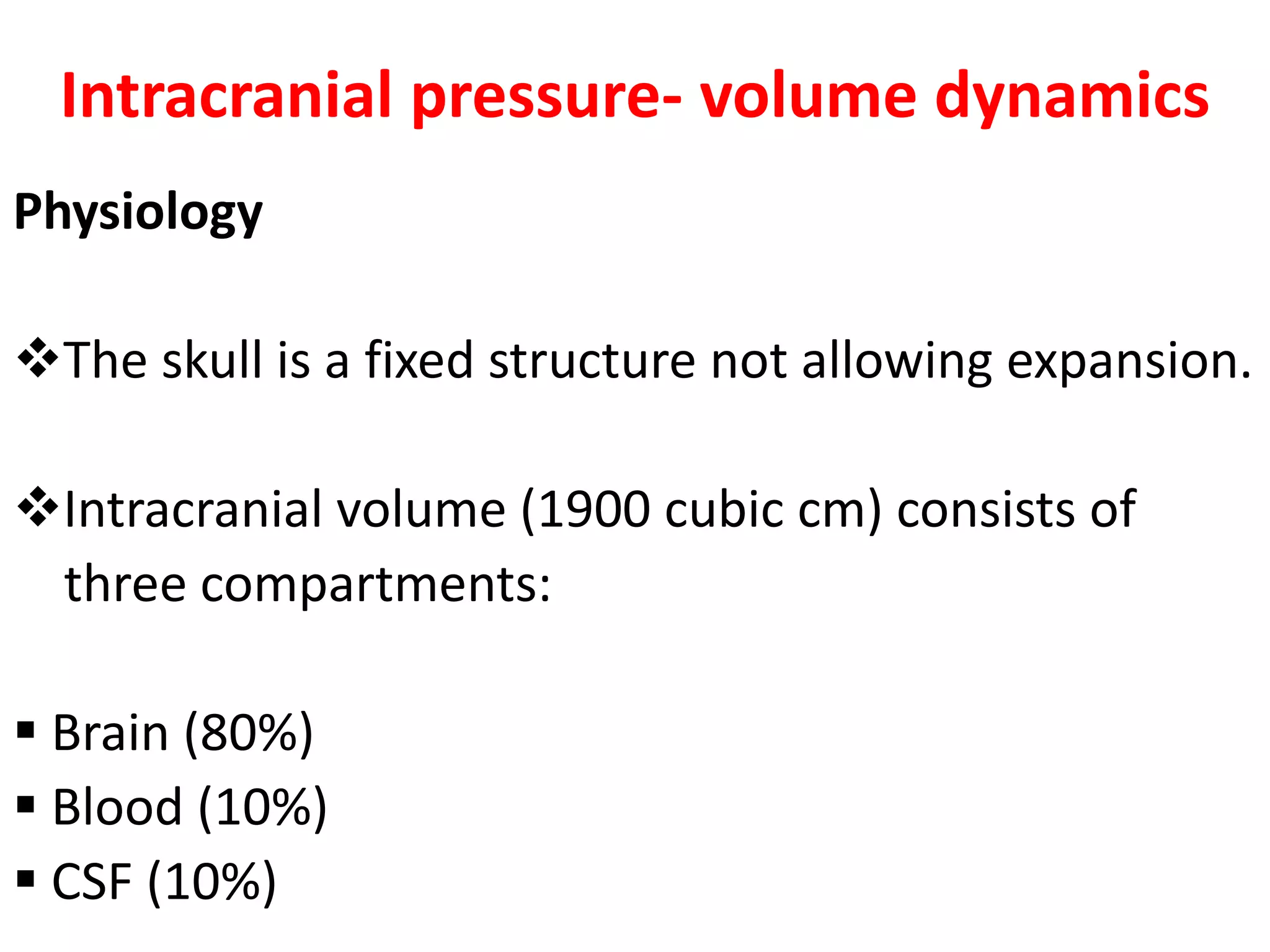
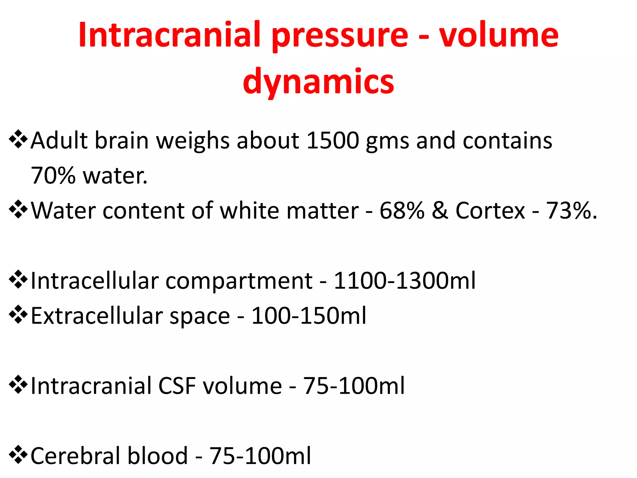
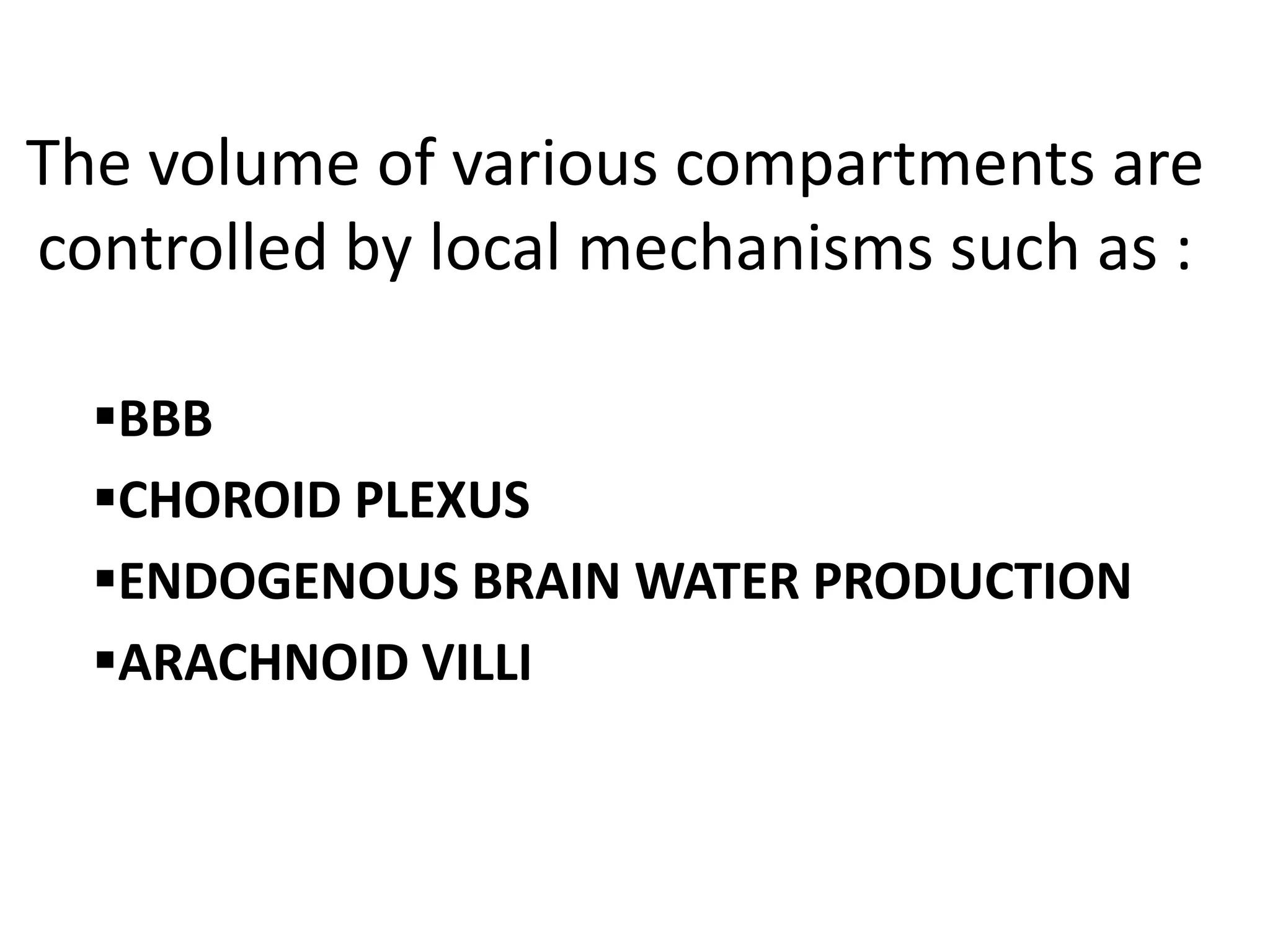
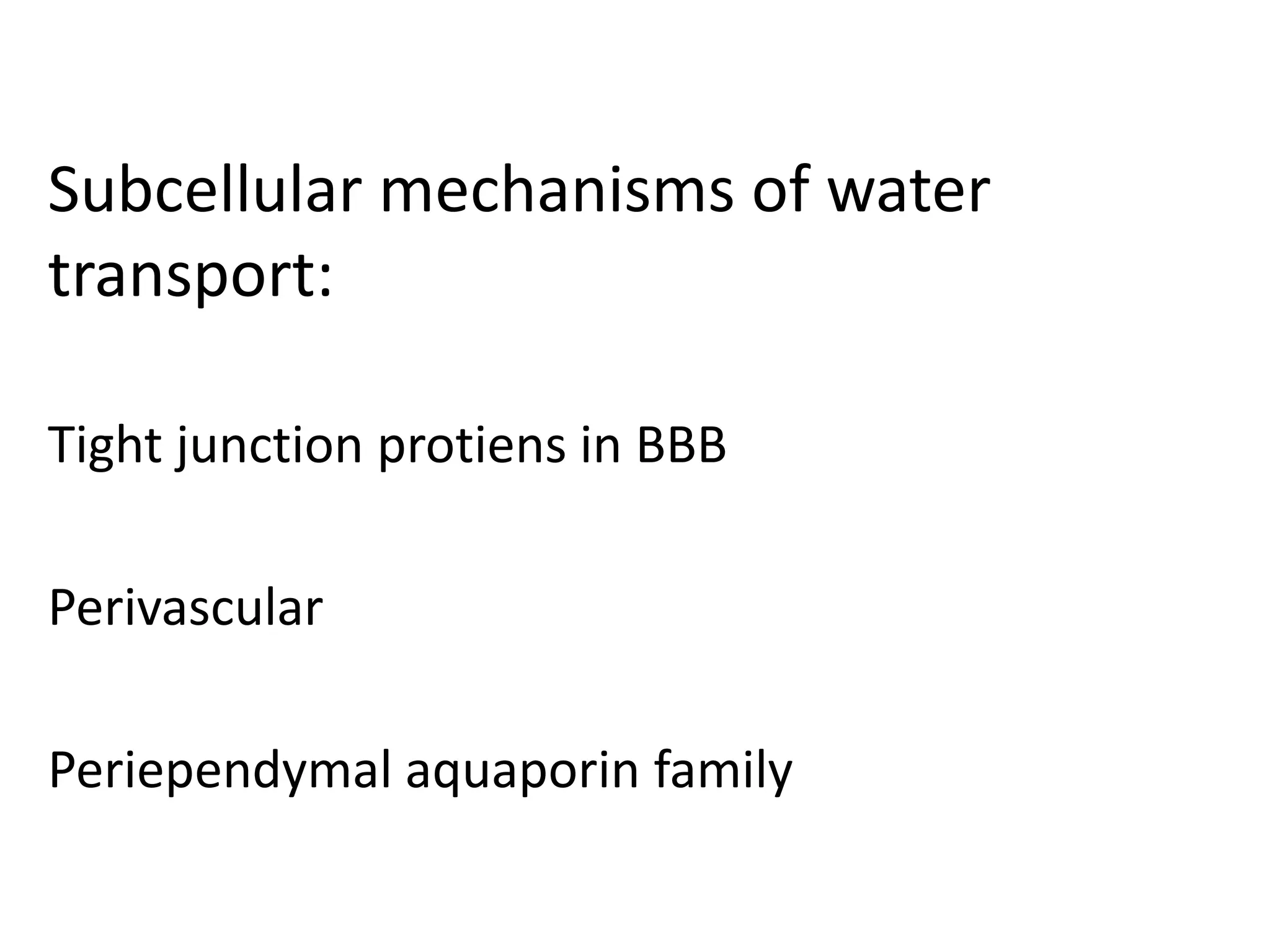
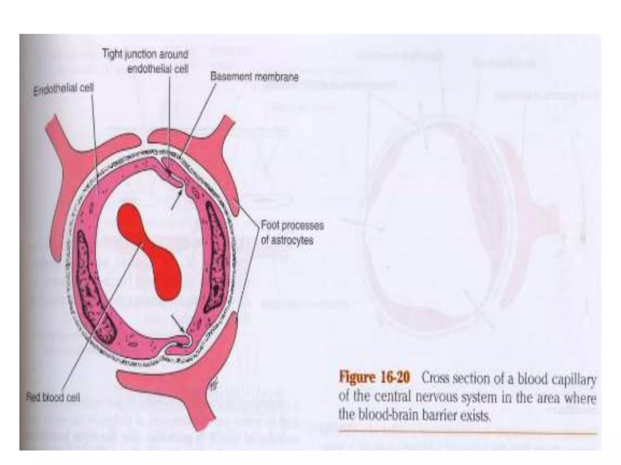
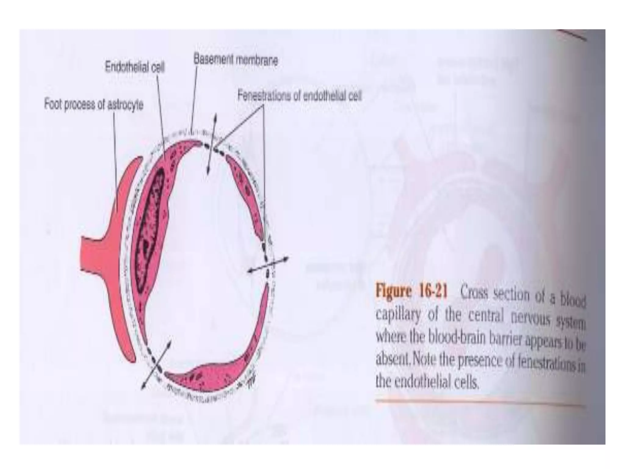
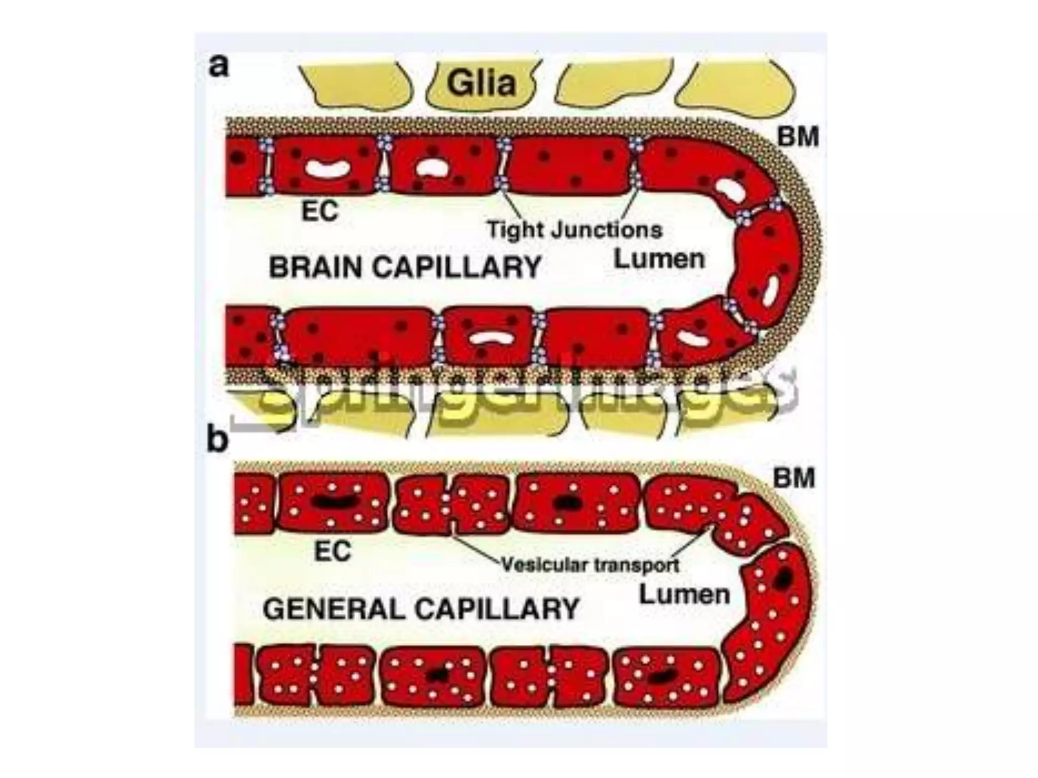
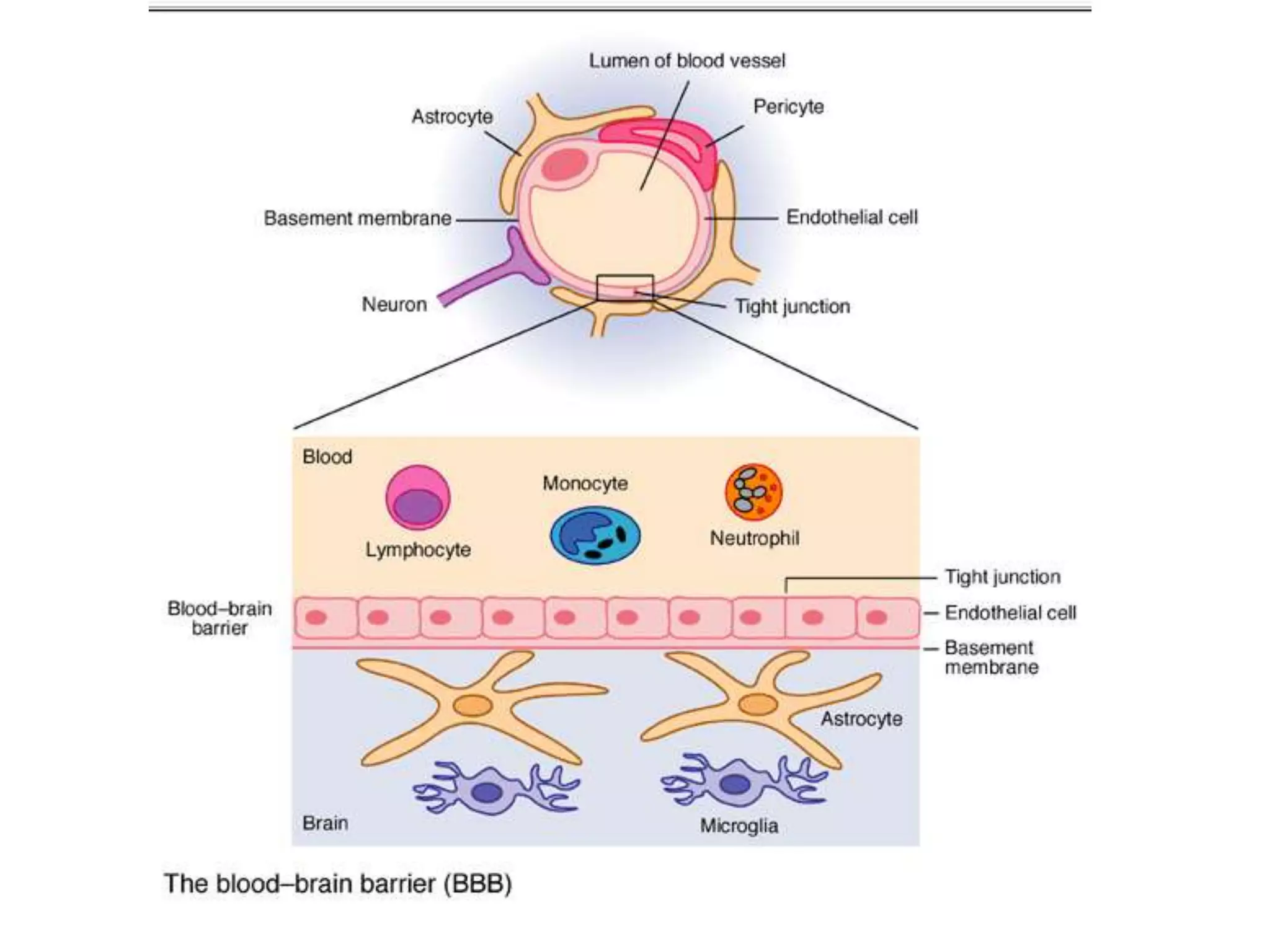
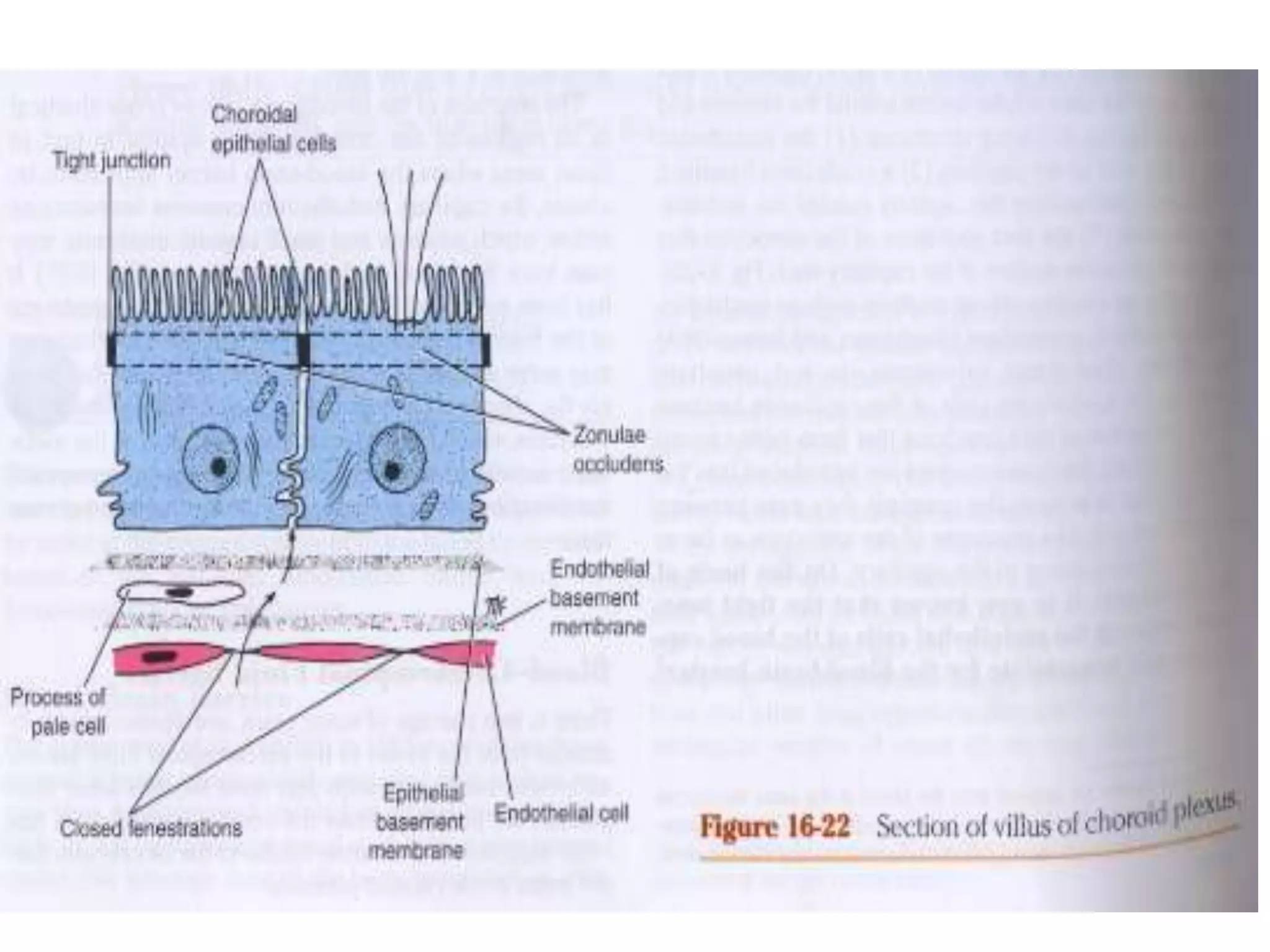
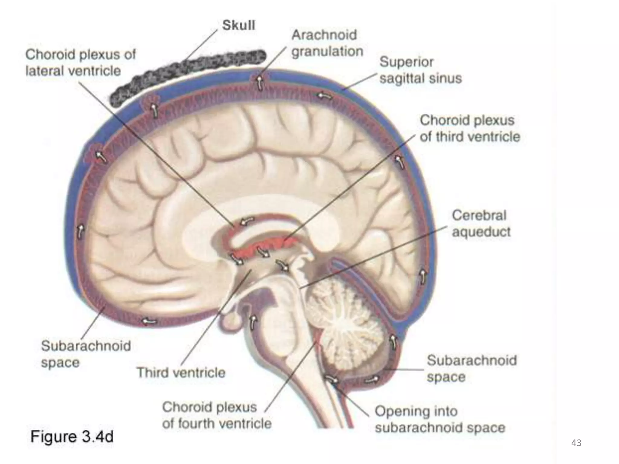
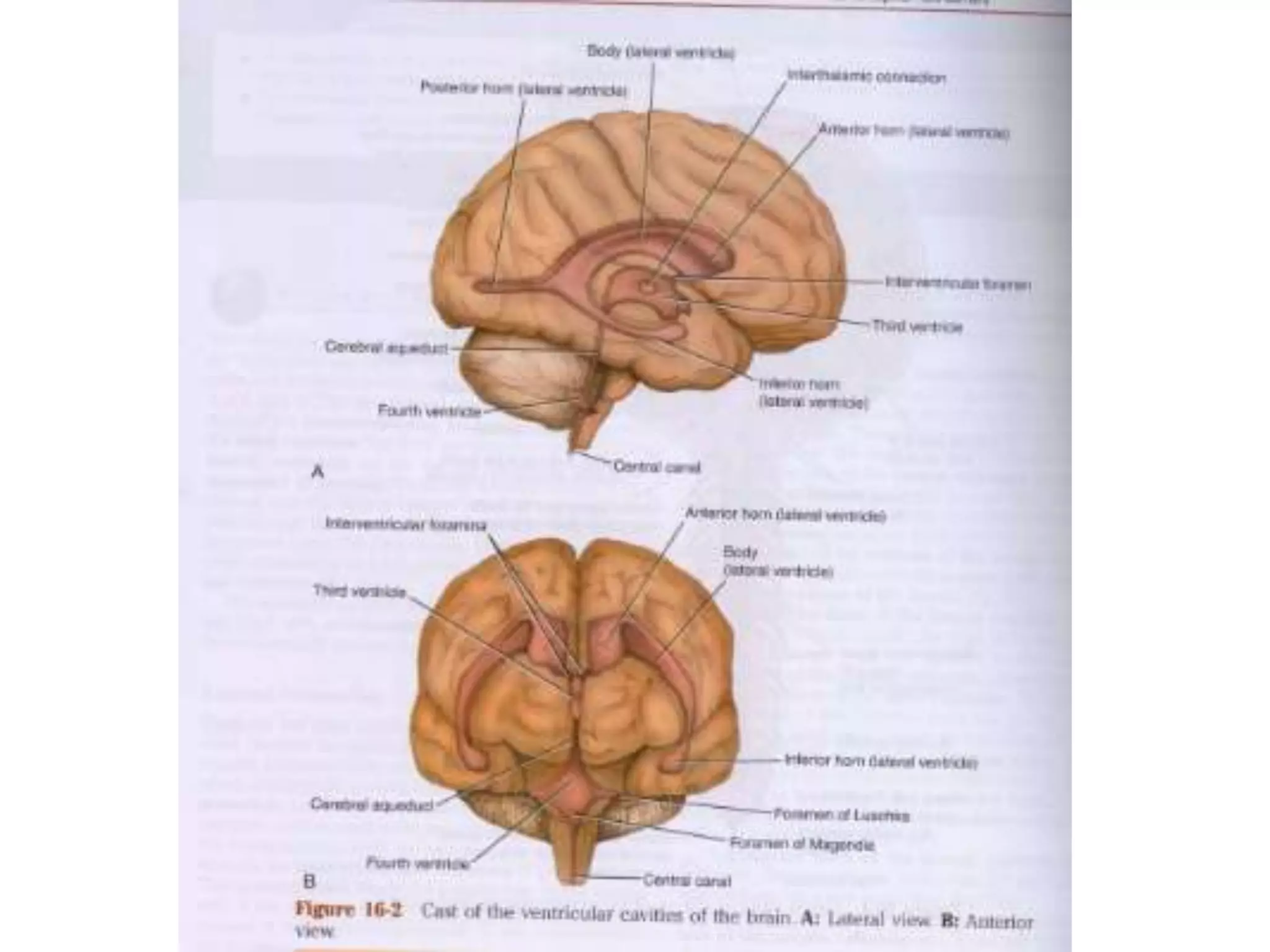
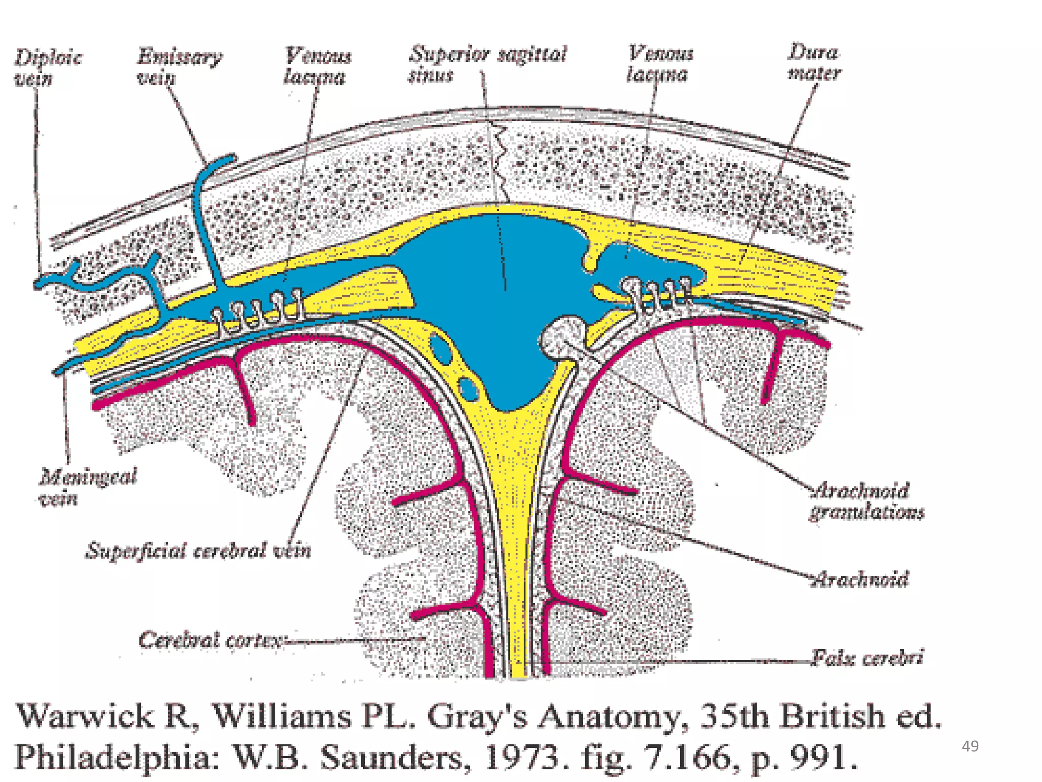
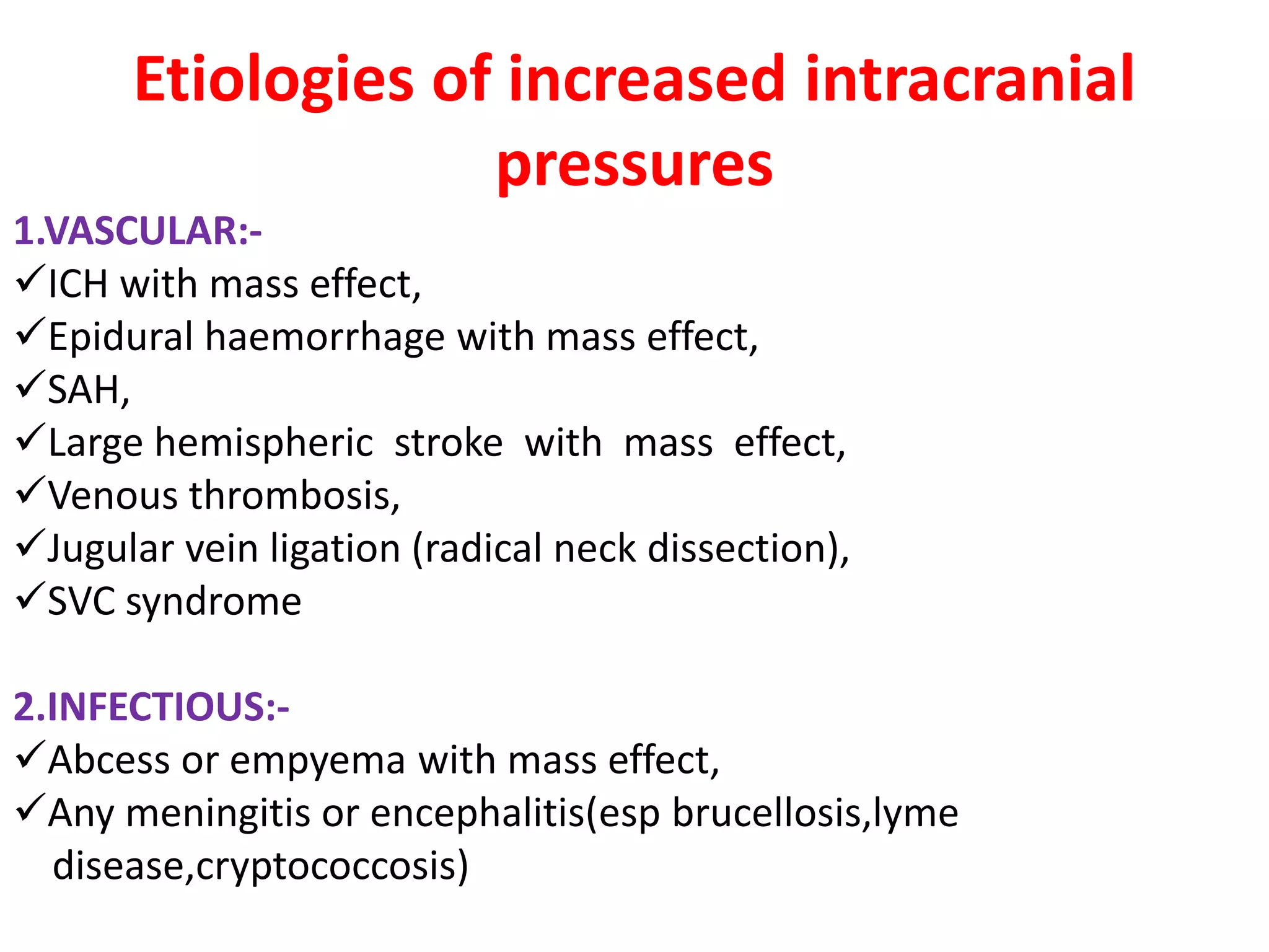
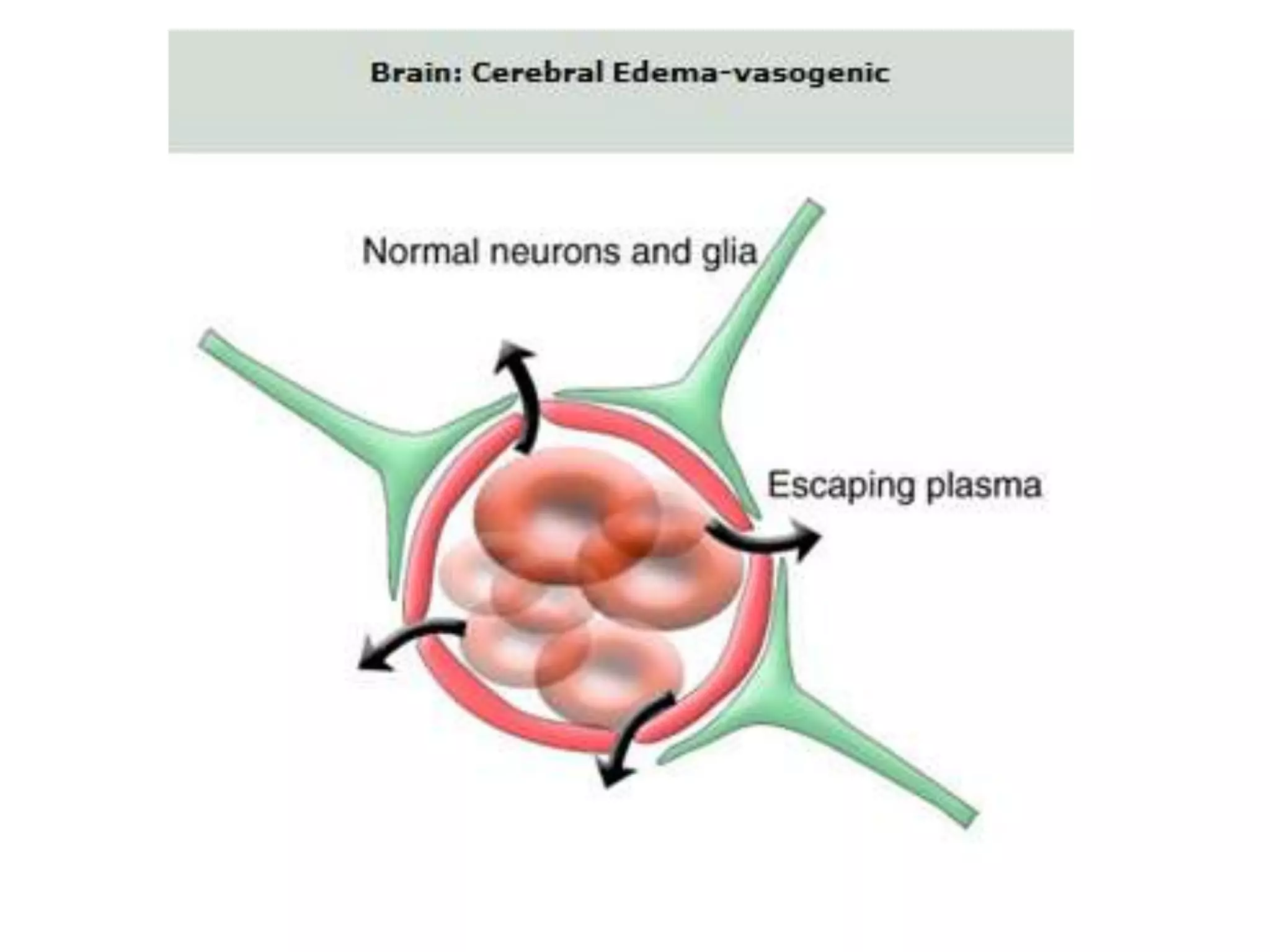
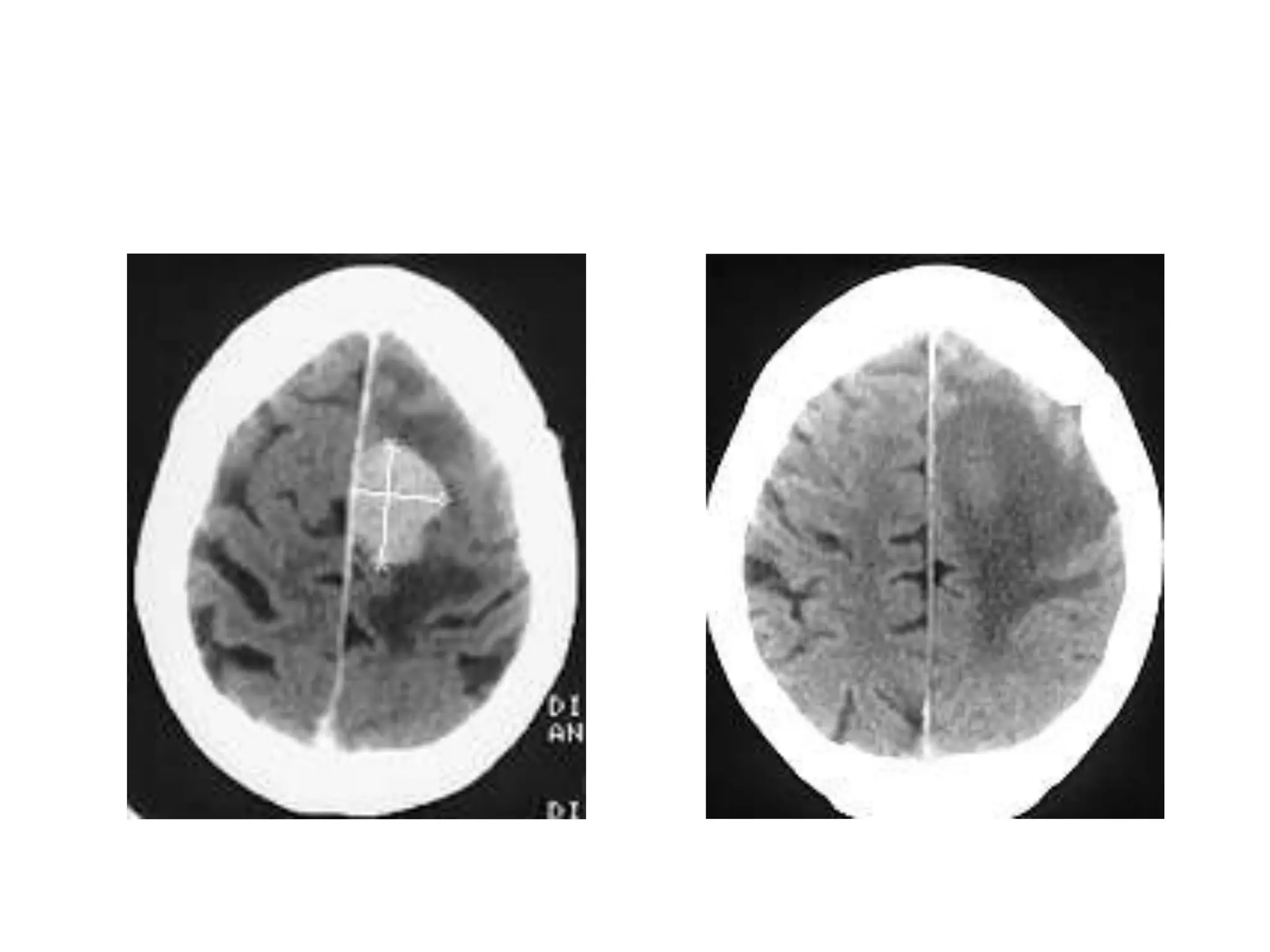
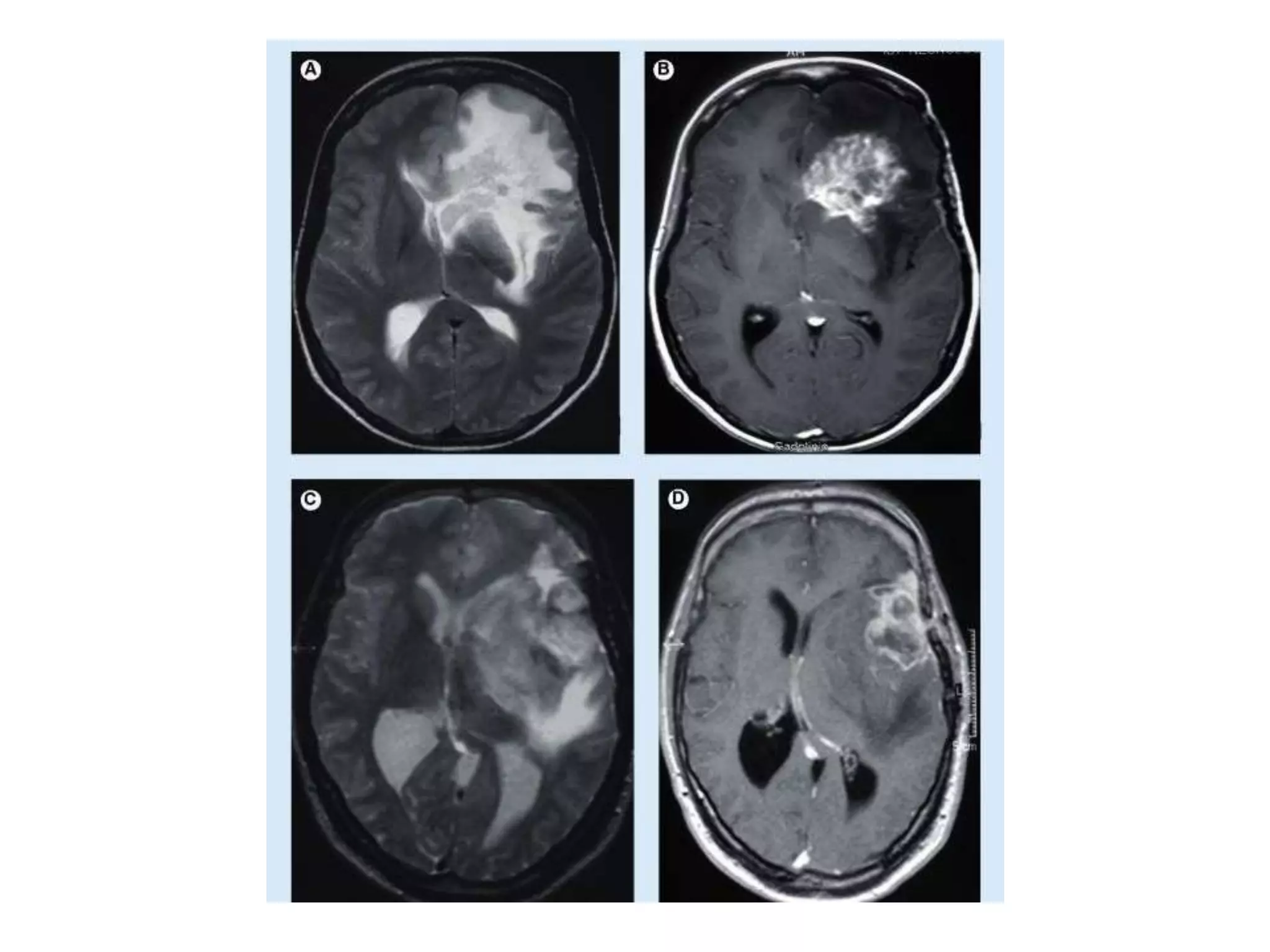
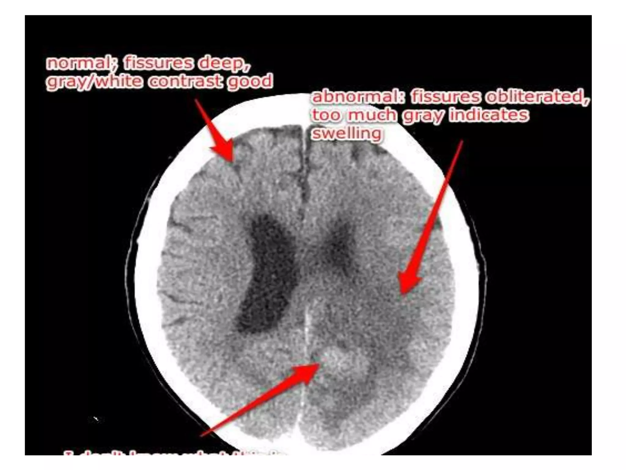
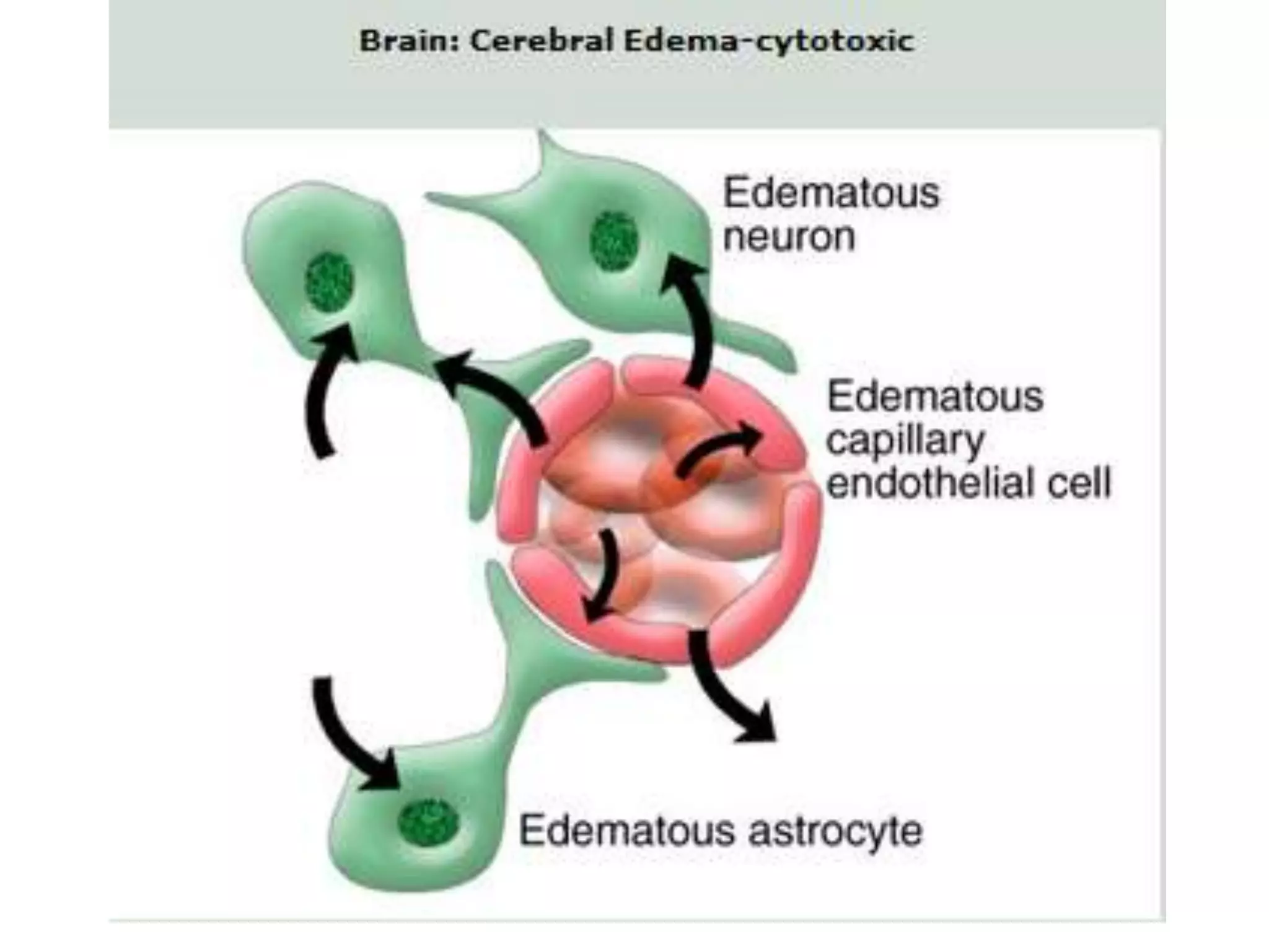
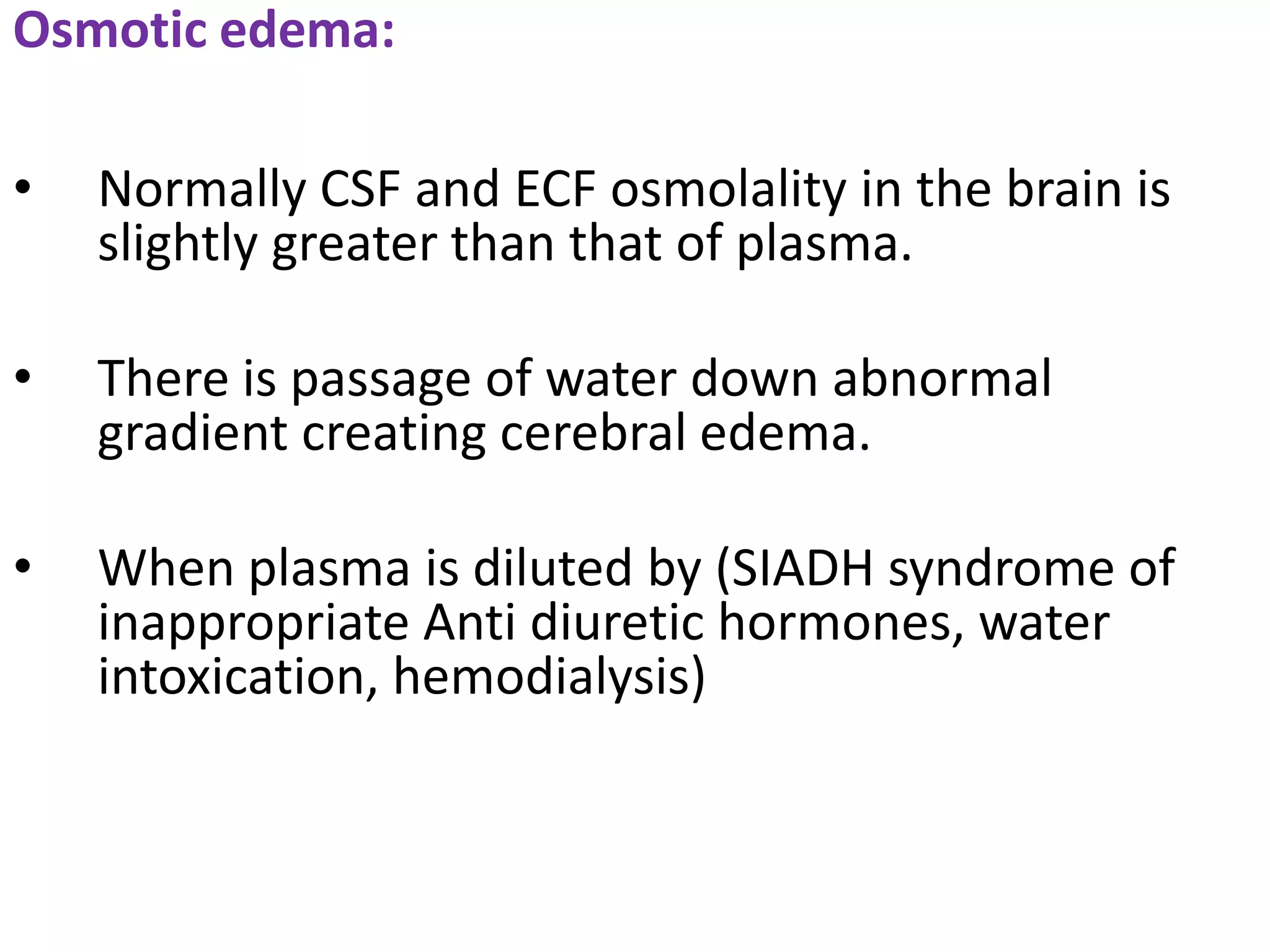
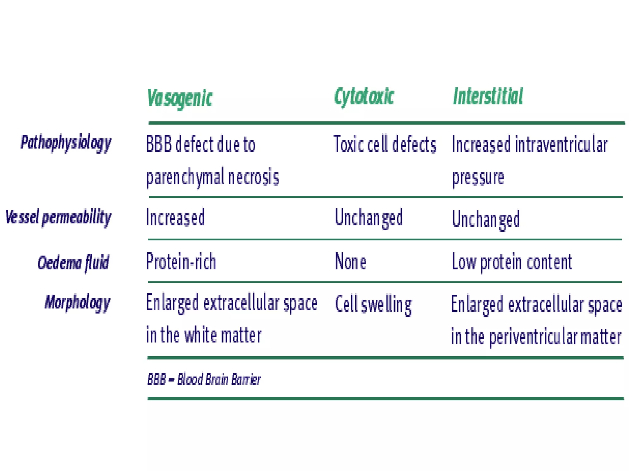
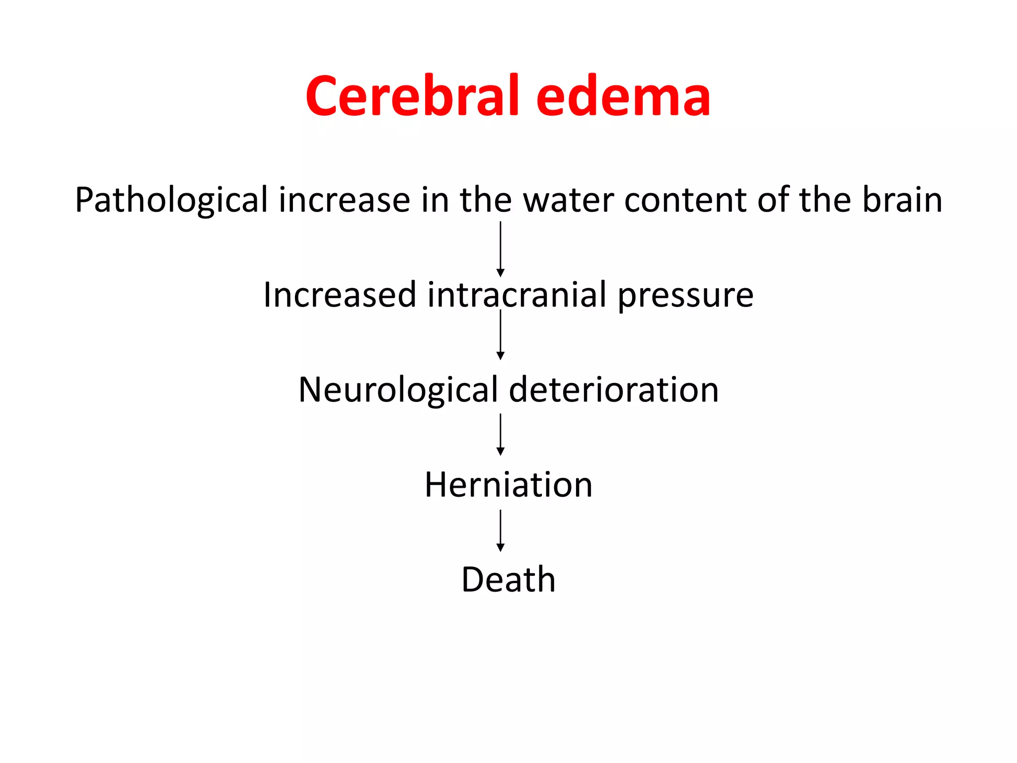
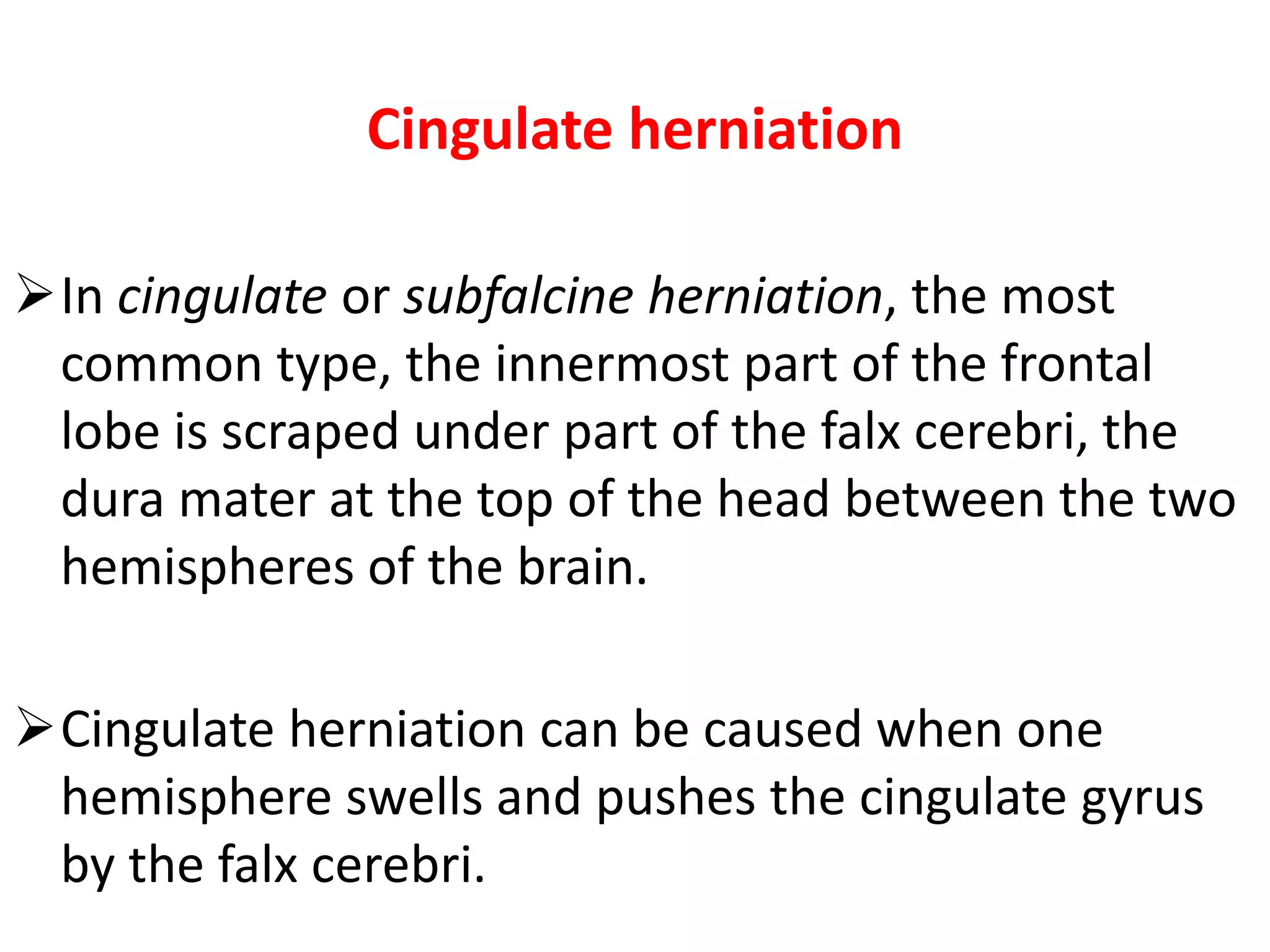
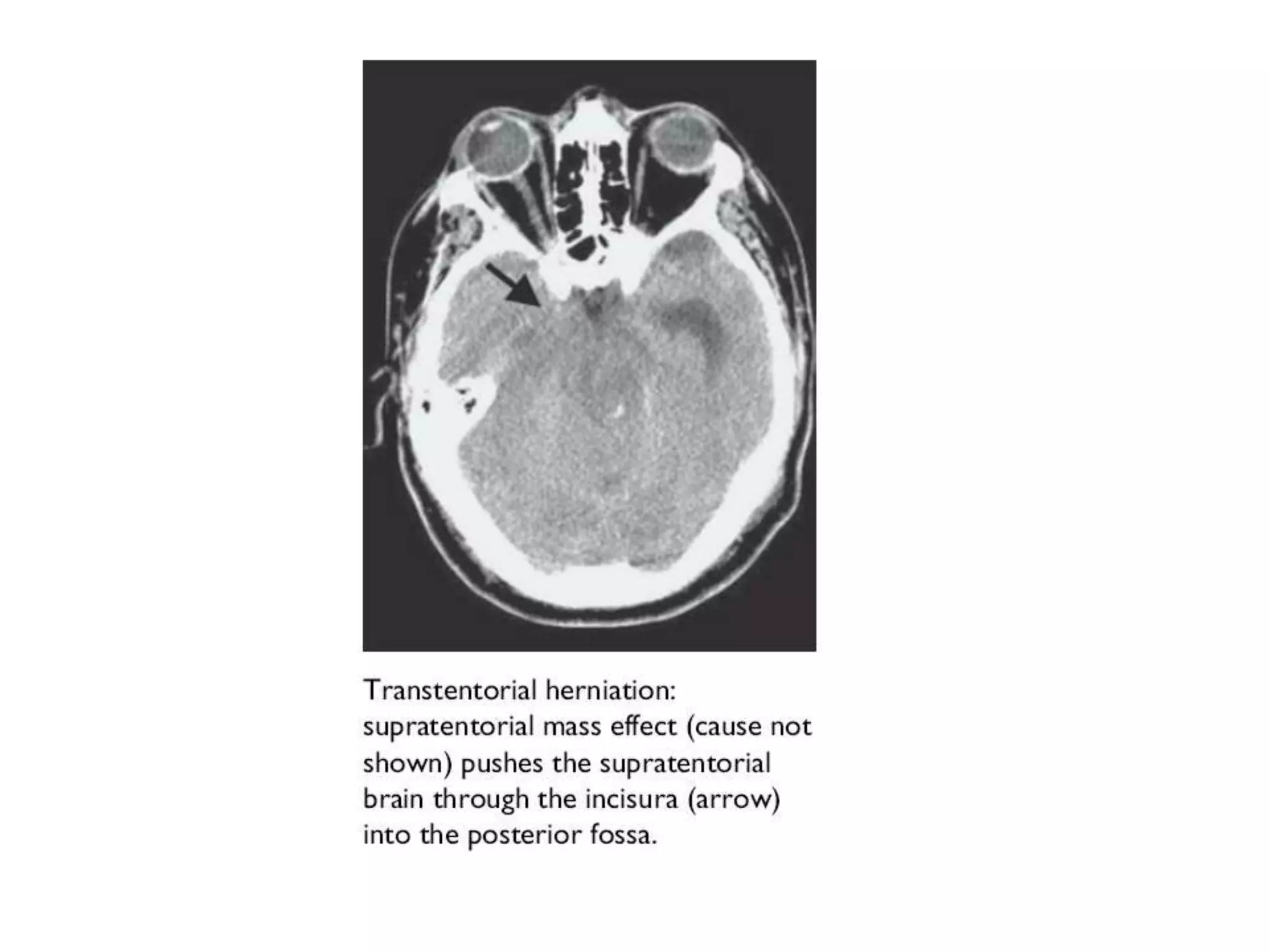
3. Intracranial pressure- volume dynamics
Physiology
The skull is a fixed structure not allowing expansion.
Intracranial volume (1900 cubic cm) consists of
three compartments:
Brain (80%)
Blood (10%)
CSF (10%)
4. Intracranial pressure - volume
dynamics
Adult brain weighs about 1500 gms and contains
70% water.
Water content of white matter - 68% & Cortex - 73%.
Intracellular compartment - 1100-1300ml
Extracellular space - 100-150ml
Intracranial CSF volume - 75-100ml
Cerebral blood - 75-100ml
5. The volume of various compartments are
controlled by local mechanisms such as :
BBB
CHOROID PLEXUS
ENDOGENOUS BRAIN WATER PRODUCTION
ARACHNOID VILLI
6. Systemic factors such as :
VASOPRESSIN
ATRIOPEPTIDES
RENIN-ANGIOTENSIN
ALDOSTERONE
7. Subcellular mechanisms of water
transport:
Tight junction protiens in BBB
Perivascular
Periependymal aquaporin family
8. These intracranial compartments are in dynamic
equilibrium due to pulsations of heart and respiratory
regulated return of venous blood from the brain.
MONROE-KELLY DOCTRINE
If the volume of one of these compartments
increases, the volume of another must decrease to
maintain normal ICP (0-20mmhg).
9. CEREBRAL BLOOD VOLUME
It is composed of arterial inflow,blood in the
capillaries and in the venous vessels.
Cerebral blood flow is generally independent of
mean arterial pressure but it is closely regulated by
arterial PaCO₂ systemically and locally by regional
factors such as release of endothelin and NO.
10. The rate of venous out flow is controlled by
intrathoracic pressure, patency of the major
venous cranial sinuses and hydrostatic pressure.
Under normal circumstances,cerebral blood
volume totals about 150ml with average blood
volumes in grey matter being 5.5ml blood per
100ml of brain and 1.4ml blood per 100ml of
brain for white matter.
11.
12. The ICP waveform can also be analysed in the time
domain - i.e. ICP waveform trend over time. This
may reveal typical Lundberg waves of ICP . A
paper chart record connected to an analogue
output from the ICP transducer often provides
better resolution than digital recording for
detection of Lundberg waves. Lundberg A waves
“or plateau waves” are steep increases in ICP
lasting for 5 to 10 minutes. They are always
pathological and represent reduced intracranial
hypertension indicative of early brain herniation.
Lundberg B waves are oscillations of ICP at a
frequency of 0.5 to 2 waves/min and are
associated with an unstable ICP. Lundberg B waves
are possibly the result of cerebral vasospasm,
because during the occurrence of these waves,
increased velocity in the middle cerebral artery
can be demonstrated on transcranial Doppler.
Lundberg C waves are oscillations with a
frequency of 4-8 waves/min. They have been
documented in healthy subjects and are probably
caused by interaction between the cardiac and
respiratory cycles.
13. Intracranial pressure
waveform recorded
.. Following surgery for
an intracranial
tumour,there are
baseline oscillations
from 0-10mm hg with
occasional c waves of
20-25mmhg.
There is also
characteristic a wave
or plateau wave that
lasts 5mts with a
pressure peak of 50-60
mmhg.Pressure waves
of this magnitude can
result in brain
herniations or
alterations in concious
state
15. Three pressure volume curves
showing the changes in the
intracranial pressure that can
occur from a baseline
pressure following a volume
insult such as intracranial
haematoma.
The curves represent
responses of 3 individuals
such as a healthy Young
person,middle aged person or
elderly person with brain
atrophy subject to the same
volume insult.For each given
unit of volume insult the
pressure response is variable
because of differences in
brain compliance.
16. RELATIONSHIP BETWEEN CEREBRAL PERFUSION PRESSURE AND
INTRACRANIAL PRESSURE
FORMULA NORMAL
ICP 0 – 20 mm Hg
CPP = MAP - ICP 70 – 100 mm Hg
CBF = CPP / CVR 20 – 70 ml/100 g/min
MAP = DBP + 1/3 PP 70 – 110 mm Hg
17. Blood brain barrier
Limits the egress of water and impedes efflux of
most ions and other compounds from the vascular
compartment to brain extracellular space.
Its role is to maintain a homeostatic environment
for neurons to function effectively and to exclude
potential toxic substances.
18. Blood-CSF barrier = Blood-brain barrier
A physiological mechanism that alters the
permeability of brain capillaries, so that some
substances, such as certain toxins and drugs, are
prevented from entering brain tissue, while other
substances are allowed to enter freely;
Physically it consists of the capillary endothelial
cells and their basement membranes and the
processes of astrocytes associated with the capillary
beds that serve the brain and spinal cord tissue.
19. Cellular elements of BBB
ENDOTHELIAL CELLS
ASTROCYTIC END FEET OR FOOT PROCESSES
PERICYTE
20. Within the structural components of bbb lie a
transporter and receptor systems that control
transmembrane and transluminal physiology.
Endothelial cells interconnected by
intermembrane protiens comprise tight junctions
21. Tight junctions consists of 3 integral
membrane protiens:
Claudin
Occludin
Junction adhesion molecules
And also a number of cytoplasmic accessory
protiens such as zona occludens-1,2,3 and cingulin.
22. These cytoplasmic protiens link membrane
protiens to the cytoskeleton protien actin in
order to maintain the structural and
functional integrity of the endothelium
23. A pericyte, also known as Rouget
cell,adventitial cell or mural cell, is
a connective tissue cell that occurs
about small blood vessels
24. As a relatively undifferentiated cell
(oligopotent), it serves to support these
vessels, but it can differentiate into
a fibroblast or a smooth muscle cell. In order
to migrate into the interstitium, the pericyte
has to break the barrier, formed by
the basement membrane, which can be
accomplished by fusion with the membrane.
Pericytes are important in blood-brain
barrier stability as well as angiogenesis.
25. Their expression of smooth muscle actin (SMA)
and vimentin (or desmin in some cases where
they are more likely to become smooth muscle
cells), and their adherence to the endovascular
cells makes them very strong candidates for
blood flow regulators in the
microvasculature, and indeed they have been
implicated in blood flow regulation at
the capillary level. After ischemia, an irreversible
constriction of pericytes may prevent brain
blood flow being restored
26. Pericytes appear to play a key role in
angiogenesis, structural integrity and
differentiation of the vessel,and formation of
endothelial tight junctions.
Astrocytic end feet release trophic factors are
critical for induction and maintenance of BBB.
27. The predominant aquaporin protien in
CNS, aquaporin-4 is a major path way for osmotically
driven water transport and is found within the
pericapillary astrocytic foot processes,the external
and sub-ependymal glial limiting membrane,and
within ependymal cells.
Aquaporin-4 facilitates movement of water into
and out of the brain since disruption of the protien
can significantly alter patterns of brain oedema and
water clearance
28.
29. Pathophysiological changes in the BBB, blood
osmolality, dysregulated blood flow, or increased
capillary pressure will influence the permeability
of BBB as well as passive diffusion of water, ions,
protiens, and other compounds in the brain.
36. choroid plexus
One of the delicate finger like
processes, consisting almost entirely of blood
vessels, which project into each of the four
ventricles of the brain which are lined by
specialized ependymal cells which secrete
cerebrospinal fluid.
37. Choroid
Plexus
CSF FORMATION AND FLOW
Formation of CSF at a pressure of
11 mmhg and at a rate of
0.3ml/mt
37
38.
39.
40. Cerebrospinal Fluid
FORMATION
• Secreted by choroid plexuses into each ventricle
• Choroid plexus are areas where the lining wall of the
ventricle is very thin and has a profusion of
capillaries
• At a pressure of 11mmHg
• At a rate of .3ml/mt
40
42. DRAINAGE
• From the roof of the 4th ventricle CSF flows through foramina into the
subarachnoid space and completely surrounds the brain and spinal
cord
• When CSF pressure is higher than venous pressure CSF passes into
the blood and when the venous pressure is higher the arachnoid villi
collapse, preventing the passage of blood constituents into the CSF
• The CSF passes back into blood through tiny diverticula of arachnoid
mater called arachnoid villi (arachnoid granulations), which project
into the venous sinuses
• Some reabsorption of CSF by cells in the walls of the ventricles occurs
53. Force of circulation
• Movement of the CSF is by pulsating blood vessels,
respiration and changes of posture
• CSF is secreted continuously at a rate of about
0.5ml per minute i.e. 720 ml per day
• Total CSF in the brain 120 ml
• CSF pressure can be measured by attaching a
vertical tube to the lumbar puncture needle – 10
cm water 53
55. Etiologies of increased intracranial
pressures
1.VASCULAR:-
ICH with mass effect,
Epidural haemorrhage with mass effect,
SAH,
Large hemispheric stroke with mass effect,
Venous thrombosis,
Jugular vein ligation (radical neck dissection),
SVC syndrome
2.INFECTIOUS:-
Abcess or empyema with mass effect,
Any meningitis or encephalitis(esp brucellosis,lyme
disease,cryptococcosis)
59. Raised cerebral venous pressure have an
affect on intracranial pressure
haemodynamics and this has been
implicated in pathogenesis of Idiopathic
intracranial hypertension.
60. INDICATIONS FOR ICP MONITORING
1. GCS <8
2. Severe head trauma
ICP WAVE FORMS
A. PLATEAU A WAVES
a. Sudden surges in ICP to 50 to 80 mm hg lasting 5 to 20 mts
b. Presence of A waves suggests failing compliance of the brain to
ICP and risk for ischaemia.
B. B WAVES:
smaller surges in ICP to 20 mm hg for 1 to 2 mts
61. MANAGEMENT OF ↑ ICP
MEASURE COMPARTMENT RECOMMENDED
General Several Head of bed at 30
Normothermic
Pain control
CSF drainage ↓ CSF External ventricular drain
Lumbar drain
Hyperventilation ↓ blood Hyperventilation to PCO2 30 mm Hg
(vasoconstriction)
Osmotic diuresis ↓ brain volume Mannitol 0.25-1 g/kg bolus, then
consider repeat q 8hr; titrate to serum
osmolality < 310
Barbiturates ↓ metabolic activity of Phenobarbital
brain & thus blood flow
Hypothermai ↓ metabolic activity of Cooling blankets
brain & thus blood flow
Surgery ↓ brain If ICP not controlled by medical mx &
surgical lesion present
62. CEREBRAL EDEMA:
Definition: Excess accumulation of water in the
intra- and/or extracellular spaces of the brain.
Previously engorgement of cerebral
vasculature was considered to be a major
factor in brain swelling associated with
neurotruma, however it is now considered
that cytotoxic brain edema is the predominant
factor causing brain swelling after neuro
trauma.
63. Classification:
Vasogenic edema:
• The disruption of the cerebral capillary provides the
underlying mechanism for vasogenic edema.
The amount of edema is greatest in the white matter (increased water and sodium in the
extracellular spaces, decreased potassium); but the same changes may take place in grey
matter but less so.
The astrocytes become swollen.
• This type of edema is seen in response to trauma, tumors,
focal inflammation, and late stages of cerebral ischemia.
69. Interstitial oedema:-
• CSF pushed into extracellular space in
preiventricular white matter in hydrocehalous
70.
71. Cytotoxic edema:
• This is due to the derangement in cellular metabolism
resulting in inadequate functioning of the sodium and
potassium pump in the glial cell membrane.
As a result there is cellular retention of sodium and water.
There are swollen astrocytes in grey and white matter.
• Cytoxotic edema is seen with various intoxications
(dinitrophenol, triethyltin, hexachlorophene, isoniazid) and
in Reye's syndrome, severe hypothermia, and early
ischemia.
72.
73.
74.
75. Osmotic edema:
• Normally CSF and ECF osmolality in the brain is
slightly greater than that of plasma.
• There is passage of water down abnormal
gradient creating cerebral edema.
• When plasma is diluted by (SIADH syndrome of
inappropriate Anti diuretic hormones, water
intoxication, hemodialysis)
76. Hydrostatic edema:
• This form of cerebral edema is seen in
acute, malignant hypertension.
• It is thought to result from direct transmission
of pressure to cerebral capillary with
transudation of fluid into the ECF.
77. High Altitude Cerebral Edema
High altitude cerebral edema (or HACE) is a
severe form of (sometimes fatal) altitude
sickness. HACE is the result of swelling of brain
tissue from leakage of fluids from the capillaries
due to the effects of hypoxia on
the mitochondria-rich endothelial cells of the
blood-brain barrier.Symptoms can include
headache, loss of coordination
(ataxia), weakness, and decreasing levels of
consciousness including disorientation, loss of
memory, hallucinations, psychotic behavior, and
coma. It generally occurs after a week or more
at high altitude.
78. Severe instances can lead to death if not
treated quickly. Immediate descent is a
necessary life-saving measure (2,000 - 4,000
feet). There are some medications
(e.g. dexamethasone) that may be prescribed
for treatment in the field, but these require
proper medical training in their use. Anyone
suffering from HACE must be evacuated to a
medical facility for proper follow-up
treatment. A gamow bag can sometimes be
used to stabilize the sufferer before transport
or descending.
88. Cingulate herniation
In cingulate or subfalcine herniation, the most
common type, the innermost part of the frontal
lobe is scraped under part of the falx cerebri, the
dura mater at the top of the head between the two
hemispheres of the brain.
Cingulate herniation can be caused when one
hemisphere swells and pushes the cingulate gyrus
by the falx cerebri.
89. This does not put as much pressure on the
brainstem as the other types of herniation, but it may
interfere with blood vessels in the frontal lobes that
are close to the site of injury (anterior cerebral
artery), or it may progress to central herniation.
Interference with the blood supply can cause
dangerous increases in ICP that can lead to more
dangerous forms of herniation.
90.
91. Symptoms for cingulate herniation are not well
defined.Usually occurring in addition to uncal
herniation, cingulate herniation may present with
abnormal posturing and coma.
Cingulate herniation is frequently believed to be a
precursor to other types of herniation.
92. Central herniation
In central herniation, the diencephalon and parts of
the temporal lobes of both of the cerebral
hemispheres are squeezed through a notch in the
tentorium cerebelli.
Transtentorial herniation can occur when the brain
moves either up or down across the tentorium, called
ascending and descending transtentorial herniation
respectively; however descending herniation is much
more common.
93.
94. Downward herniation can stretch branches of the
basilar artery (pontine arteries), causing them to tear and
bleed, known as a Duret hemorrhage. The result is usually
fatal.
Radiographically, downward herniation is characterized
by obliteration of the suprasellar cistern from temporal
lobe herniation into the tentorial hiatus with associated
compression on the cerebral peduncles.
Upwards herniation, on the other hand, can be
radiographically characterized by obliteration of the
quadrigeminal cistern. Intracranial hypotension syndrome
has been known to mimic downwards transtentorial
herniation.
95. Uncal herniation
In uncal herniation, a common subtype of
transtentorial herniation, the innermost part of the
temporal lobe, the uncus, can be squeezed so much
that it goes by the tentorium and puts pressure on the
brainstem, most notably the midbrain.
The tentorium is a structure within the skull formed
by the meningeal layer of the dura mater. Tissue may
be stripped from the cerebral cortex in a process
called decortication
96.
97. The uncus can squeeze the third cranial nerve, which may affect
the parasympathetic input to the eye on the side of the affected
nerve, causing the pupil of the affected eye to dilate and fail to
constrict in response to light as it should.
Pupillary dilation often precedes the somatic motor effects of
cranial nerve III compression, which present as deviation of the eye
to a "down and out" position due to loss of innervation to all ocular
motility muscles except for the lateral rectus (innervated by cranial
nerve VI) and the superior oblique (innervated by cranial nerve IV).
The symptoms occur in this order because the parasympathetic
fibers surround the motor fibers of CNIII and are hence compressed
first.
98. Compression of the ipsilateral posterior cerebral artery will
result in ischemia of the ipsilateral primary visual cortex and
contralateral visual field deficits in both eyes (contralateral
homonymous hemianopsia).
Another important finding is a false localizing sign, the so
called Kernohan's notch, which results from compression of the
contralateral cerebral crus containing descending corticospinal
and some corticobulbar tract fibers.
This leads to contralateral (opposite as herniation)
hemiparesis. Since the corticospinal tract predominately
innervates flexor muscles, extension of the leg may also be seen
99. With increasing pressure and progression of the hernia there will be
distortion of the brainstem leading to Duret hemorrhages (tearing of small
vessels in the parenchyma) in the median and paramedian zones of the
mesencephalon and pons.
The rupture of these vessels leads to linear or flamed shaped hemorrhages.
The disrupted brainstem can lead to decorticate posture, respiratory center
depression and death. Other possibilities resulting from brain stem distortion
include lethargy, slow heart rate, and pupil dilation.
Uncal herniation may advance to central herniation.
A complication of an uncal herniation is a Duret hemorrhage. This results in
the midbrain and pons compression, possibly causing damage to the reticular
formation. If untreated, death will ensue
100. Transcalvarial herniation
In transcalvarial herniation, the brain squeezes
through a fracture or a surgical site in the skull.
Also called "external herniation", this type of
herniation may occur during craniectomy, surgery in
which a flap of skull is removed, preventing the piece
of skull from being replaced.
103. Upward herniation
Increased pressure in the posterior fossa can cause
the cerebellum to move up through the tentorial
opening in upward, or cerebellar herniation.
The midbrain is pushed through the tentorial notch.
106. Tonsillar herniation
In tonsillar herniation, also called downward
cerebellar herniation, or "coning", the cerebellar
tonsils move downward through the foramen
magnum possibly causing compression of the lower
brainstem and upper cervical spinal cord as they pass
through the foramen magnum.
Increased pressure on the brainstem can result in
dysfunction of the centers in the brain responsible for
controlling respiratory and cardiac function.
107.
108.
109. Tonsillar herniation of the cerebellum is also known as a Chiari
Malformation (CM), or previously as Arnold Chiari Malformation
(ACM).
There are at least three types of Chiari malformation that are
widely recognized, and they represent very different disease
processes with different symptoms and prognosis.
These conditions can be found in asymptomatic patients as an
incidental finding, or can be so severe as to be life-threatening.
This condition is now being diagnosed more frequently by
radiologists, as more and more patients undergo MRI scans of
their heads
110. Cerebellar ectopia is a term used by radiologists to describe
cerebellar tonsils that are "low lying" but that do not meet the
radiographic criteria for definition as a Chiari malformation.
The currently accepted radiographic definition for a Chiari
malformation is that cerebellar tonsils lie at least 5mm below
the level of the foramen magnum.
Some clinicians have reported that some patients appear to
experience symptoms consistent with a Chiari malformation
without radiographic evidence of tonsillar herniation.
Sometimes these patients are described as having a 'Chiari
[type] 0'.
111. There are many suspected causes of tonsillar
herniation including:
Decreased or malformed posterior fossa (the lower,
back part of the skull) not providing enough room for
the cerebellum,
Hydrocephalus or abnormal CSF volume pushing
the tonsils out,
Connective tissue disorders, such as Ehlers Danlos
Syndrome.