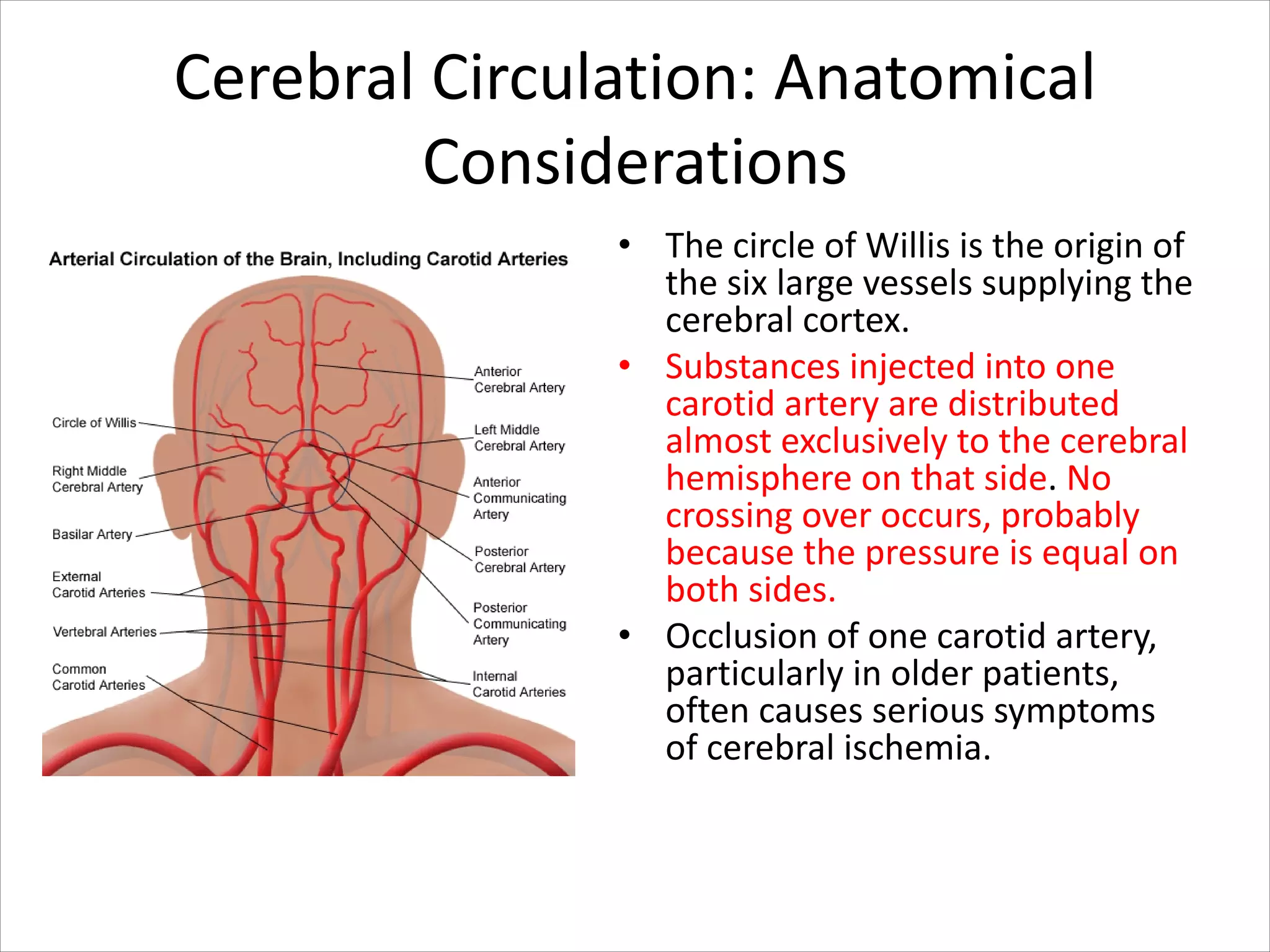
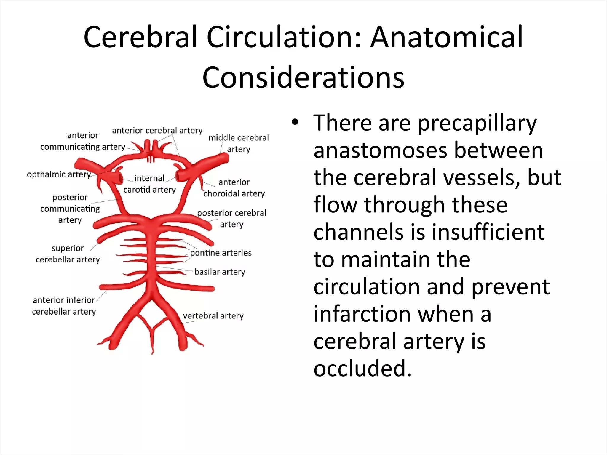
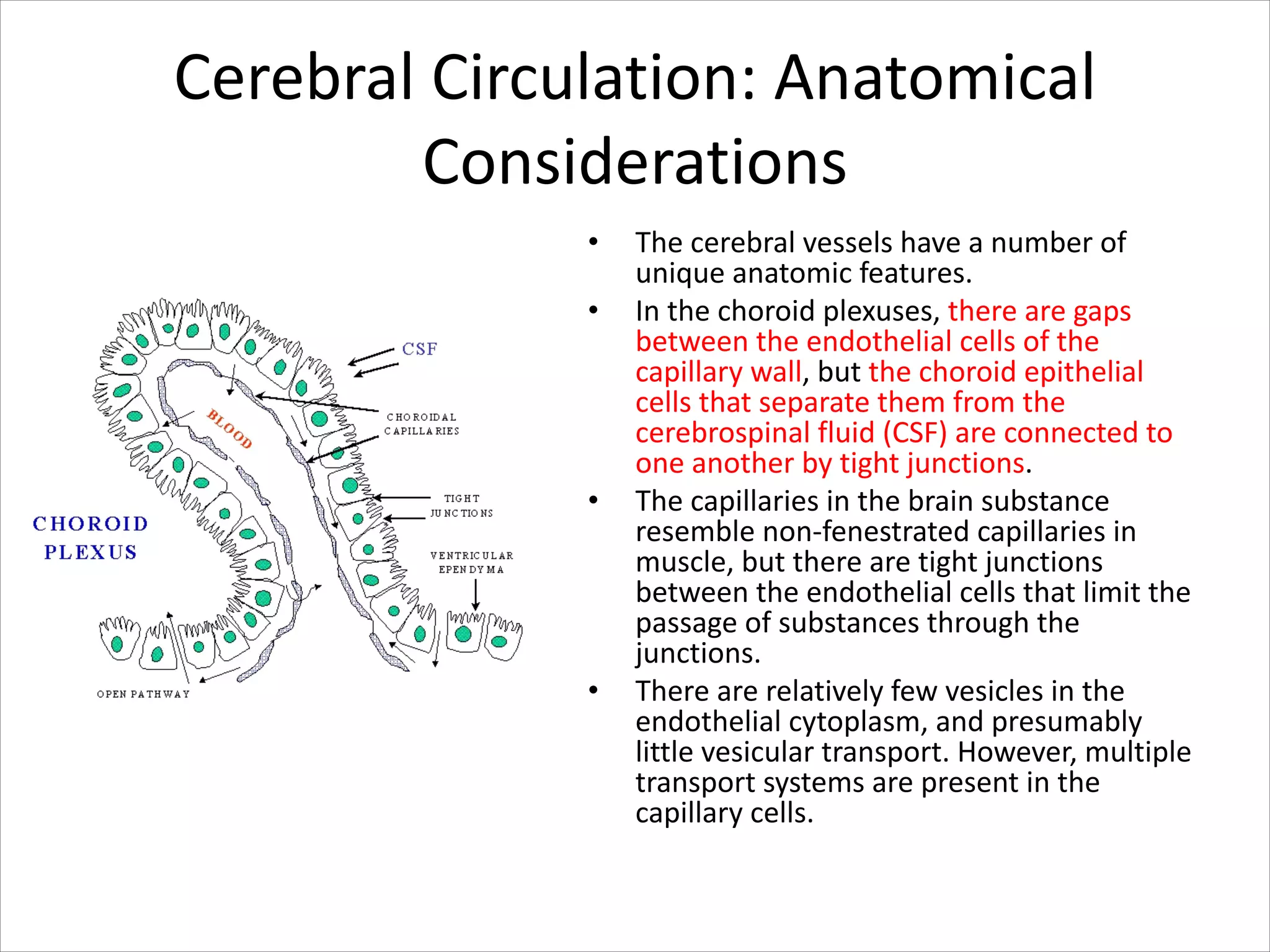
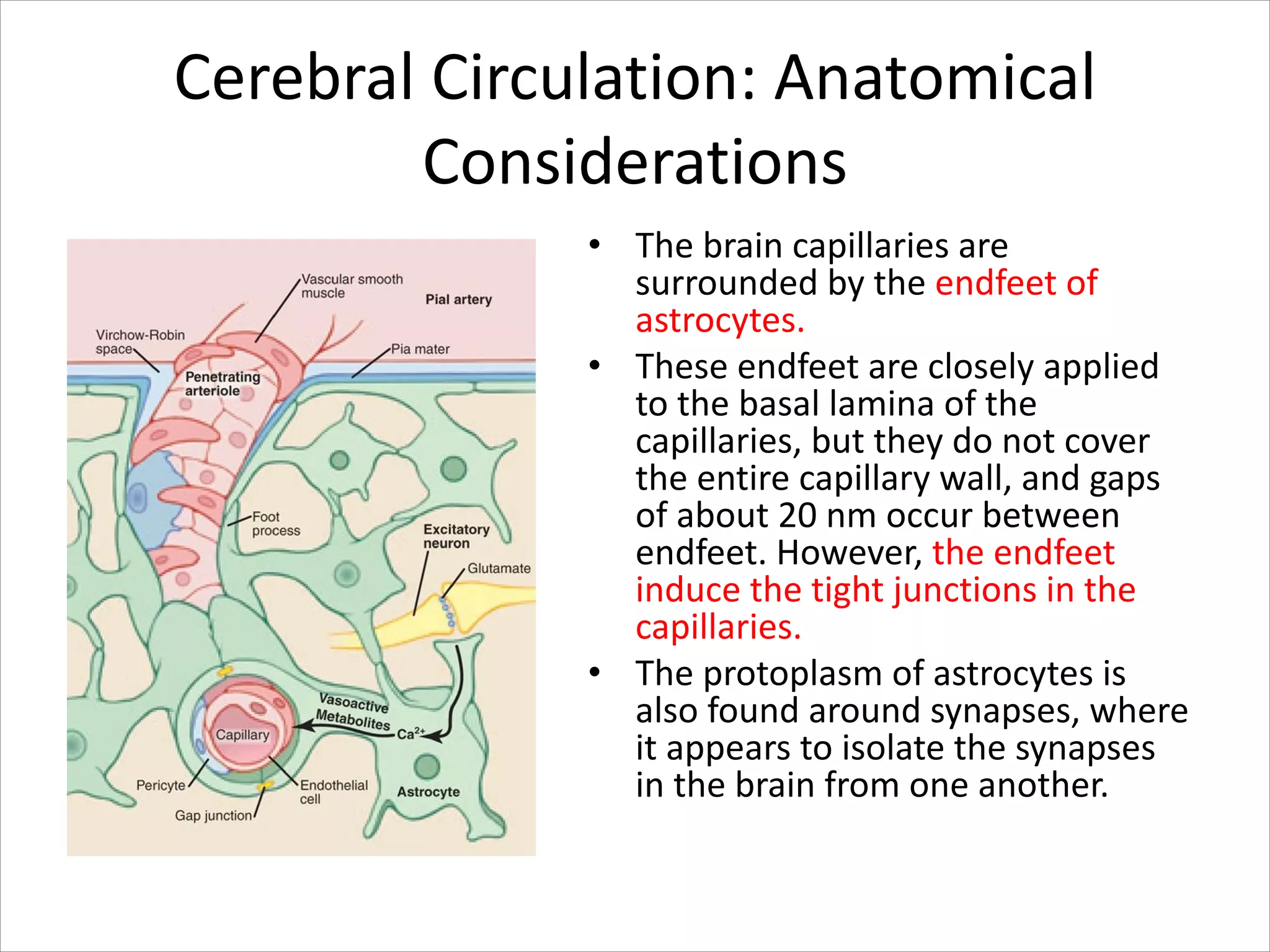
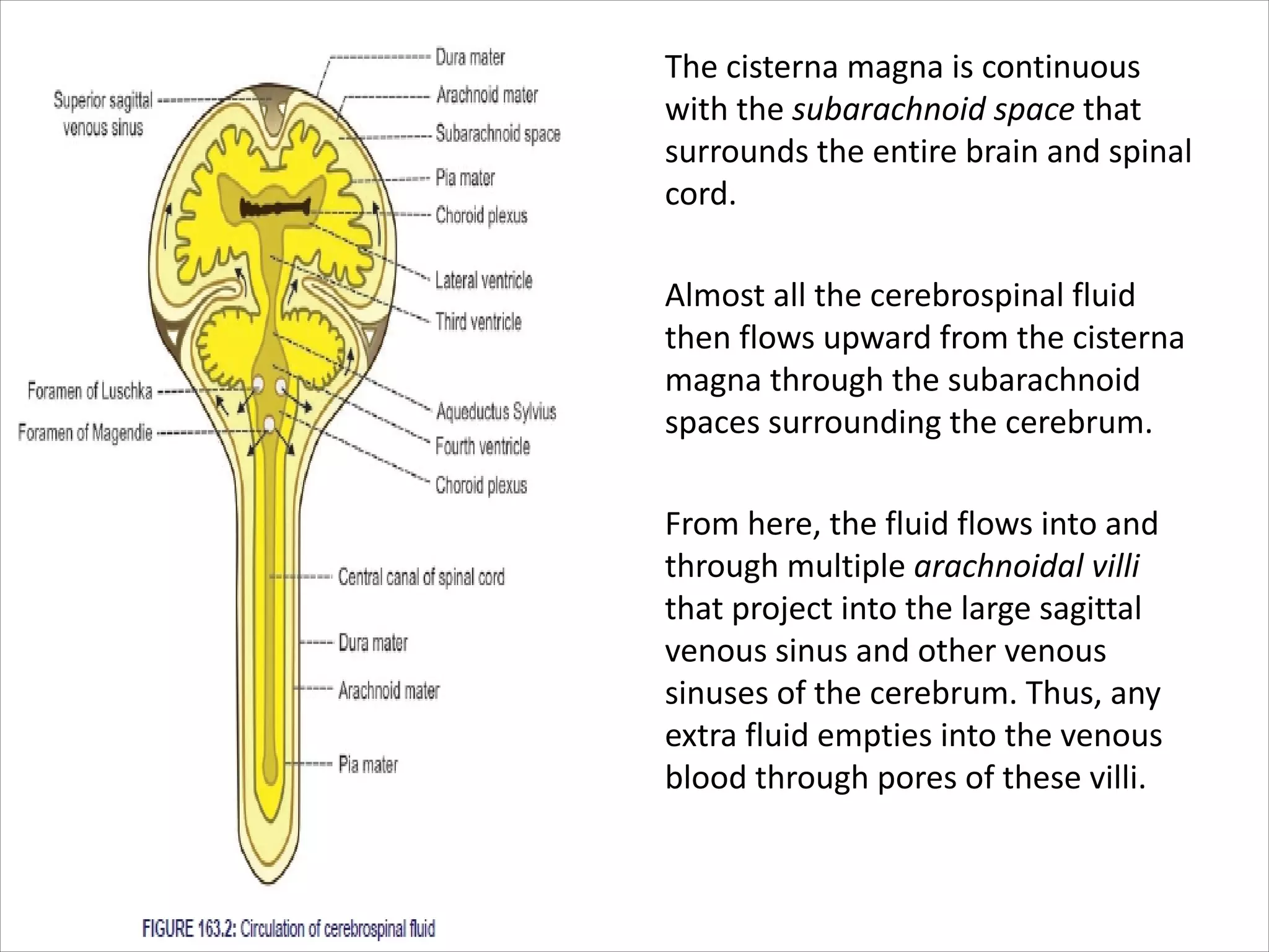
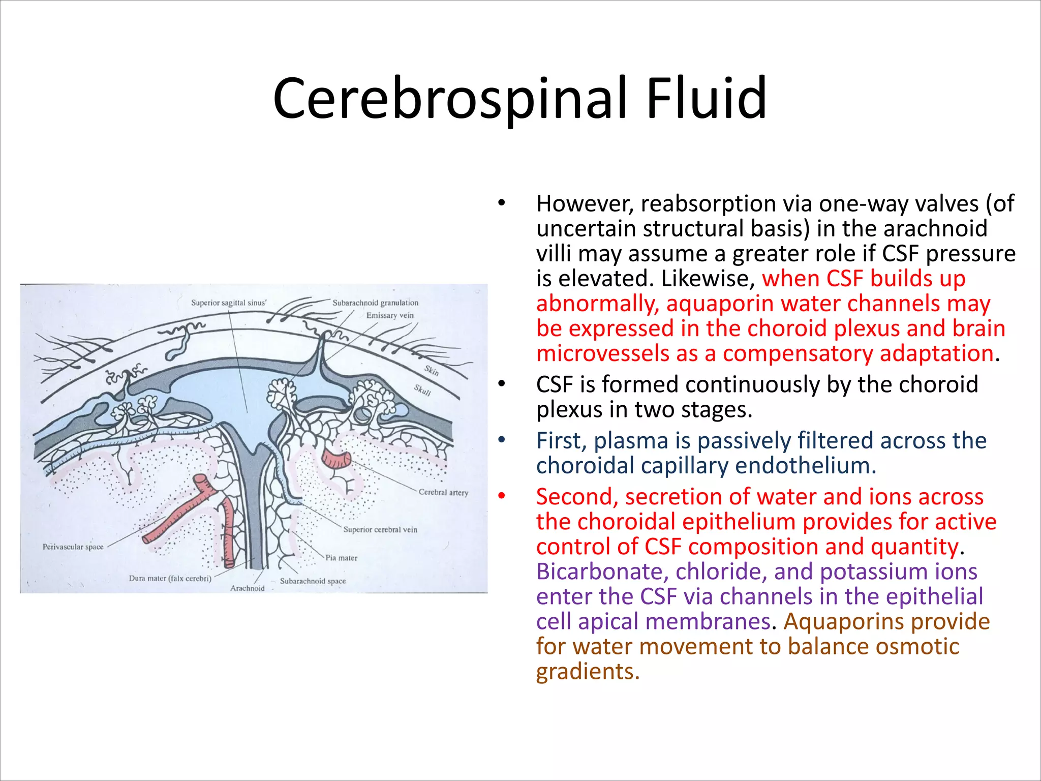
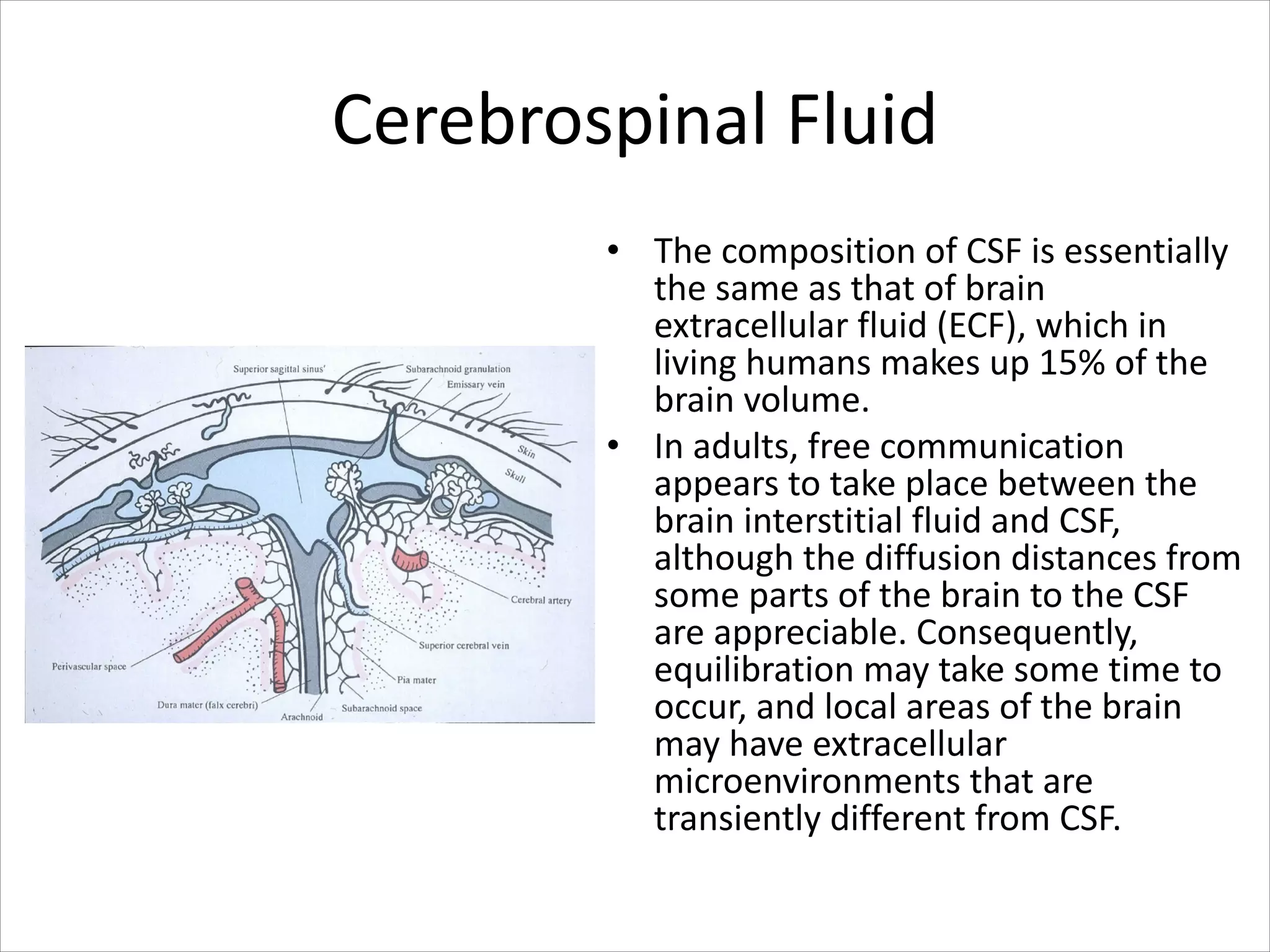
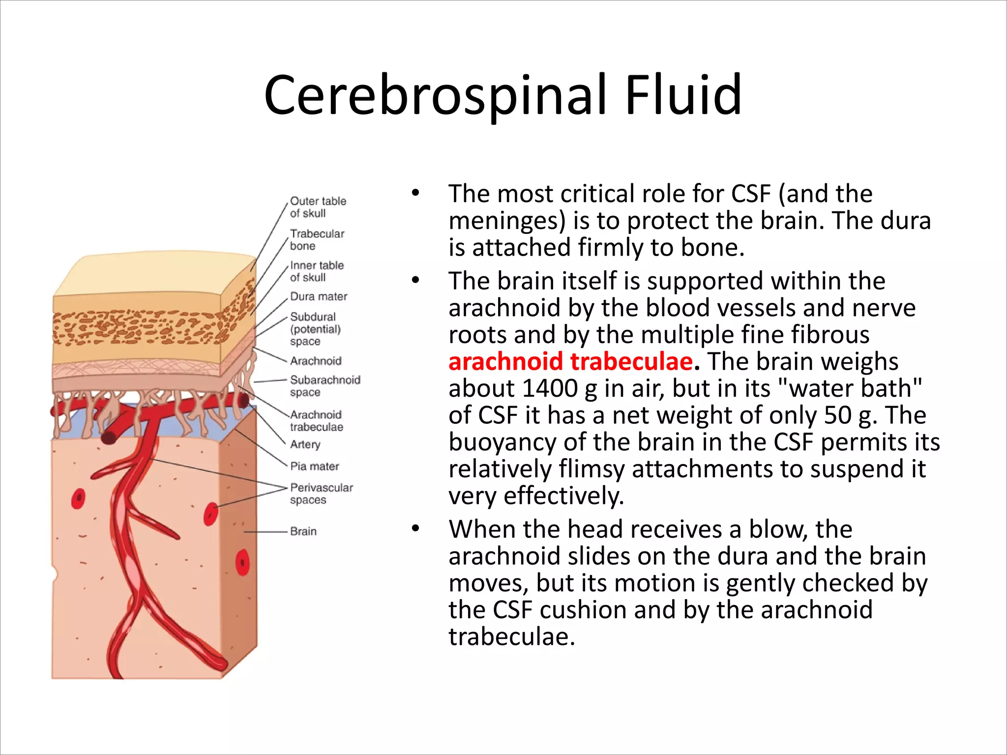
This document discusses circulation in special regions of the body including the brain, heart, skin, and fetus. It focuses on the unique anatomical and physiological features of cerebral circulation, including the protective role of cerebrospinal fluid and the blood-brain barrier. Specifically, it describes the formation and reabsorption of cerebrospinal fluid, the tight junctions between endothelial cells that limit substance entry into the brain, and how cerebral blood flow is regulated by innervation.


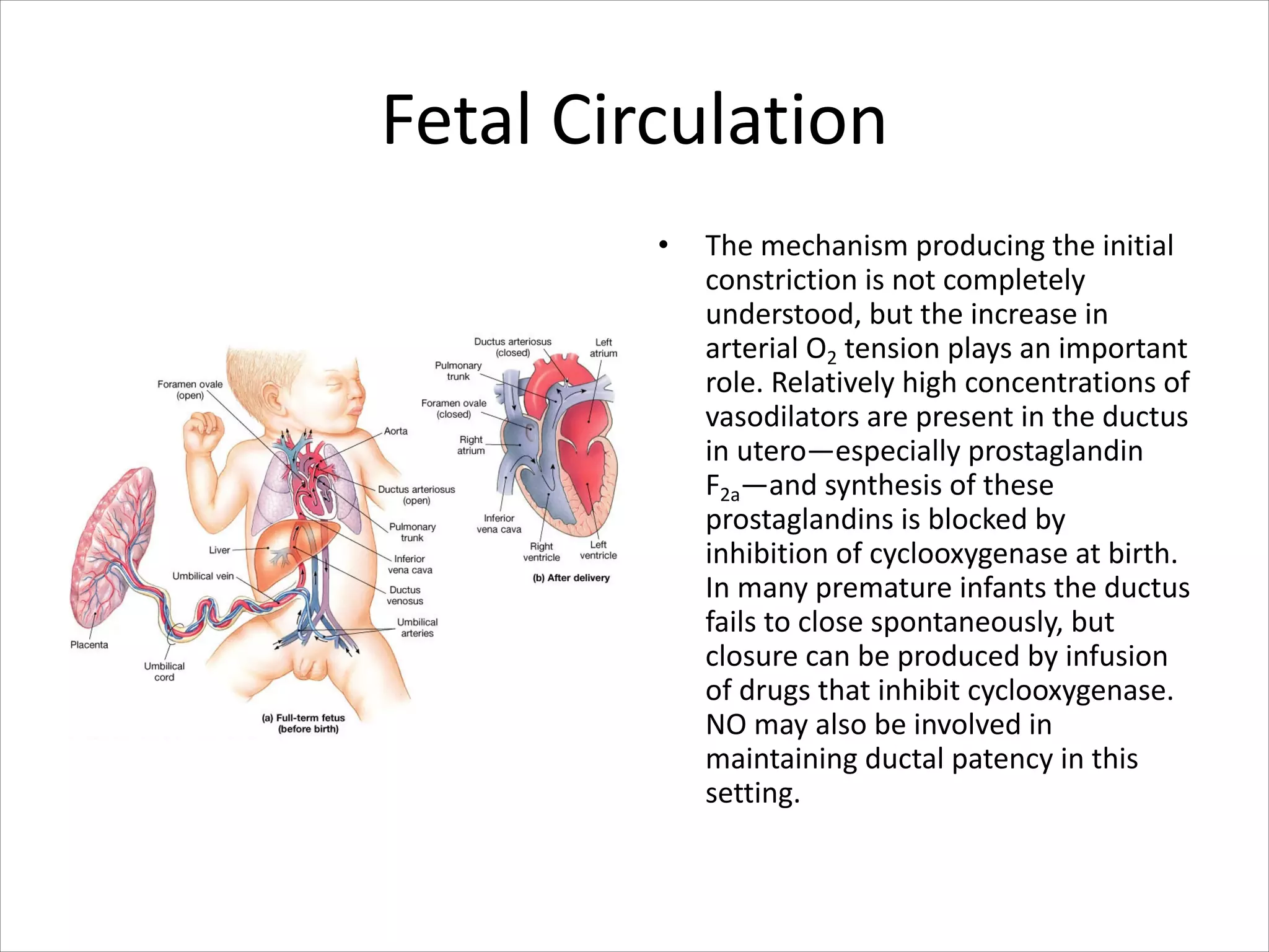
![Fetal Circulation
• Blood from the placenta is carried to
the fetus by the umbilical vein. Less
than a third of this enters the fetal
ductus venosus and is carried to the
inferior vena cava,[2] while the rest
enters the liver proper from the inferior
border of the liver.
• The branch of the umbilical vein that
supplies the right lobe of the liver first
joins with the portal vein. The blood
then moves to the right atrium of the
heart. In the fetus, there is an opening
between the right and left atrium (the
foramen ovale), and most of the blood
flows through this hole directly into the
left atrium from the right atrium, thus
bypassing pulmonary circulation.](https://image.slidesharecdn.com/specialcirculation-210701230211/75/Special-circulation-59-2048.jpg)
![Fetal Circulation
• The continuation of this blood flow is
into the left ventricle, and from there it
is pumped through the aorta into the
body. Some of the blood moves from
the aorta through the internal iliac
arteries to the umbilical arteries, and
re-enters the placenta, where carbon
dioxide and other waste products from
the fetus are taken up and enter the
maternal circulation.[1]
• Some of the blood entering the right
atrium does not pass directly to the left
atrium through the foramen ovale, but
enters the right ventricle and is pumped
into the pulmonary artery. In the fetus,
there is a special connection between
the pulmonary artery and the aorta,
called the ductus arteriosus, which
directs most of this blood away from
the lungs (which are not being used for
respiration at this point )](https://image.slidesharecdn.com/specialcirculation-210701230211/75/Special-circulation-60-2048.jpg)


![Apporach to lung biopsy [Auto-saved].pptx latest](https://cdn.slidesharecdn.com/ss_thumbnails/apporachtolungbiopsyauto-saved-251211225655-93258539-thumbnail.jpg?width=640&height=640&fit=bounds)


