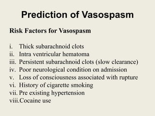
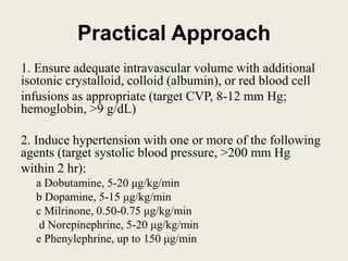
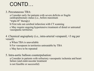
CEREBRAL VASOSPASM
ROSHAN KHATIWADA
Senior Resident
Neurosurgery
INTRODUCTION
Cerebral vasospasm is a prolonged, sometimes
severe, reversible narrowing of the cerebral
arteries that begins few days after subarachnoid
hemorrhage (SAH).
Blood clots in the subarachnoid space and
ventricles
Causes : rupture of a aneurysm, traumatic SAH,
vascular malformations or tumor bleed
Types
• Angiographic vasospasm : Vascular imaging :
focal, segmental, or diffuse;
– mild (<25%),
– moderate (25% to 50%)
– severe (>50%)
50-90% of aneurysmal SAH
• Clinical vasospasm : narrowing causing cerebral
ischemia with corresponding symptoms and signs
(delayed ischemic neurological deficits)
Only half of the patient with angiographic vasospasm
Prediction of Vasospasm
Risk Factors for Vasospasm
i. Thick subarachnoid clots
ii. Intra ventricular hematoma
iii. Persistent subarachnoid clots (slow clearance)
iv. Poor neurological condition on admission
v. Loss of consciousness associated with rupture
vi. History of cigarette smoking
vii. Pre existing hypertension
viii.Cocaine use
Modified Fisher Scale and
Risk for Vasospasm
ļ‚§ 1 : Focal or diffuse thin layer of SAH, no IVH:
low risk
ļ‚§ 2: Focal or diffuse thin layer of SAH, IVH
present: moderate risk
ļ‚§ 3: Focal or diffuse thick layer of SAH, no
IVH: high risk
ļ‚§ 4: Focal or diffuse thick layer of SAH, IVH
present: very high risk
Pathogenesis
• Smooth muscle contraction:
– Hemoglobin ---- entry and release of calcium ---
activation of calcium/calmodulin -dependent
myosin light-chain kinase --- phosphorylation of
the myosin light chain ---- actin and myosin cross-
linkage and mechanical shortening (smooth muscle
contraction).
• Endothelial Injury, decreased Nitric Oxide,
and increase Endothelin-1
Theories on the Pathogenesis
of Vasospasm
ļ‚§ Prolonged vasoconstriction
ļ‚§ Endothelin-1 overproduction (vasoconstrictor)
ļ‚§ Nitric oxide underproduction (vasodilator)
ļ‚§ Inflammation-mediated ā€œremodelingā€ and
narrowing of the arterial wall.
Symptoms and Signs
• Diminished attention, changes in verbal
output, or a slight but new pronator drift of the
upper extremity.
• Increased headache and either agitation or
somnolence, change in patient behavior
ļ‚§ Monoparesis , hemiparesis , aphasia confusion,
drowsiness, poverty of speech, and abulia.
Differential Diagnosis
• Increased edema surrounding hematomas,
contusions, or infarcts
• Re-bleeding of aneurysms or aneurysmal
remnant
• Hydrocephalus
• Infection, ventriculitis
• Hyponatremia
• Hypoxemia
Diagnosis
• Pathologic increases in TCD velocities; there is a high
probability of significant large-artery vasospasm with
velocities >200 cm/sec
• Pathologic reductions in CBF detected by
Perfusion CT scanning
SPECT
Xenon-enhanced CT
Thermal diffusion flowmetry
• Detection of cerebral ischemia by
– Diffusion-weighted MRI
– Microdialysis
– Jugular venous oxygen saturation
• Vascular imaging by
– Catheter-based angiography
– CT angiography
Prevention of Vasospasm and
Cerebral Protection
General Measures
– Maintain euvolemia (at least 3 L of isotonic fluids
daily)
– Prevent anemia (hemoglobin <9 g/dL)
– Maintain normal to high systemic blood pressure
– Prevent high ICP (and maintain CPP) with
external ventricular drainage as necessary
– Nimodipine 60 mg by mouth or nasogastric tube
every 4 hr.
Investigational Treatments
Endothelin receptor antagonists
HMG-CoA inhibitors (statins)
Magnesium sulfate (intravenous infusion)
Intrathecal vasodilators (slow-release implants)
– nicardipine implants
CONTD…
• Augmentation of Subarachnoid Clot
Clearance
Clot lysis
Lamina terminalis fenestration
Lumbar CSF drainage
Combined treatment
Reversal of Vasospasm and
Cerebral Ischemia: Rescue
Treatments
• Triple-H Therapy: Hypervolemia, Hypertension, and
Hemodilution
– central venous pressure between 8 and 10 mm Hg or a
– pulmonary capillary wedge pressure in the range of 14 to 16 mm Hg
– Systolic blood pressure of 200 mm Hg or higher or CPP greater than 80 mm Hg
• Intra-aortic Balloon Counterpulsation
– placement of a transfemoral aortic balloon that inflates on aortic valve closure
with each cardiac cycle and augments diastolic flow proximally to the coronary
and cranial arteries and distally to the peripheral circulation .
Endovascular Reversal of
Vasospasm
Chemical Angioplasty:
Papaverine
Verapamil (ā‰ˆ3 mg per vessel or ā‰ˆ20 mg per
patient)
nicardipine and nimodipine
Balloon Angioplasty:
Practical Approach
1. Ensure adequate intravascular volume with additional
isotonic crystalloid, colloid (albumin), or red blood cell
infusions as appropriate (target CVP, 8-12 mm Hg;
hemoglobin, >9 g/dL)
2. Induce hypertension with one or more of the following
agents (target systolic blood pressure, >200 mm Hg
within 2 hr):
a Dobutamine, 5-20 μg/kg/min
b Dopamine, 5-15 μg/kg/min
c Milrinone, 0.50-0.75 μg/kg/min
d Norepinephrine, 5-20 μg/kg/min
e Phenylephrine, up to 150 μg/min
CONTD…
3. Percutaneous TBA:
a Consider early for patients with severe deficits or fragile
cardiopulmonary status (i.e., before maximum
ā€œtriple-Hā€ therapy)
b First rule out cerebral infarction with CT scanning
c May require ongoing hypertensive treatment of distal or untreated
vasospastic territories
4 .Chemical angioplasty (i.e., intra-arterial verapamil, ā‰ˆ3 mg per
vessel):
a When TBA is unavailable
b For vasospasm in territories untreatable by TBA
c May have to be repeated
5. Intra-aortic balloon counterpulsation:
a Consider in patients with refractory vasospastic ischemia and heart
failure (and endovascular treatment
is not feasible or successful)
Question 1
• What is the percentage of people who develop
clinical vasospasm after a cerebrovascular
aneurysm rupture?
ANSWER 1
• 20-30%
Question 2
• What is the early onset cerebral vasospasm?
Answer 2
• Within 48hrs
Question 3
• How long can vasospasm last?
Answer 3
• upto 4weeks
Question 4
• What is the most common screening tool for
catching early vasospasm signs?
Answer 4
• TCD
THANK YOU

Cerebral vasospasm

  • 1.
  • 2.
    INTRODUCTION Cerebral vasospasm isa prolonged, sometimes severe, reversible narrowing of the cerebral arteries that begins few days after subarachnoid hemorrhage (SAH). Blood clots in the subarachnoid space and ventricles Causes : rupture of a aneurysm, traumatic SAH, vascular malformations or tumor bleed
  • 3.
    Types • Angiographic vasospasm: Vascular imaging : focal, segmental, or diffuse; – mild (<25%), – moderate (25% to 50%) – severe (>50%) 50-90% of aneurysmal SAH • Clinical vasospasm : narrowing causing cerebral ischemia with corresponding symptoms and signs (delayed ischemic neurological deficits) Only half of the patient with angiographic vasospasm
  • 4.
    Prediction of Vasospasm RiskFactors for Vasospasm i. Thick subarachnoid clots ii. Intra ventricular hematoma iii. Persistent subarachnoid clots (slow clearance) iv. Poor neurological condition on admission v. Loss of consciousness associated with rupture vi. History of cigarette smoking vii. Pre existing hypertension viii.Cocaine use
  • 5.
    Modified Fisher Scaleand Risk for Vasospasm ļ‚§ 1 : Focal or diffuse thin layer of SAH, no IVH: low risk ļ‚§ 2: Focal or diffuse thin layer of SAH, IVH present: moderate risk ļ‚§ 3: Focal or diffuse thick layer of SAH, no IVH: high risk ļ‚§ 4: Focal or diffuse thick layer of SAH, IVH present: very high risk
  • 6.
    Pathogenesis • Smooth musclecontraction: – Hemoglobin ---- entry and release of calcium --- activation of calcium/calmodulin -dependent myosin light-chain kinase --- phosphorylation of the myosin light chain ---- actin and myosin cross- linkage and mechanical shortening (smooth muscle contraction). • Endothelial Injury, decreased Nitric Oxide, and increase Endothelin-1
  • 7.
    Theories on thePathogenesis of Vasospasm ļ‚§ Prolonged vasoconstriction ļ‚§ Endothelin-1 overproduction (vasoconstrictor) ļ‚§ Nitric oxide underproduction (vasodilator) ļ‚§ Inflammation-mediated ā€œremodelingā€ and narrowing of the arterial wall.
  • 8.
    Symptoms and Signs •Diminished attention, changes in verbal output, or a slight but new pronator drift of the upper extremity. • Increased headache and either agitation or somnolence, change in patient behavior ļ‚§ Monoparesis , hemiparesis , aphasia confusion, drowsiness, poverty of speech, and abulia.
  • 9.
    Differential Diagnosis • Increasededema surrounding hematomas, contusions, or infarcts • Re-bleeding of aneurysms or aneurysmal remnant • Hydrocephalus • Infection, ventriculitis • Hyponatremia • Hypoxemia
  • 10.
    Diagnosis • Pathologic increasesin TCD velocities; there is a high probability of significant large-artery vasospasm with velocities >200 cm/sec • Pathologic reductions in CBF detected by Perfusion CT scanning SPECT Xenon-enhanced CT Thermal diffusion flowmetry
  • 11.
    • Detection ofcerebral ischemia by – Diffusion-weighted MRI – Microdialysis – Jugular venous oxygen saturation • Vascular imaging by – Catheter-based angiography – CT angiography
  • 12.
    Prevention of Vasospasmand Cerebral Protection General Measures – Maintain euvolemia (at least 3 L of isotonic fluids daily) – Prevent anemia (hemoglobin <9 g/dL) – Maintain normal to high systemic blood pressure – Prevent high ICP (and maintain CPP) with external ventricular drainage as necessary – Nimodipine 60 mg by mouth or nasogastric tube every 4 hr.
  • 13.
    Investigational Treatments Endothelin receptorantagonists HMG-CoA inhibitors (statins) Magnesium sulfate (intravenous infusion) Intrathecal vasodilators (slow-release implants) – nicardipine implants
  • 14.
    CONTD… • Augmentation ofSubarachnoid Clot Clearance Clot lysis Lamina terminalis fenestration Lumbar CSF drainage Combined treatment
  • 15.
    Reversal of Vasospasmand Cerebral Ischemia: Rescue Treatments • Triple-H Therapy: Hypervolemia, Hypertension, and Hemodilution – central venous pressure between 8 and 10 mm Hg or a – pulmonary capillary wedge pressure in the range of 14 to 16 mm Hg – Systolic blood pressure of 200 mm Hg or higher or CPP greater than 80 mm Hg • Intra-aortic Balloon Counterpulsation – placement of a transfemoral aortic balloon that inflates on aortic valve closure with each cardiac cycle and augments diastolic flow proximally to the coronary and cranial arteries and distally to the peripheral circulation .
  • 16.
    Endovascular Reversal of Vasospasm ChemicalAngioplasty: Papaverine Verapamil (ā‰ˆ3 mg per vessel or ā‰ˆ20 mg per patient) nicardipine and nimodipine Balloon Angioplasty:
  • 17.
    Practical Approach 1. Ensureadequate intravascular volume with additional isotonic crystalloid, colloid (albumin), or red blood cell infusions as appropriate (target CVP, 8-12 mm Hg; hemoglobin, >9 g/dL) 2. Induce hypertension with one or more of the following agents (target systolic blood pressure, >200 mm Hg within 2 hr): a Dobutamine, 5-20 μg/kg/min b Dopamine, 5-15 μg/kg/min c Milrinone, 0.50-0.75 μg/kg/min d Norepinephrine, 5-20 μg/kg/min e Phenylephrine, up to 150 μg/min
  • 18.
    CONTD… 3. Percutaneous TBA: aConsider early for patients with severe deficits or fragile cardiopulmonary status (i.e., before maximum ā€œtriple-Hā€ therapy) b First rule out cerebral infarction with CT scanning c May require ongoing hypertensive treatment of distal or untreated vasospastic territories 4 .Chemical angioplasty (i.e., intra-arterial verapamil, ā‰ˆ3 mg per vessel): a When TBA is unavailable b For vasospasm in territories untreatable by TBA c May have to be repeated 5. Intra-aortic balloon counterpulsation: a Consider in patients with refractory vasospastic ischemia and heart failure (and endovascular treatment is not feasible or successful)
  • 19.
    Question 1 • Whatis the percentage of people who develop clinical vasospasm after a cerebrovascular aneurysm rupture?
  • 20.
  • 21.
    Question 2 • Whatis the early onset cerebral vasospasm?
  • 22.
  • 23.
    Question 3 • Howlong can vasospasm last?
  • 24.
  • 25.
    Question 4 • Whatis the most common screening tool for catching early vasospasm signs?
  • 26.
  • 27.