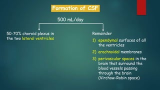
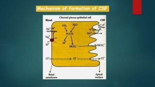
Cerebrospinal Fluid
and
Blood Brain Barrier
DR. KARISHMA R. PANDEY
ASSISTANT PROFESSOR
DEPARTMENT OF BASIC AND CLINICAL
PHYSIOLOGY
Topics
1) Brain ventricular system
1) Cerebrospinal fluid (formation, circulation, aborption and
functions)
2) Blood brain barrier
• Structure
• Functions
The cavities of the brain
are known as ventricles and
the cavity of the spinal
cord is called the central
canal.
Both spaces are filled with
CSF.
500 ml of CSF is formed
each day by the choroid
plexus in the cerebral
ventricles.
Choroid plexus consists of
networks of capillaries
surrounded by an
epithelial layer.
Formation of CSF
500 mL/day
50-70% choroid plexus in
the two lateral ventricles
Remainder
1) ependymal surfaces of all
the ventricles
2) arachnoidal membranes
3) perivascular spaces in the
brain that surround the
blood vessels passing
through the brain
(Virchow-Robin space)
Mechanism of formation of CSF
Composition of CSF and plasma
Circulation of CSF
 The CSF is absorbed from the
subarachnoid space by the
arachnoid villi into the
subduaral venous sinuses.
 Main driving force: colloid
osmotic pressure of plasma,
which is 25 mmHg higher than
CSF.
 Additional force: hydrostatic
pressure of CSF is 0.5-5
mmHg higher than subdural
venous sinuses.
Absorption of CSF
Functions of CSF
1) It regulates the extracellular environment of nerve cells, as it is in direct contact with
the extracellular fluid of the brain, it helps to provide a stable ionic environment for
neuronal function.
2) it acts as a hydraulic buffer to cushion the brain against damage resulting from
movements of the head.
3) The brain weighs about 1400 g in air, but in its "water bath" of CSF it has a net weight of
only 50 g. The buoyancy of the brain in the CSF permits its relatively flimsy attachments
to suspend it very effectively.
4) Removes proteins which has leaked in the interstitial fluid of the brain
5) Homeostatic role: increased hydrogen ions due to local hypoxia --- communicate to the
CSF---- transmit the change to chemosensitive respiratoy neurons as well as cerebral
blood vessels.
Brain injury: countrecoup phenomenon
Because of inertia
CSF EXAMINATION
Lumbar
puncture
CSF pressure
measurement
Lumbar CSF pressure =70-180 mm CSF.
Avg normal CSF pressure = 112 mm CSF.
 damage the brain (hemorrhage, head injury, inflammation of the
aqueduct, or the growth of a tumor)
 Meningitis
 lumbar puncture --- risky ----- because the sudden release of
pressure by the puncture may suck the brainstem downwards.
 The raised CSF pressure may be detected during examination of
the fundus of the eye. (Monro Kellie doctrine)
Effect of raised CSF pressure
Hydrocephalus
Increase in the volume of CSF
Leading to enlargement of cerebral ventricles
Causes:
1) Increased rate of formation of CSF (rare)
2) Reduced rate of absorption of CSF (communicating type)
3) Obstruction to the flow of CSF due to blockage of foramina of
Magendie and Luschka (non-communicating type)
If the Fomamina of Magendie and Luschka are blocked the entire
ventricular system gets distended.
If the aqueduct of sylvius is occluded : the lateral ventricles and
the 3rd ventricles get distended.
 If one of the foramina of Monro is blocked: the corresponding
lateral ventricle gets distended.
Secretion and Absorption of CSF
Volume of CSF = 150 ml.
CSF is replaced about
3.7 times every day.
Blood Brain Barrier
 Capillaries in the brain do not have pores between adjacent endothelial cells
 tight junctions :
1. endothelial cells of brain capillaries
2. the epithelial cells in the choroid plexus
 Effectively prevent proteins from entering the brain in adults and slow the
penetration of smaller molecules.
 This uniquely limited exchange of substances into the brain is referred to as
the blood–brain barrier.
 Water, CO2, and O2 and lipid-soluble free forms of steroid hormones :
easy penetration
 Glucose : major ultimate source of energy for nerve cells.
GLUT 1
 molecules within brain capillaries must be moved through the endothelial
cells by diffusion and active transport, as well as by endocytosis and
exocytosis
 brain cannot obtain molecules from the blood plasma by a nonspecific
filtering process
Circum Ventricular Organs: brain areas outside the
blood brain barrier
NH:The neurohypophysis,
OVLT: organum vasculosum of the lamina terminalis
SFO: subfornical organ,
AP: area postrema
(PI: pineal, SCO: subcommissural organ)
• Capillaries are fenestrated
• Allows relatively free exchange between the plasma
and the interstitial fluid of the neural space
Function :
• sensory receptors that respond to specific changes in
the body fluids, such as changes in osmolality and in
glucose concentration,
• receptors for peptide hormones that regulate thirst,
such as angiotensin II.
Functions of BBB
1) maintains the constancy of the environment of the neurons in
the central nervous system.
2) protection of the brain from endogenous and exogenous toxins in
the blood and
3) prevention of the escape of neurotransmitters into the general
circulation.
Clinical Considerations
1) BBB presents difficulties in the chemotherapy of brain diseases because
drugs that could enter other organs may not be able to enter the brain.
2) In treating infections such as meningitis, only those antibiotics that can cross
the blood-brain barrier are used.
3) BBB is broken down in areas of infection or injury.
4) Tumors develop new blood vessels --- capillaries that are formed lack contact
with normal astrocytes--- no tight junctions-----vessels fenestrated---- helps
in identifying the location of tumors---- substances such as radioactive iodine-
labeled albumin penetrate normal brain tissue very slowly, but they enter
tumor tissue----tumor stand out as an island of radioactivity in the
surrounding normal brain.
Cerebrospinal fluid

Cerebrospinal fluid

  • 1.
    Cerebrospinal Fluid and Blood BrainBarrier DR. KARISHMA R. PANDEY ASSISTANT PROFESSOR DEPARTMENT OF BASIC AND CLINICAL PHYSIOLOGY
  • 2.
    Topics 1) Brain ventricularsystem 1) Cerebrospinal fluid (formation, circulation, aborption and functions) 2) Blood brain barrier • Structure • Functions
  • 3.
    The cavities ofthe brain are known as ventricles and the cavity of the spinal cord is called the central canal. Both spaces are filled with CSF.
  • 5.
    500 ml ofCSF is formed each day by the choroid plexus in the cerebral ventricles. Choroid plexus consists of networks of capillaries surrounded by an epithelial layer.
  • 6.
    Formation of CSF 500mL/day 50-70% choroid plexus in the two lateral ventricles Remainder 1) ependymal surfaces of all the ventricles 2) arachnoidal membranes 3) perivascular spaces in the brain that surround the blood vessels passing through the brain (Virchow-Robin space)
  • 7.
  • 8.
  • 9.
  • 10.
     The CSFis absorbed from the subarachnoid space by the arachnoid villi into the subduaral venous sinuses.  Main driving force: colloid osmotic pressure of plasma, which is 25 mmHg higher than CSF.  Additional force: hydrostatic pressure of CSF is 0.5-5 mmHg higher than subdural venous sinuses. Absorption of CSF
  • 11.
    Functions of CSF 1)It regulates the extracellular environment of nerve cells, as it is in direct contact with the extracellular fluid of the brain, it helps to provide a stable ionic environment for neuronal function. 2) it acts as a hydraulic buffer to cushion the brain against damage resulting from movements of the head. 3) The brain weighs about 1400 g in air, but in its "water bath" of CSF it has a net weight of only 50 g. The buoyancy of the brain in the CSF permits its relatively flimsy attachments to suspend it very effectively. 4) Removes proteins which has leaked in the interstitial fluid of the brain 5) Homeostatic role: increased hydrogen ions due to local hypoxia --- communicate to the CSF---- transmit the change to chemosensitive respiratoy neurons as well as cerebral blood vessels.
  • 12.
    Brain injury: countrecoupphenomenon Because of inertia
  • 13.
    CSF EXAMINATION Lumbar puncture CSF pressure measurement LumbarCSF pressure =70-180 mm CSF. Avg normal CSF pressure = 112 mm CSF.
  • 14.
     damage thebrain (hemorrhage, head injury, inflammation of the aqueduct, or the growth of a tumor)  Meningitis  lumbar puncture --- risky ----- because the sudden release of pressure by the puncture may suck the brainstem downwards.  The raised CSF pressure may be detected during examination of the fundus of the eye. (Monro Kellie doctrine) Effect of raised CSF pressure
  • 15.
    Hydrocephalus Increase in thevolume of CSF Leading to enlargement of cerebral ventricles Causes: 1) Increased rate of formation of CSF (rare) 2) Reduced rate of absorption of CSF (communicating type) 3) Obstruction to the flow of CSF due to blockage of foramina of Magendie and Luschka (non-communicating type)
  • 16.
    If the Fomaminaof Magendie and Luschka are blocked the entire ventricular system gets distended. If the aqueduct of sylvius is occluded : the lateral ventricles and the 3rd ventricles get distended.  If one of the foramina of Monro is blocked: the corresponding lateral ventricle gets distended.
  • 17.
    Secretion and Absorptionof CSF Volume of CSF = 150 ml. CSF is replaced about 3.7 times every day.
  • 18.
    Blood Brain Barrier Capillaries in the brain do not have pores between adjacent endothelial cells  tight junctions : 1. endothelial cells of brain capillaries 2. the epithelial cells in the choroid plexus  Effectively prevent proteins from entering the brain in adults and slow the penetration of smaller molecules.  This uniquely limited exchange of substances into the brain is referred to as the blood–brain barrier.
  • 19.
     Water, CO2,and O2 and lipid-soluble free forms of steroid hormones : easy penetration  Glucose : major ultimate source of energy for nerve cells. GLUT 1  molecules within brain capillaries must be moved through the endothelial cells by diffusion and active transport, as well as by endocytosis and exocytosis  brain cannot obtain molecules from the blood plasma by a nonspecific filtering process
  • 21.
    Circum Ventricular Organs:brain areas outside the blood brain barrier NH:The neurohypophysis, OVLT: organum vasculosum of the lamina terminalis SFO: subfornical organ, AP: area postrema (PI: pineal, SCO: subcommissural organ) • Capillaries are fenestrated • Allows relatively free exchange between the plasma and the interstitial fluid of the neural space Function : • sensory receptors that respond to specific changes in the body fluids, such as changes in osmolality and in glucose concentration, • receptors for peptide hormones that regulate thirst, such as angiotensin II.
  • 22.
    Functions of BBB 1)maintains the constancy of the environment of the neurons in the central nervous system. 2) protection of the brain from endogenous and exogenous toxins in the blood and 3) prevention of the escape of neurotransmitters into the general circulation.
  • 23.
    Clinical Considerations 1) BBBpresents difficulties in the chemotherapy of brain diseases because drugs that could enter other organs may not be able to enter the brain. 2) In treating infections such as meningitis, only those antibiotics that can cross the blood-brain barrier are used. 3) BBB is broken down in areas of infection or injury. 4) Tumors develop new blood vessels --- capillaries that are formed lack contact with normal astrocytes--- no tight junctions-----vessels fenestrated---- helps in identifying the location of tumors---- substances such as radioactive iodine- labeled albumin penetrate normal brain tissue very slowly, but they enter tumor tissue----tumor stand out as an island of radioactivity in the surrounding normal brain.

Editor's Notes

  • #5 the brain and spinal cord lie within a bony case formed by the skull and vertebral canal of the spinal column and are covered by three membranes called the meninges. Immediately beneath the skull lies a tough membrane of dense connective tissue called the dura mater (or dura) that envelops the whole brain and extends in the form of a tube over the spinal cord. Attached to the inner face of the dura is the arachnoid membrane. Beneath the arachnoid lies the highly vascularized pia mater (or pia) which is attached to the surface of the brain and spinal cord, following every contour. The narrow space between the pia and arachnoid membranes is filled with a clear fluid called the cerebrospinal fluid (CSF) that is actively secreted by the choroid plexuses.
  • #12 Space between the skull and the brain is filled with CSF, the brain floats in a fluid-filled container. This arrangement restricts the displacement of the brain within the skull during movements of the head and limits the stresses on the blood vessels and the cranial nerves. When the head receives a blow, the arachnoid slides on the dura and the brain moves, but its motion is gently checked by the CSF cushion and by the arachnoid trabeculae. If the brain rested heavily on the floor of the cranium, the pressure would kill the nervous tissue.
  • #15 As with the circulation of the blood, the circulation of the CSF is driven by the difference in pressure between the cerebral ventricles (the site of production) and the venous sinuses. The hydrostatic pressure of the CSF can be measured by lumbar puncture and is normally about 0.5–1.5 kPa (approximately 4–12 mmHg). Obstruction of the meninges become inflamed, leading to a potentially life-threatening condition. In meningitis, the intracranial pressure is increased due to restricted drainage of the CSF, giving rise to characteristic clinical signs including headache, vomiting, photophobia, and rigidity of the neck. In severe cases convulsions and coma may occur. Because the CSF flows freely around the brain and spinal cord, a sample of CSF obtained by lumbar puncture can be used to ascertain the nature of the invading organism. As the brain is encased in the skull, which provides a rigid enclosure except in very young children, anything that increases the volume of the brain tissue, such as a cerebral tumor or a P.306 CSF drainage leads to an increase in intracranial pressure. This can occur following hemorrhage, head injury, inflammation of the aqueduct, or the growth of a tumor. In an adult, the raised intracranial pressure elicits the Cushing's reflex discussed above. In babies and young children (where the skull sutures are not fused), the increased pressure leads to a disproportionate increase in the size of the head known as hydrocephalus. If this is severe and remains untreated, brain tissue may be damaged, resulting in mental impairment. A further cause of raised intracranial pressure is meningitis in which the membranes covering the brain (the meninges) are invaded by viruses or bacteria. Under these circumstances, the cerebral hemorrhage, will increase the pressure within the skull (the intracranial pressure). This will tend to reduce the blood flow to the brain and force the brainstem into the opening of the skull through which the spinal cord passes (the foramen magnum). The resulting compression of the brainstem elicits a marked rise in arterial blood pressure, which acts to offset the fall in cerebral blood flow. This is known as Cushing's reflex (sometimes also called Cushing's response). In addition to the rise in blood pressure, there is a fall in heart rate mediated by the baroreceptor reflex. This combination of a marked rise in blood pressure and a slow heart rate is an important clinical indicator of a space-occupying lesion within the skull. If the increase in intracranial pressure is very high, cerebral blood flow will decline and the sufferer will experience mental confusion, leading to coma and death, unless prompt action is taken. Because brain tissue and spinal fluid are essentially incompressible, the volume of blood, spinal fluid, and brain in the cranium at any time must be relatively constant (Monro–Kellie doctrine). More importantly, the cerebral vessels are compressed whenever the intracranial pressure rises. Any change in venous pressure promptly causes a similar change in intracranial pressure. Thus, a rise in venous pressure decreases cerebral blood flow both by decreasing the effective perfusion pressure and by compressing the cerebral vessels. This relationship helps to compensate for changes in arterial blood pressure at the level of the head.
  • #18 Lumbar CSF pressure is normally 70–180 mm CSF. Up to pressures well above this range, the rate of CSF formation is independent of intraventricular pressure. However, absorption, which takes place largely by bulk flow, is proportionate to the pressure (Figure 32–3). At a pressure of 112 mm CSF, which is the average normal CSF pressure, filtration and absorption are equal. Below a pressure of approximately 68 mm CSF, absorption stops. Large amounts of fluid accumulate when the reabsorptive capacity of the arachnoid villi is decreased (external hydrocephalus, communicating hydrocephalus). Fluid also accumulates proximal to the block and distends the ventricles when the foramens of Luschka and Magendie are blocked or there is obstruction within the ventricular system (internal hydrocephalus, noncommunicating hydrocephalus).
  • #22 The blood-brain barrier is breached at a few sites along the mid-line of the brain. These sites are located along the third and fourth cerebral ventricles and are known as the circumventricular organs. Here the capillaries are fenestrated, allowing relatively free exchange between the plasma and the interstitial fluid of the neural space. The function of these areas is still being elucidated, but they appear to play a significant role in the regulation of fluid balance. four small areas in or near the brainstem stain like the tissues outside the brain. These areas are (1) the posterior pituitary (neurohypophysis) and the adjacent ventral part of the median eminence of the hypothalamus, (2) the area postrema, (3) the organum vasculosum of the lamina terminalis (OVLT, supraoptic crest), and (4) the subfornical organ (SFO). All have fenestrated capillaries, and because of their permeability they are said to be "outside the blood–brain barrier." Some of them function as neurohemal organs, ie, areas in which polypeptides secreted by neurons enter the circulation; for example, oxytocin and vasopressin enter the general circulation in the posterior pituitary, and hypothalamic hypophysiotropic hormones enter the portal hypophysial circulation in the median eminence. Other circumventricular organs contain receptors for many different peptides and other substances, and they function as chemoreceptor zones in which substances in the circulating blood can act to trigger changes in brain function without penetrating the blood–brain barrier. The area postrema is a chemoreceptor trigger zone that initiates vomiting in response to chemical changes in the plasma (see Chapter 14: Central Regulation of Visceral Function). It is also concerned with cardiovascular control, and in many species circulating angiotensin II acts on the area postrema to produce a neurally mediated increase in blood pressure. Angiotensin II also acts on the SFO and possibly on the OVLT to increase water intake. In addition, it appears that the OVLT is the site of the osmoreceptor controlling vasopressin secretion (see Chapter 14: Central Regulation of Visceral Function), and evidence suggests that circulating IL-1 produces fever by acting on this circumventricular organ.
  • #23 These neurons are so dependent on the concentrations of K+, Ca2+, Mg2+, H+, and other ions in the fluid bathing them that even minor variations have far-reaching consequences. The constancy of the composition of the ECF in all parts of the body is maintained by multiple homeostatic mechanisms (see Chapters 1 and 39), but because of the sensitivity of the cortical neurons to ionic change, it is not surprising that an additional defense has evolved to protect them.
  • #24 amines dopamine and serotonin penetrate brain tissue to a very limited degree but their corresponding acid precursors, L-dopa and 5-hydroxytryptophan, respectively, enter with relative ease Another important clinical consideration is the fact that the blood–brain barrier tends to break down in areas of infection or injury. Tumors develop new blood vessels, and the capillaries that are formed lack contact with normal astrocytes. Therefore, there are no tight junctions, and the vessels may even be fenestrated. The lack of a barrier helps in identifying the location of tumors; substances such as radioactive iodine-labeled albumin penetrate normal brain tissue very slowly, but they enter tumor tissue, making the tumor stand out as an island of radioactivity in the surrounding normal brain. The blood–brain barrier can also be temporarily disrupted by sudden marked increases in blood pressure or by intravenous injection of hypertonic fluids.