Download as PDF, PPTX
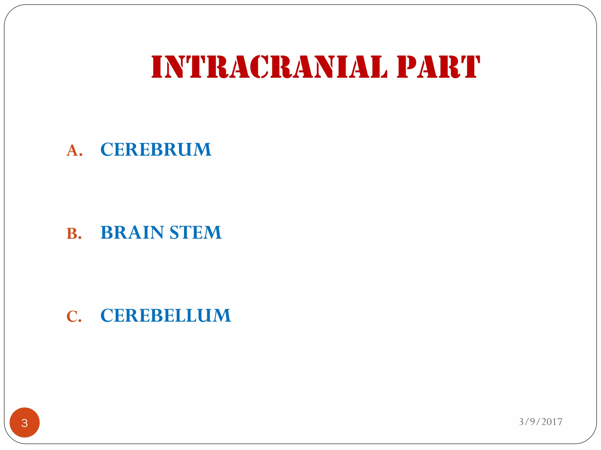
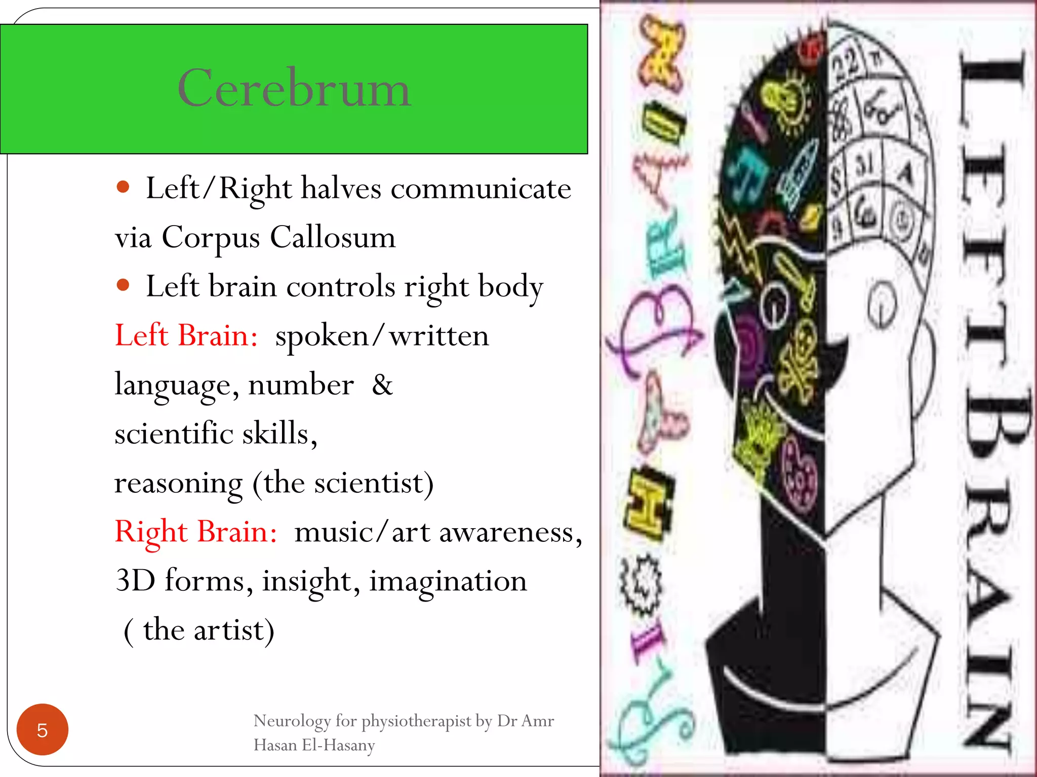

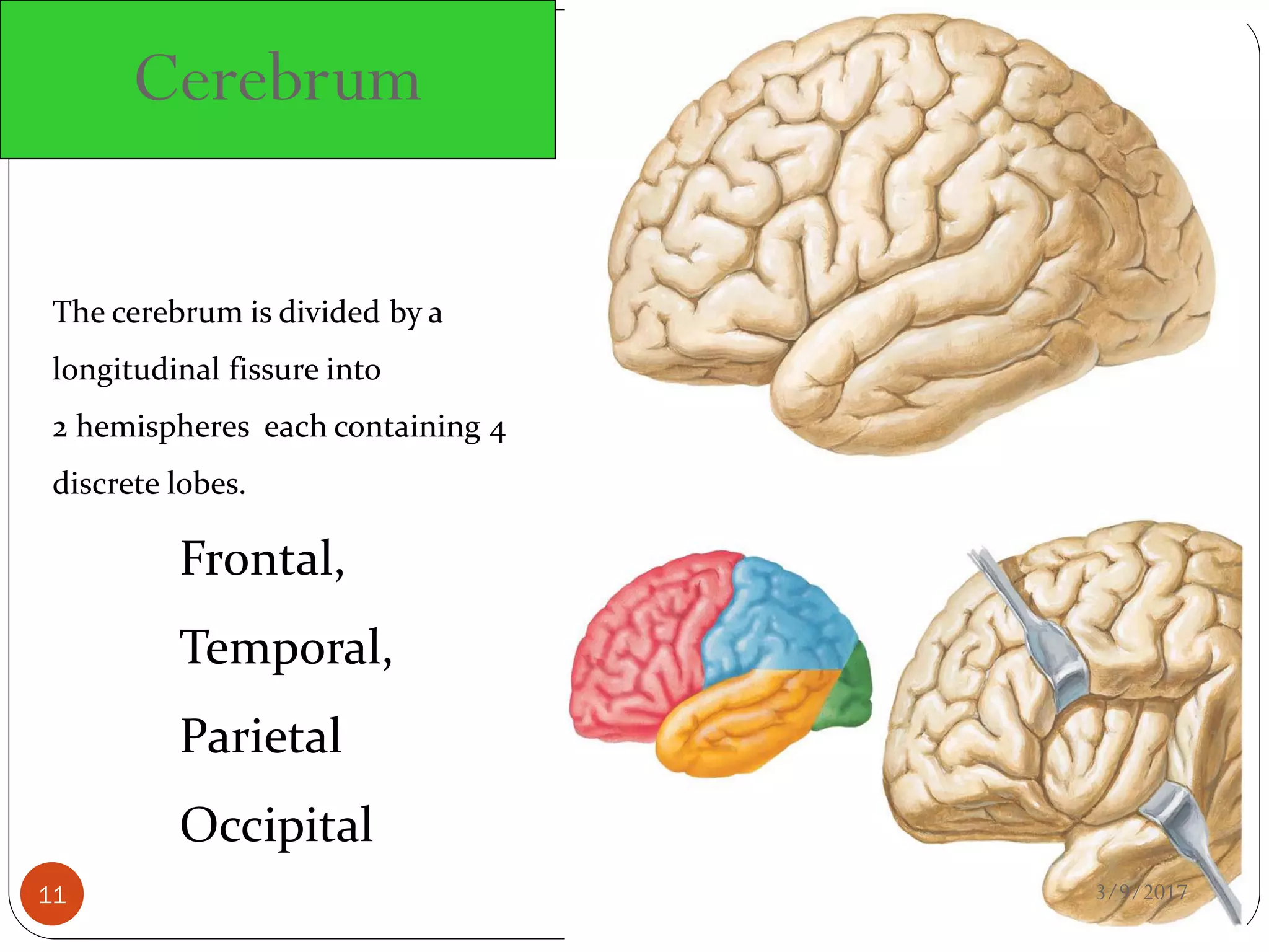
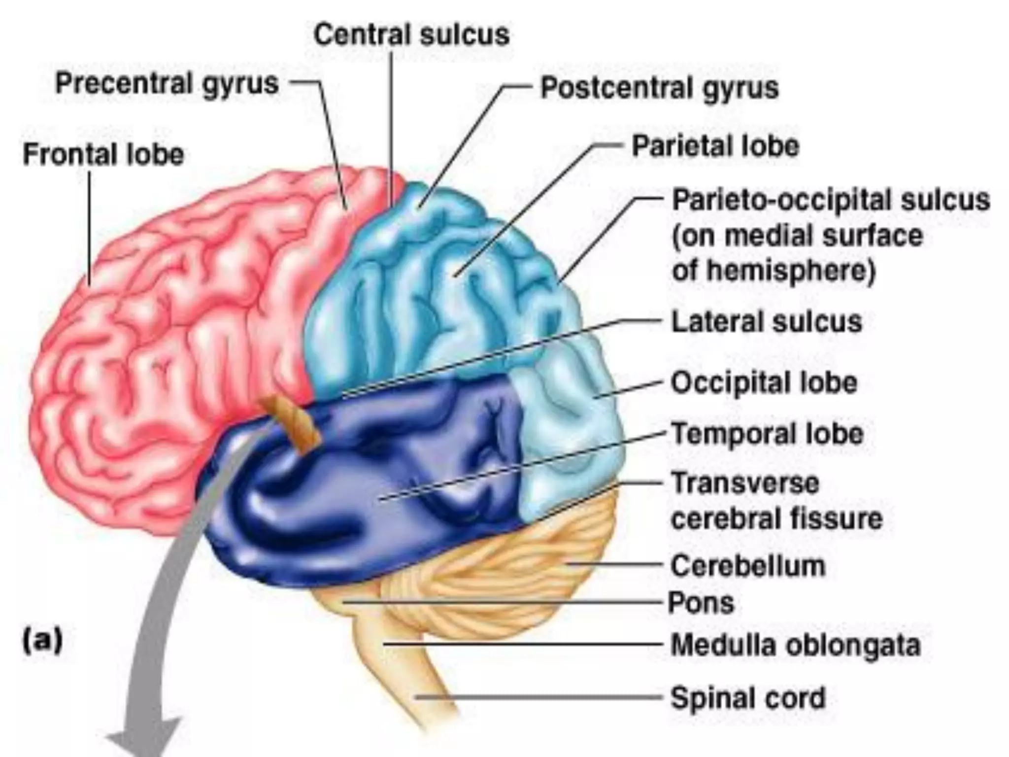
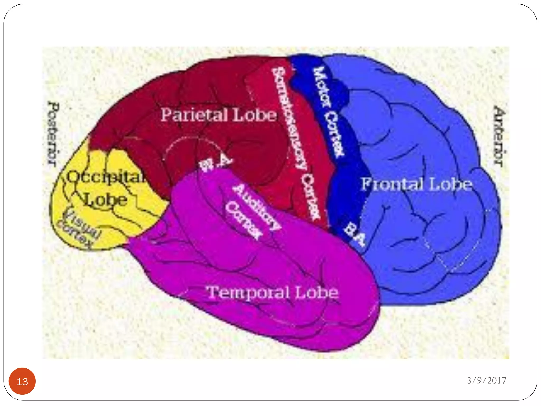
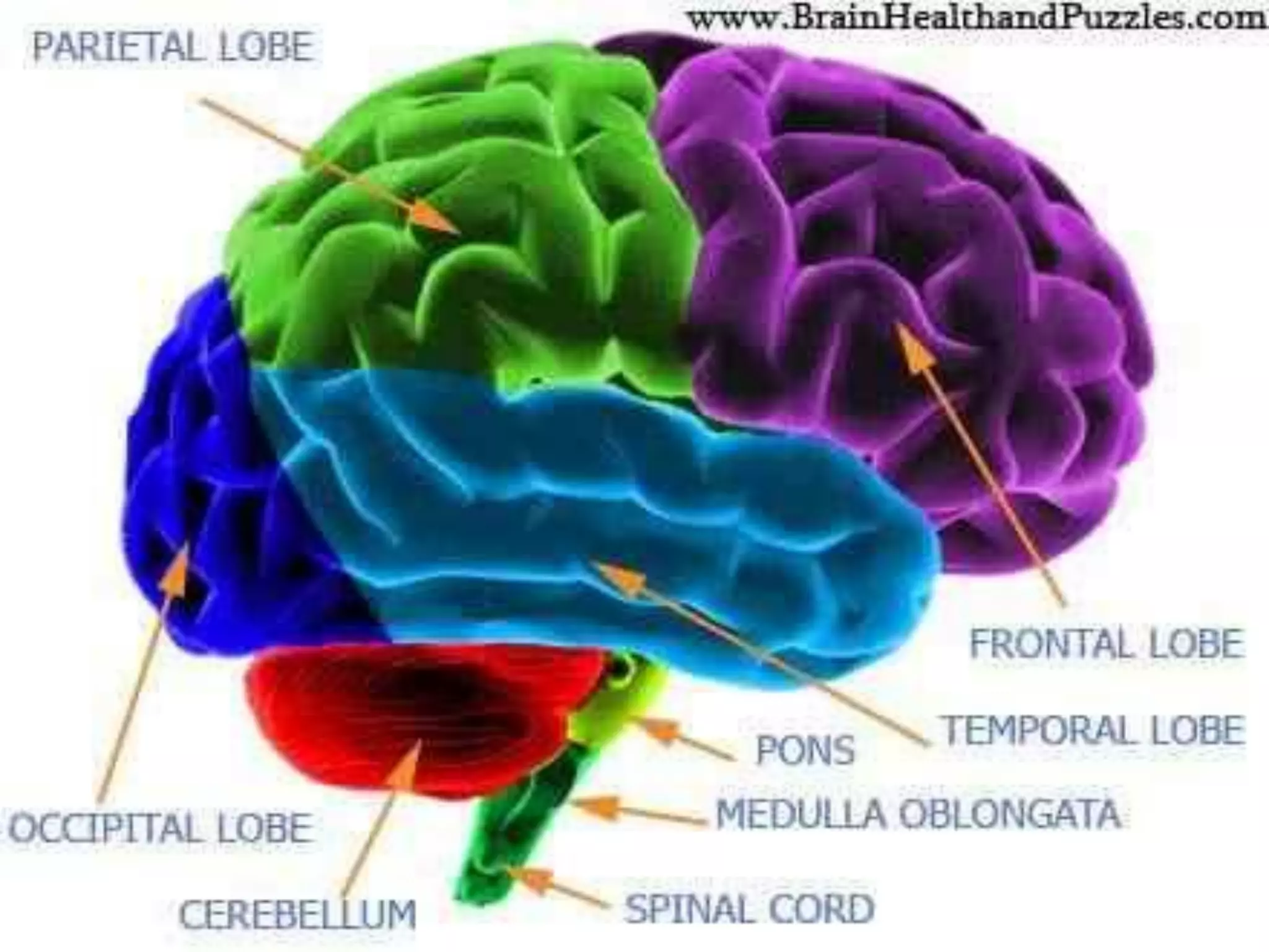
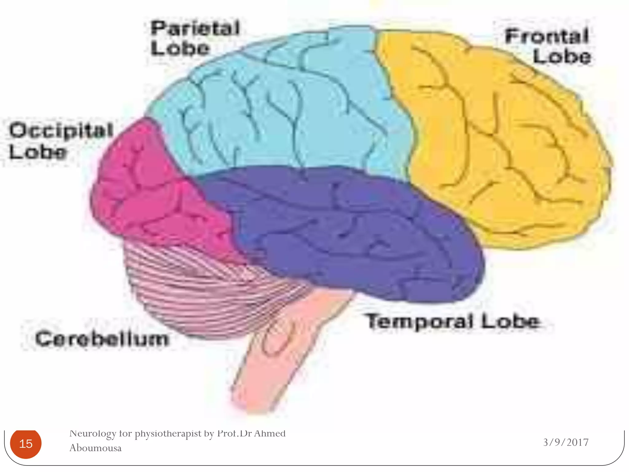
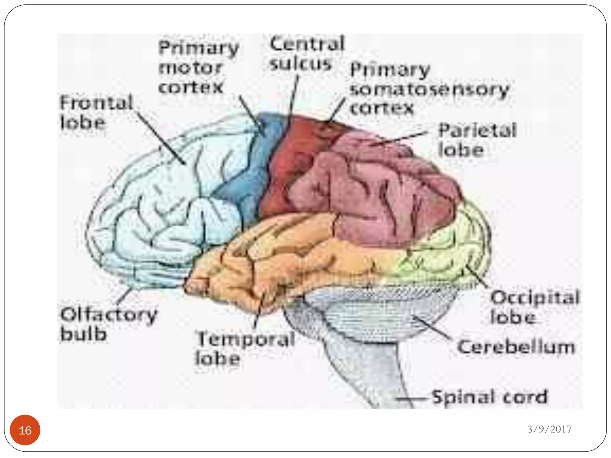
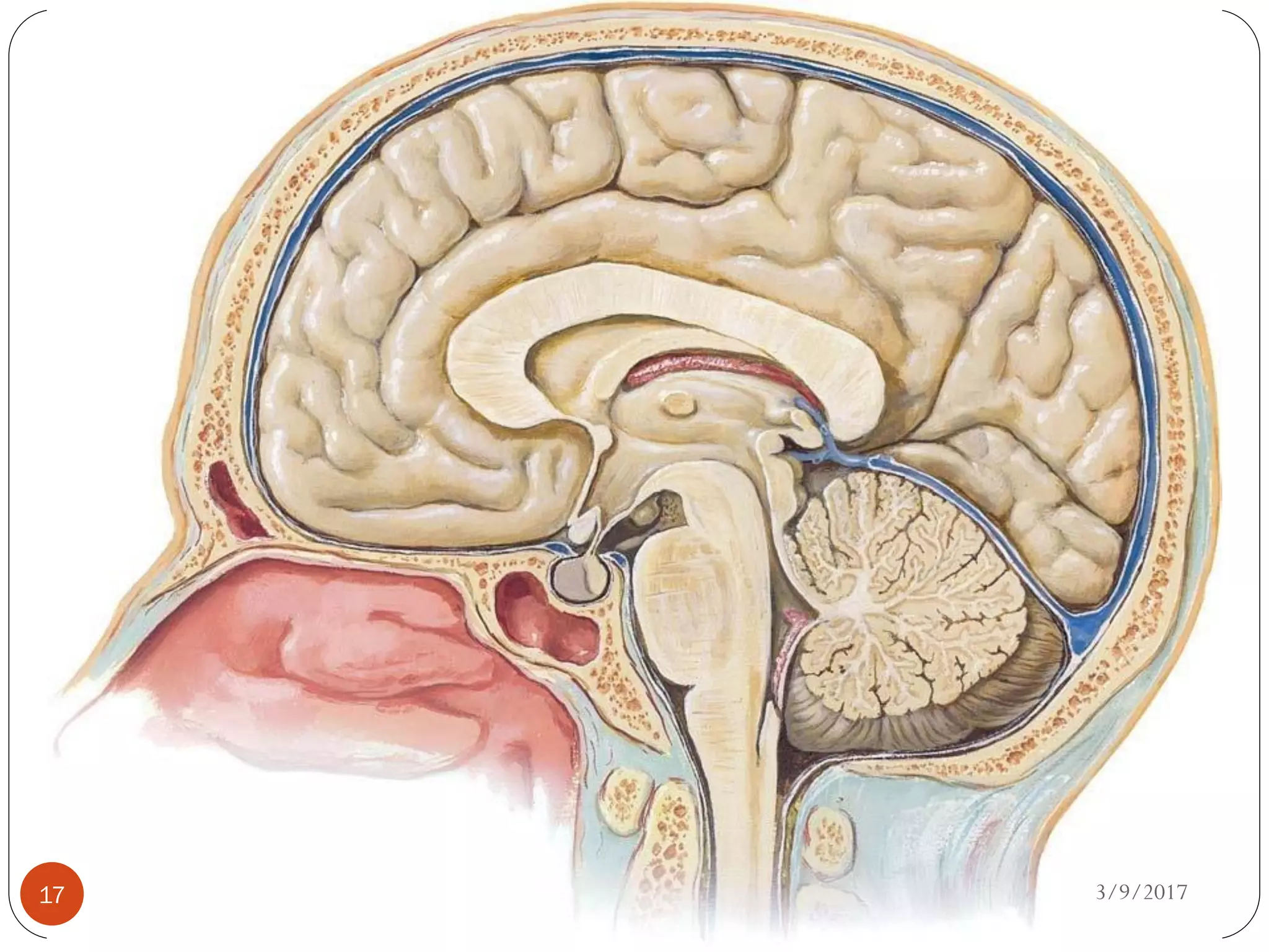
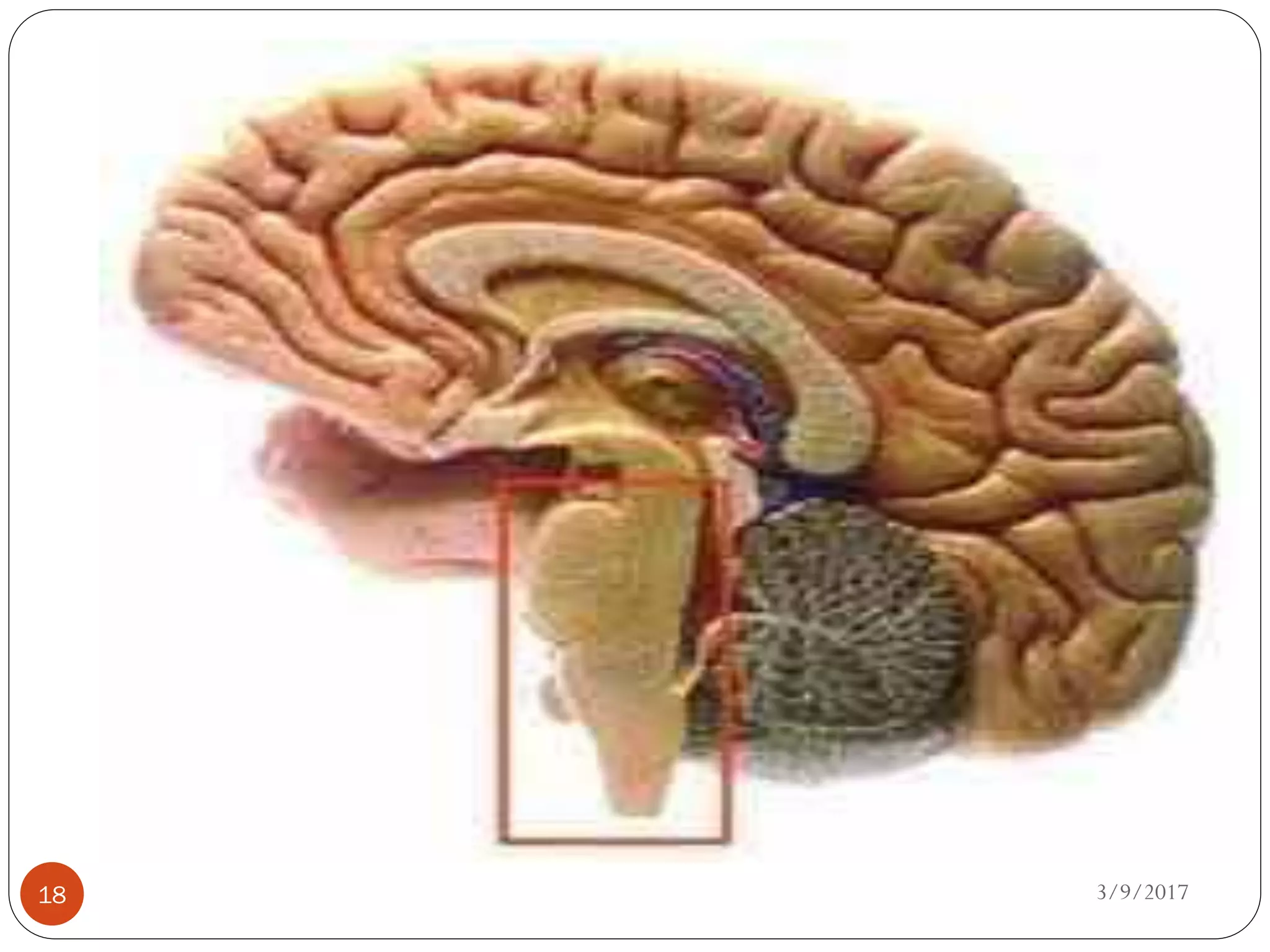
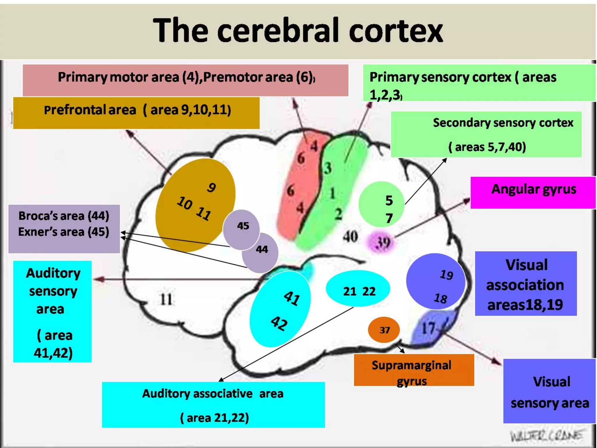
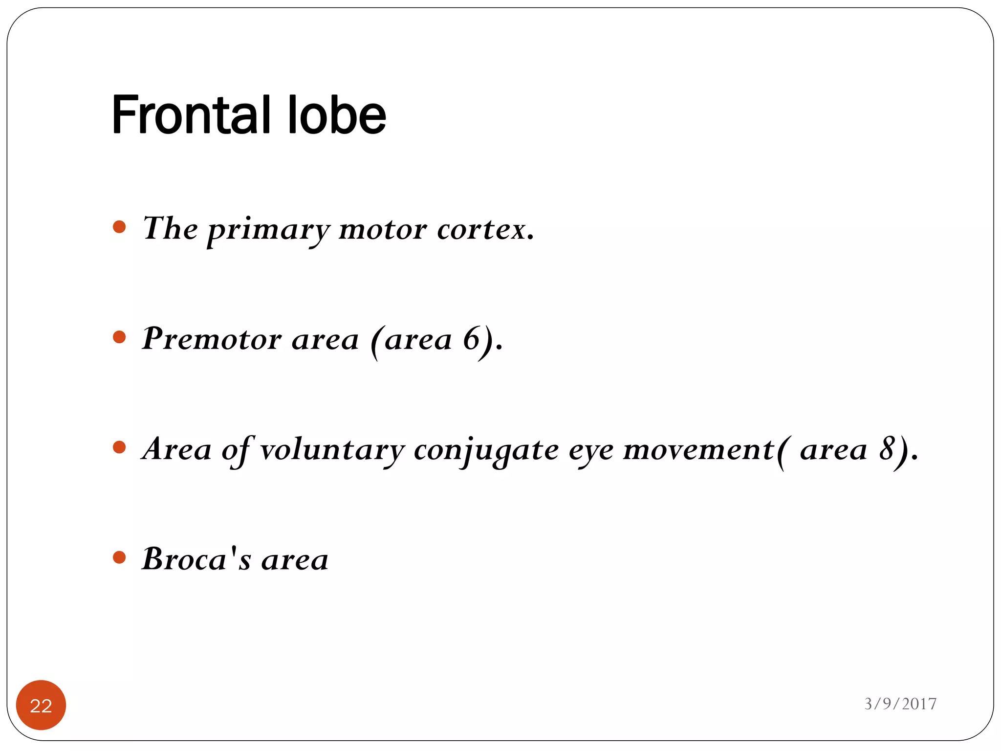
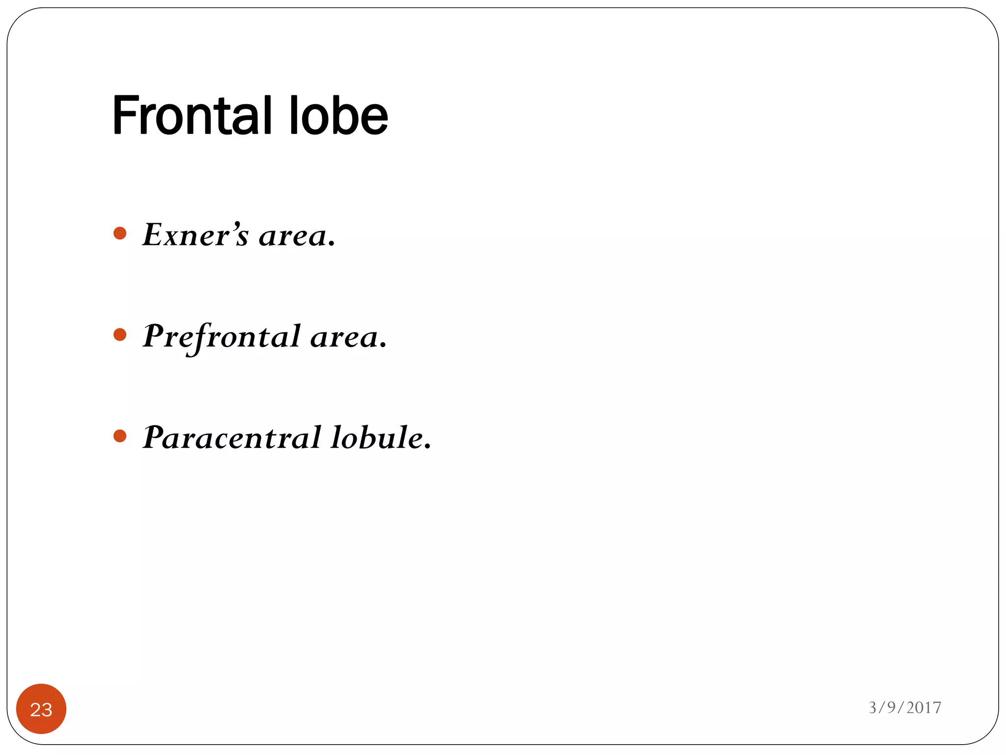

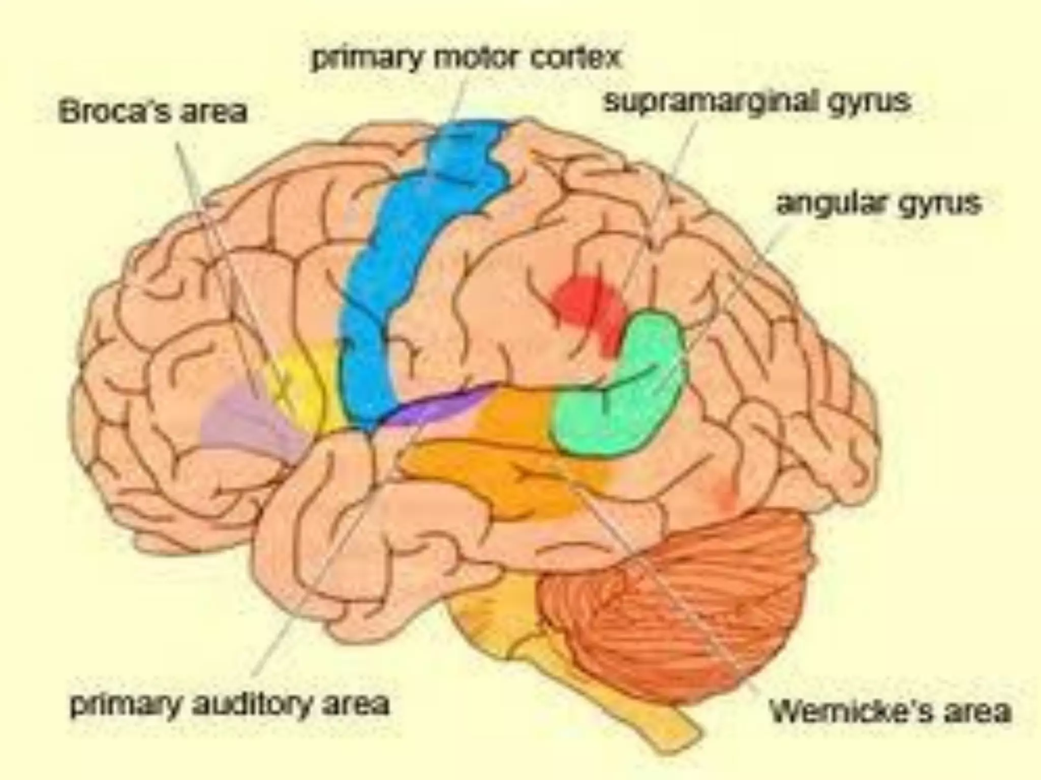
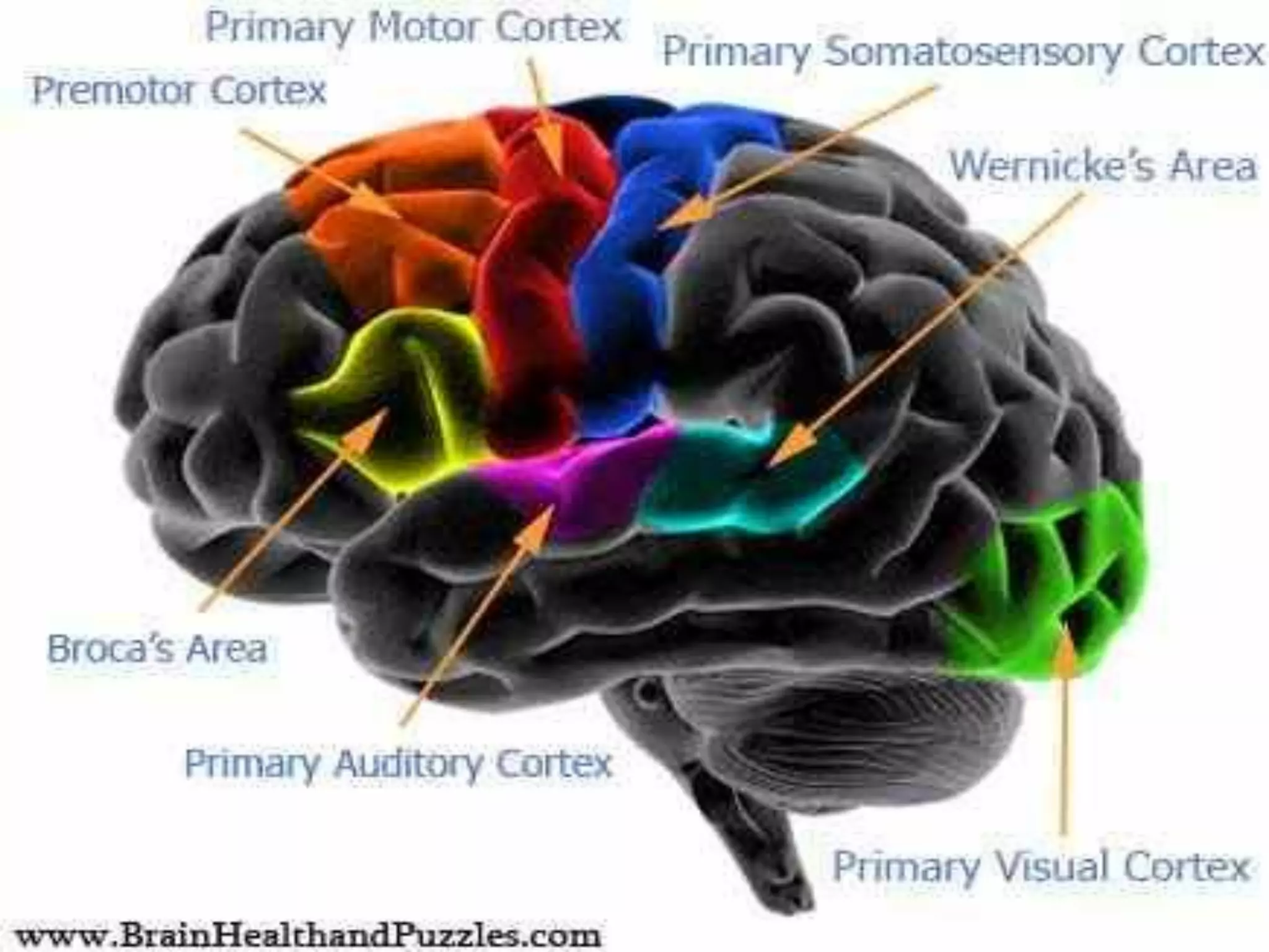
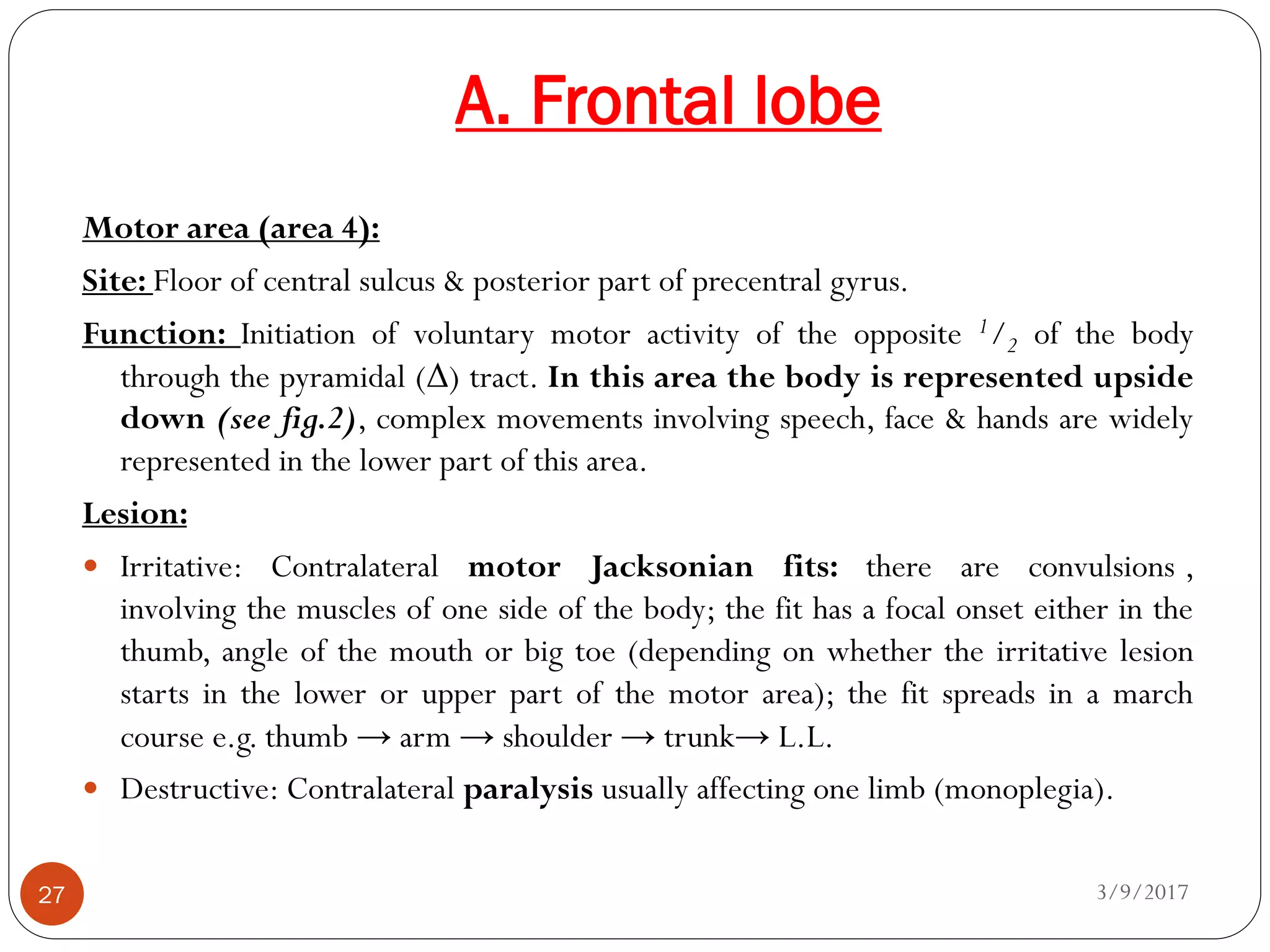

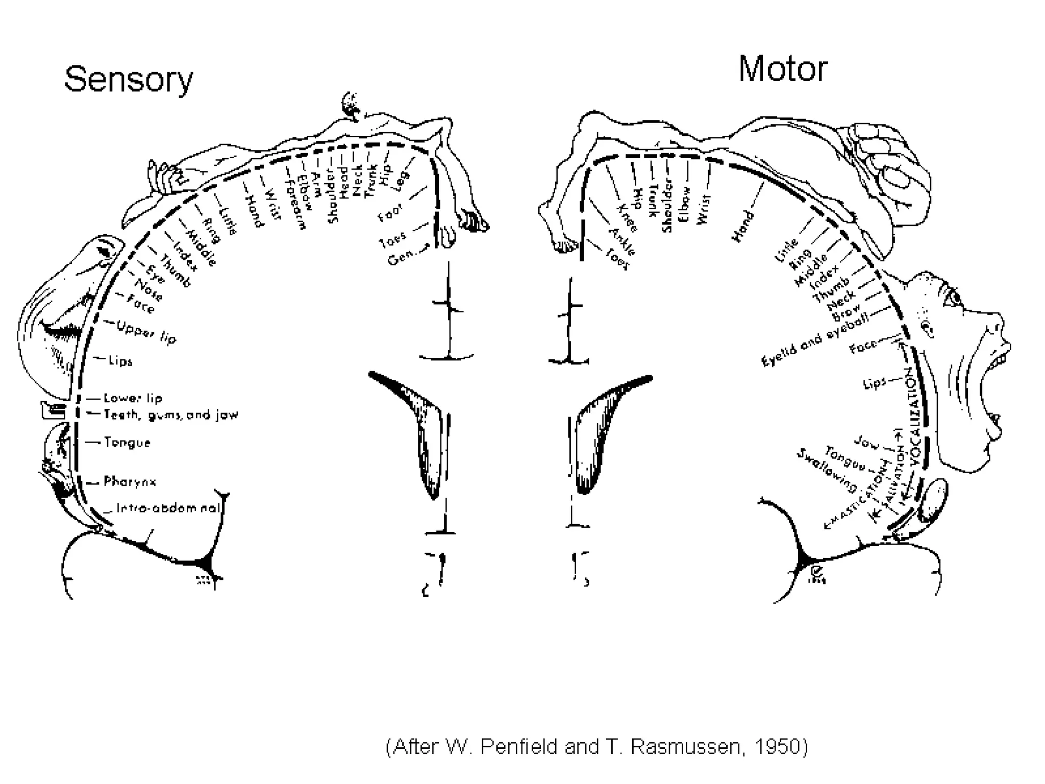
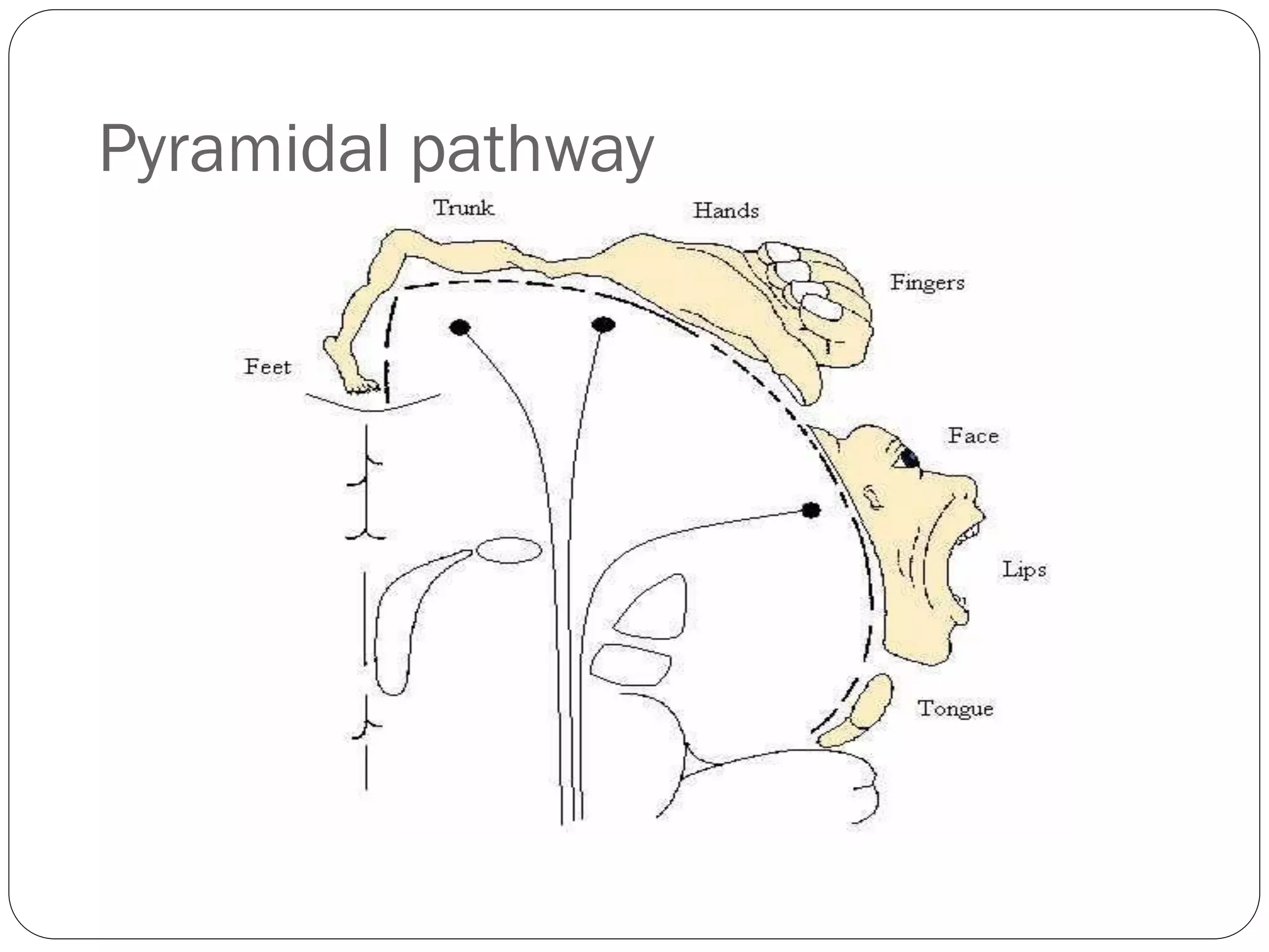
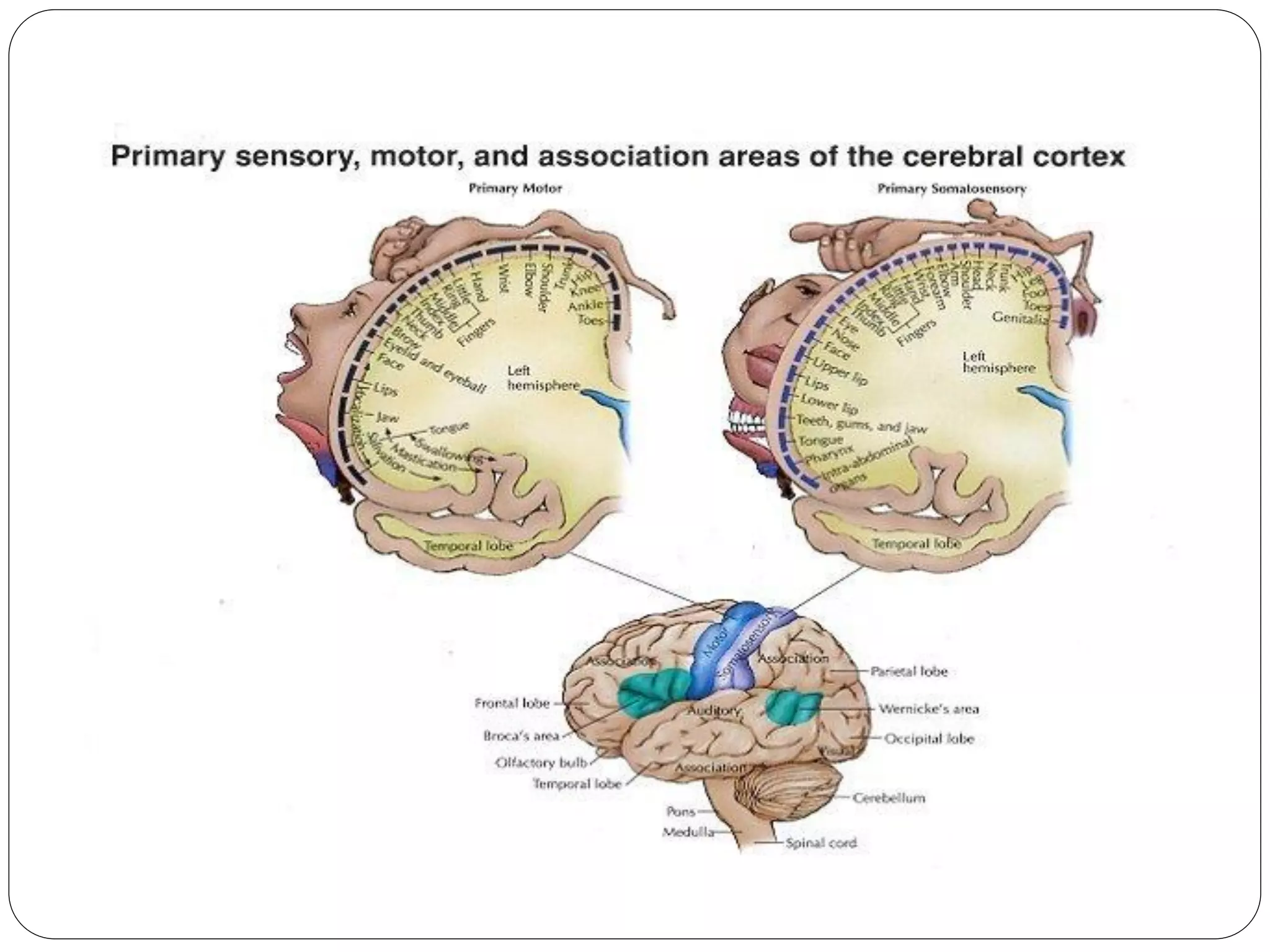


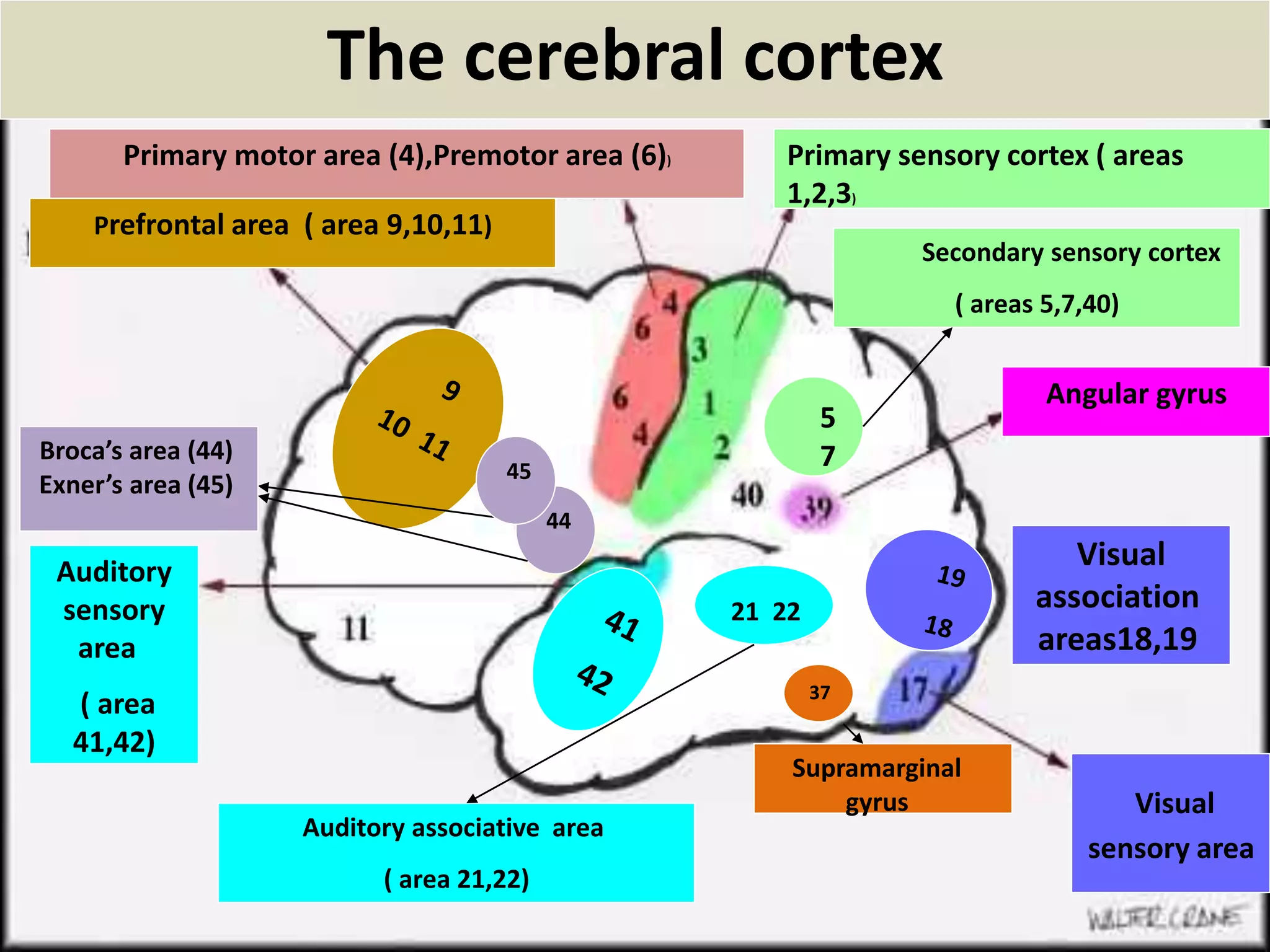
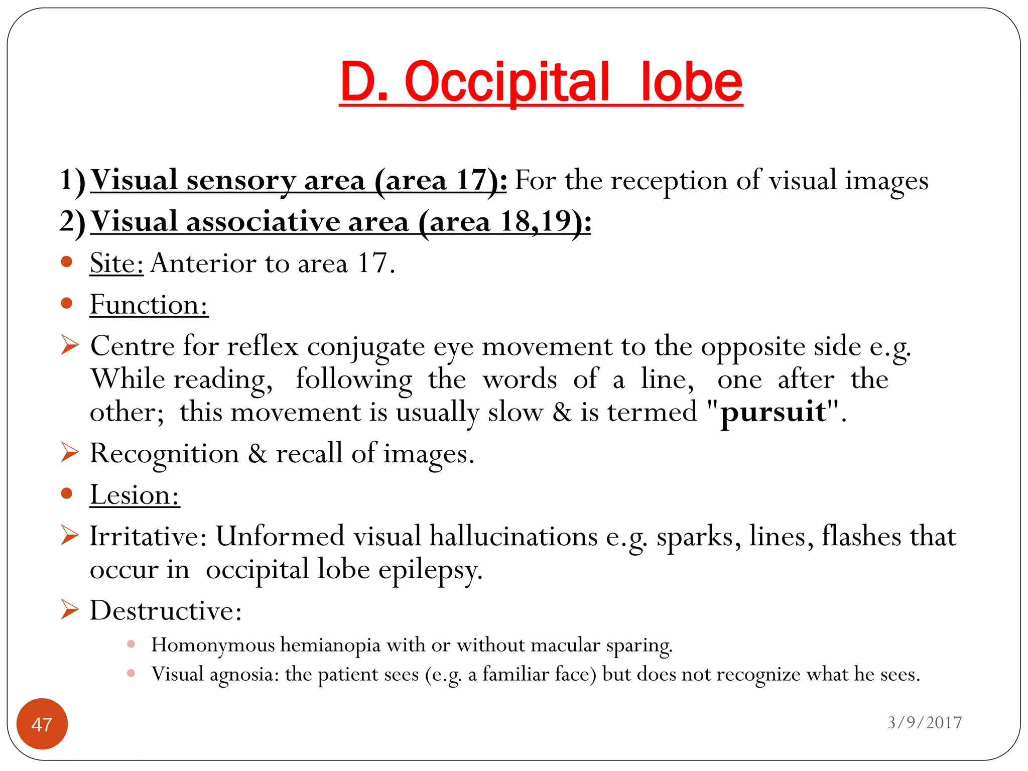


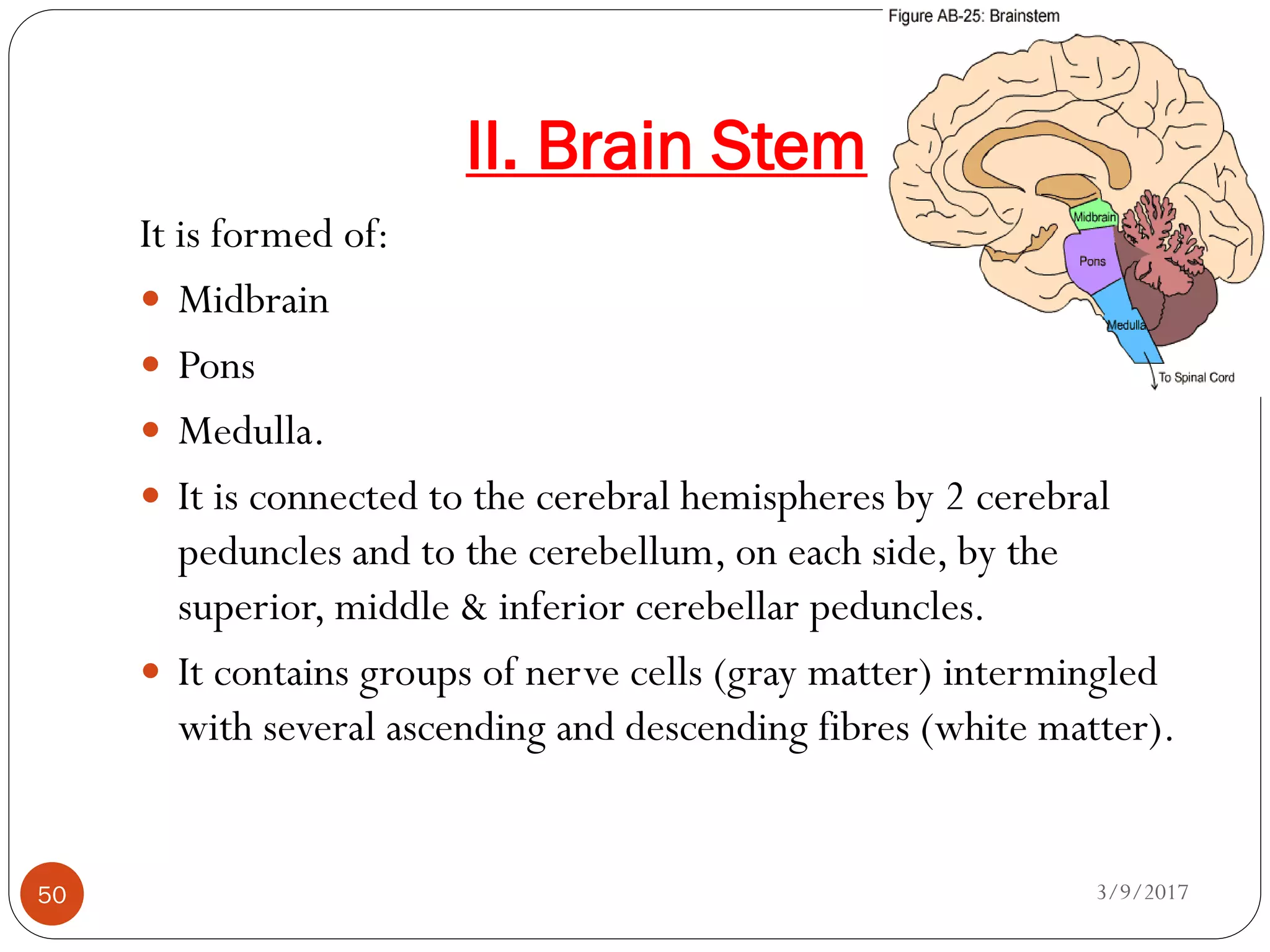
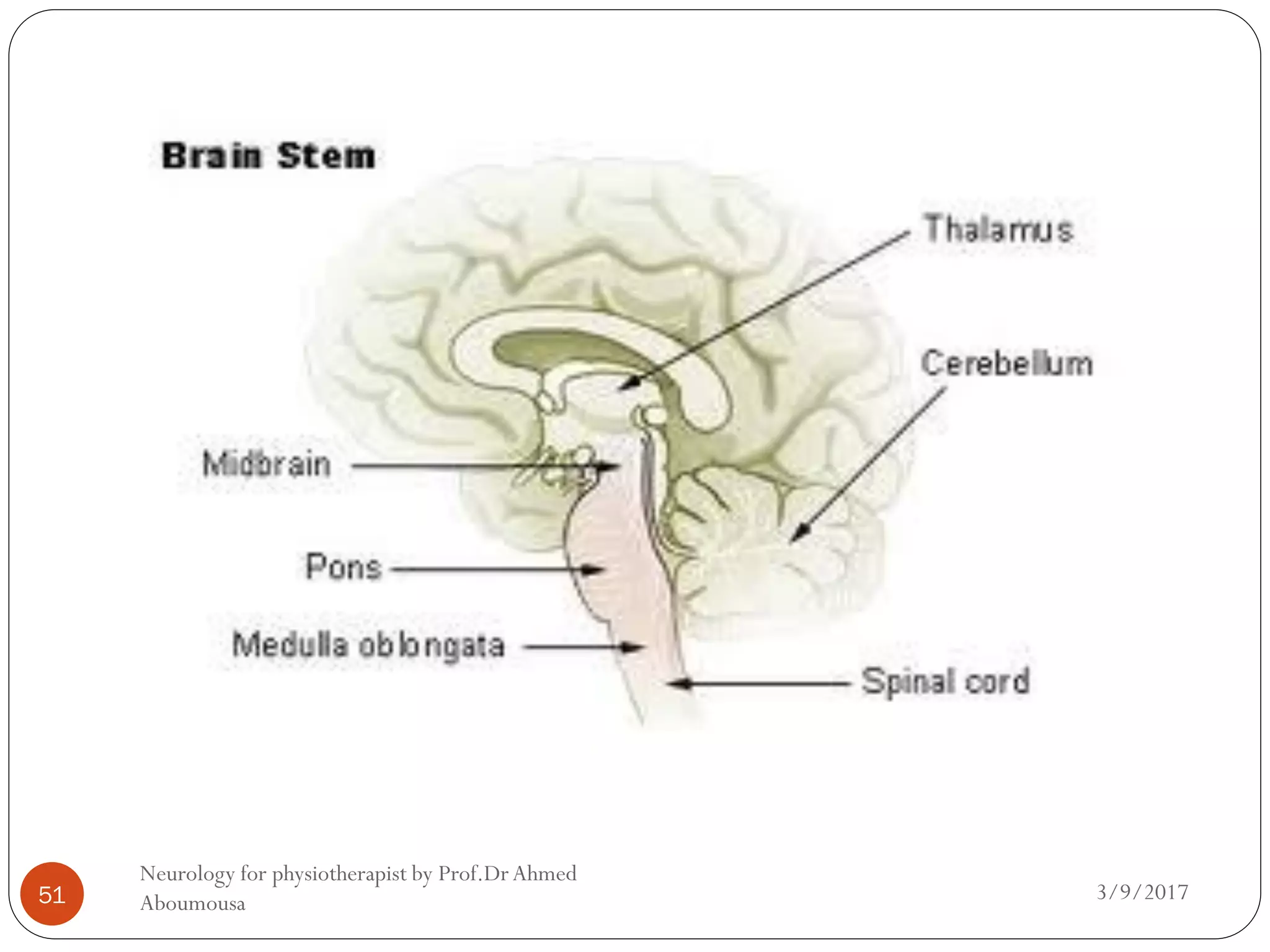


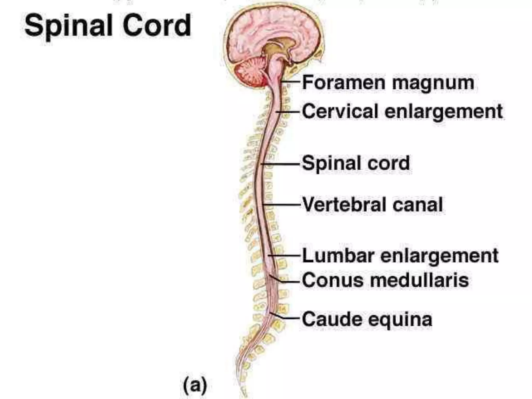
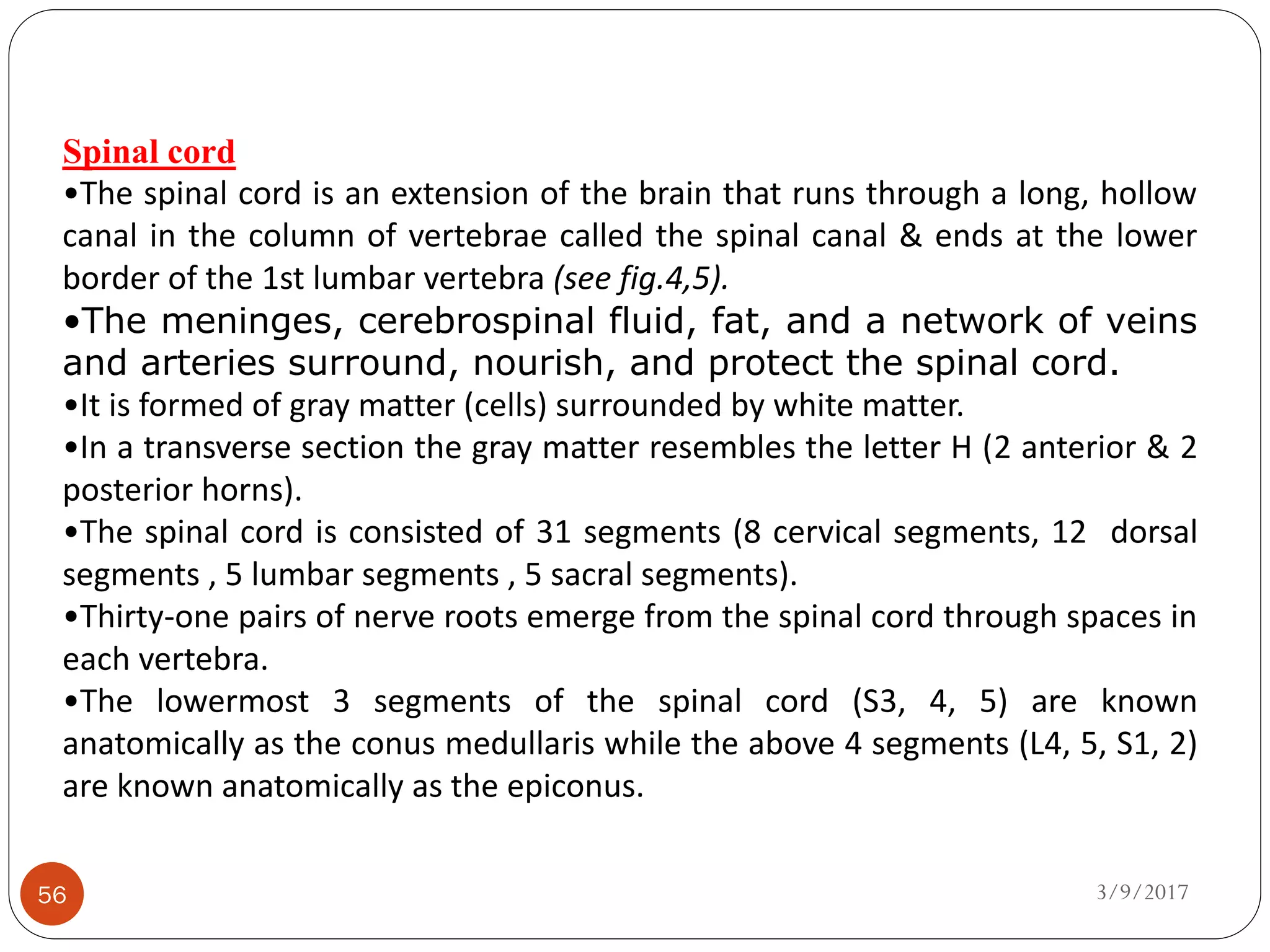
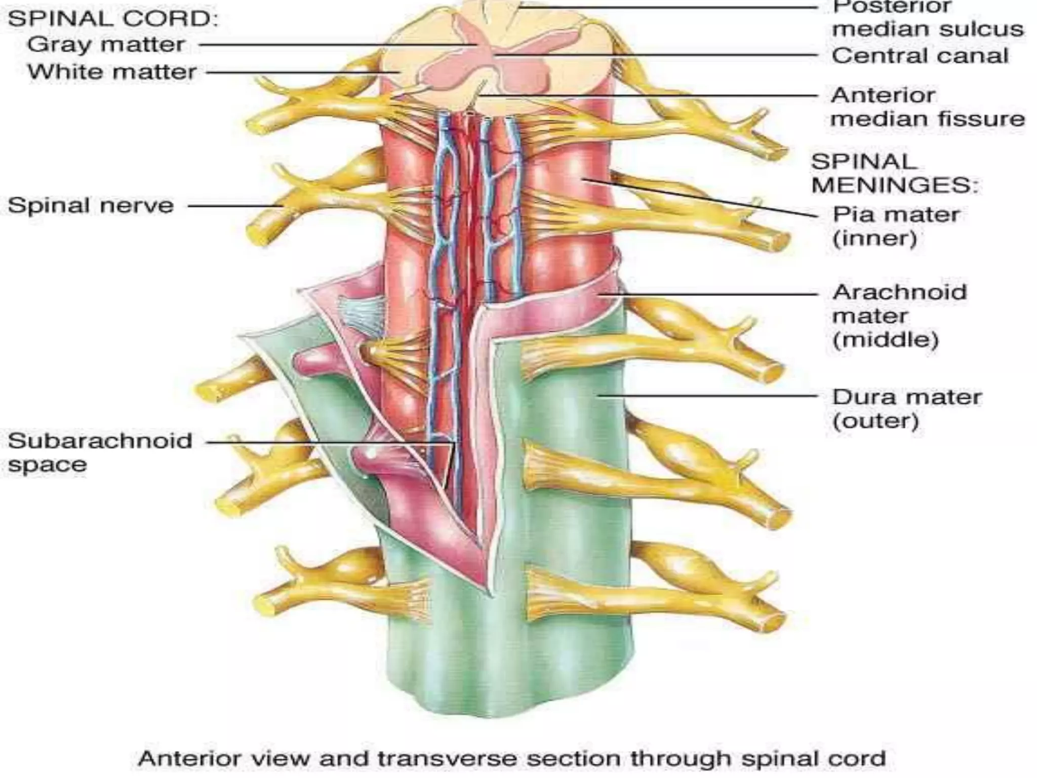
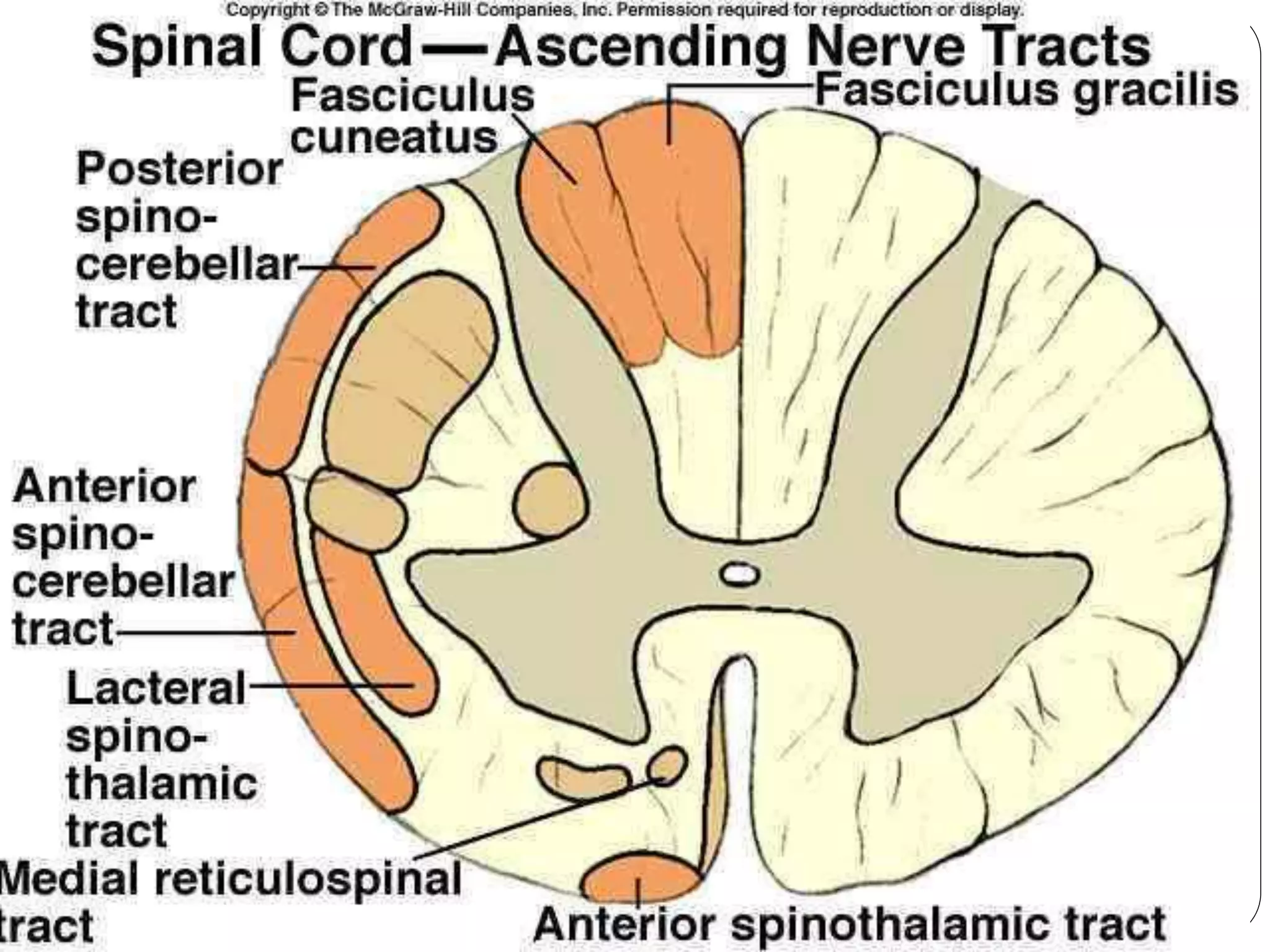
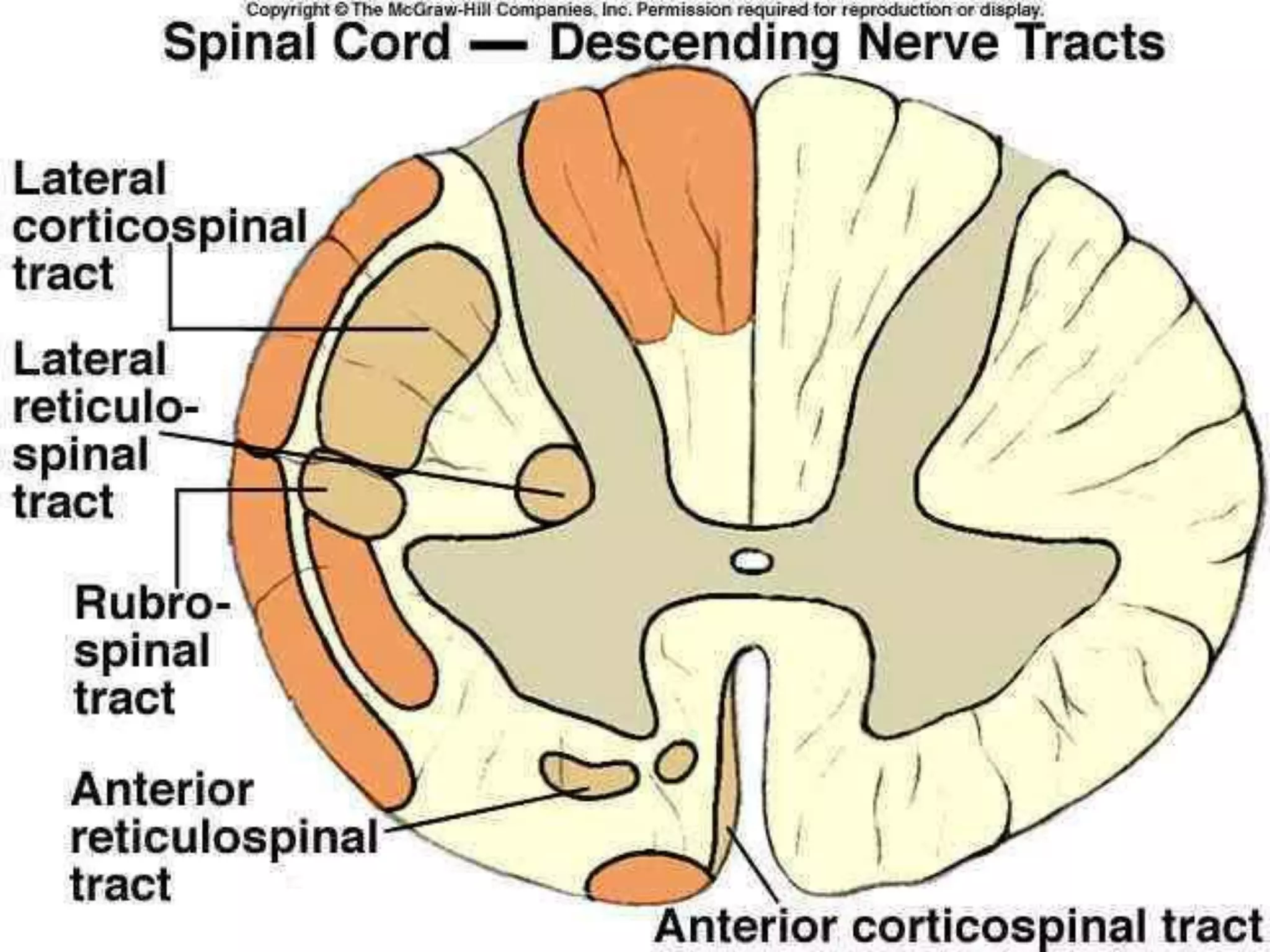


The document provides a detailed overview of the cerebral cortex and its various components, including the cerebrum, brain stem, and spinal cord. It explains the functions and lesions associated with different areas of the brain, notably detailing the roles of the frontal, parietal, temporal, and occipital lobes. Additionally, it discusses the structure and function of the spinal cord and its connectivity to the brain.