Download as PDF, PPTX


![• AION (Ischemic Optic Neuropathy)
• Vasculitic Disorders (i.e. SLE)
• Hereditary (i.e. Leber’s)
• Toxic/Nutritional (ETOH)
• Infectious (i.e.Bartonella, Lyme)
• Inflammatory (i.e. Sarcoid)
• Neoplastic/Paraneoplastic (i.e. lymphoma)
• Compressive (i.e.Tumours, Grave’s orbitopathy)
• Multiple Evanescent White Dot Syndrome [MEWDS]
• Acute Idiopathic Blind Spot Enlargement Syndrome
[AIBSE]).
Optic Neuritis: D.D.](https://image.slidesharecdn.com/cis-170112190731/85/clinically-isolated-syndromes-20-320.jpg)


![MRI brain and cervical cord (1)
with Gd
Abnormal
[conversion rate 80%] (2)
Wait till
CDMS
DMT
Normal
[conversion rate 20%] (2)
Follow up
39
Conversion of CIS to CDMS](https://image.slidesharecdn.com/cis-170112190731/85/clinically-isolated-syndromes-39-320.jpg)


![46
The ONTT was a prospective, randomized, multicenter placebo-
controlled clinical trial designed to compare the benefits of
treatment with
(1) intravenous methylprednisolone (IVMP) (250 mg administered
every 6 h for 3 days followed by oral prednisone [1 mg/kg/day]
for 11 days);
(2) oral prednisone (1 mg/kg/day); or
(3) oral placebo in 457 patients with acute optic neuritis.
Beck RW, Cleary PA, Anderson MM Jr, Keltner JL, Shults WT, Kaufman DI. A randomized, controlled trial of corticosteroids in the treatment of acute optic neuritis. The
Optic Neuritis Study Group. N Engl J Med. 1992 Feb 27. 326(9):581-8.
PLEASE](https://image.slidesharecdn.com/cis-170112190731/85/clinically-isolated-syndromes-46-320.jpg)

![Kasr Alaini Protocol of Manangement of CIS
48
MRI brain and cervical cord (1) with Gd
Abnormal
[conversion rate 80%] (2)
Offer DMT (3)
Normal
[conversion rate 20%] (2)
Positive radiology and/or
CSFNegative results
Follow up: Clinical (q 3 mo)
and Radiological (q 6m or
whenever indicated)
Offer DMT
Comprehensive Approach:
1- Exclude mimics
2- Further assessment: (fMRI (DTI) , CSF
analysis, cognitive assessment (4))
3- Neurophysiological](https://image.slidesharecdn.com/cis-170112190731/85/clinically-isolated-syndromes-48-320.jpg)


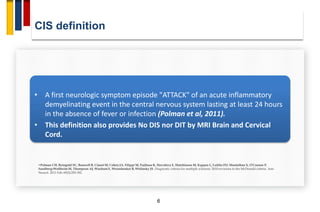
Clinically isolated syndromes (CIS) refer to the first clinical episodes of neurological symptoms suggestive of multiple sclerosis. The document discusses CIS in three parts: definition and clinical features of CIS, risk factors for conversion from CIS to multiple sclerosis, and management of CIS. Regarding clinical features, optic neuritis, transverse myelitis, and brainstem syndromes are highlighted as common presentations of CIS. MRI abnormalities, younger age of onset, smoking, and vitamin D deficiency are identified as risk factors for progression to multiple sclerosis. The management section outlines acute treatment with corticosteroids, use of disease-modifying therapies based on MRI findings, and consideration of vitamin D supplementation.


