The cerebrum : external appearance and internalstructures.pptx
1.
The cerebrum :
theexternal appearance
and internal structures
By
Dr.Abdelraouf Abheiri
MbChb ,MSc Neurosurgery
2025
2.
General Appearance ofthe Cerebral
Hemispheres
• Largest part of the brain
• they are separated by a deep midline sagittal fissure, the longitudinal cerebral
fissure :
• Falx cerebri: dura mater
• The anterior cerebral arteries.
• Corpus callosum
• Tentorium cerebelli : Asecond horizontal fold of dura mater
separates the cerebral hemispheres from the cerebellum
Folds or gyri, which are separated from each other by sulci or fissures
The central and parieto-occipital sulci and the lateral and calcarine sulci are
boundaries used for the division of the cerebral hemisphere into frontal, parietal,
temporal, and occipital lobes.
3.
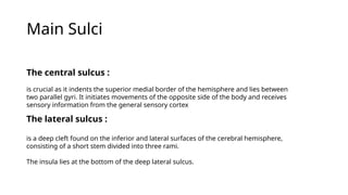
Main Sulci
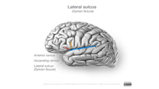
The lateralsulcus :
is a deep cleft found on the inferior and lateral surfaces of the cerebral hemisphere,
consisting of a short stem divided into three rami.
The insula lies at the bottom of the deep lateral sulcus.
The central sulcus :
is crucial as it indents the superior medial border of the hemisphere and lies between
two parallel gyri. It initiates movements of the opposite side of the body and receives
sensory information from the general sensory cortex
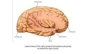
Lateral view ofthe right cerebral hemisphere dissected
to reveal the right Insula.
10.
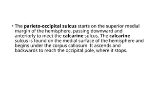
• The parieto-occipitalsulcus starts on the superior medial
margin of the hemisphere, passing downward and
anteriorly to meet the calcarine sulcus. The calcarine
sulcus is found on the medial surface of the hemisphere and
begins under the corpus callosum. It ascends and
backwards to reach the occipital pole, where it stops.
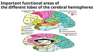
11.
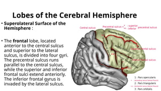
Lobes of theCerebral Hemisphere
• Superolateral Surface of the
Hemisphere :
• The frontal lobe, located
anterior to the central sulcus
and superior to the lateral
sulcus, is divided into four gyri.
The precentral sulcus runs
parallel to the central sulcus,
while the superior and inferior
frontal sulci extend anteriorly.
The inferior frontal gyrus is
invaded by the lateral sulcus.
12.
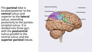
• The parietallobe is
located posterior to the
central sulcus and
superior to the lateral
sulcus, extending
posteriorly to the parieto-
occipital sulcus. It is
divided into three gyri,
with the postcentral
sulcus parallel to the
central sulcus and the
superior parietal lobule.
13.
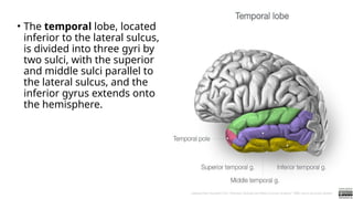
• The temporallobe, located
inferior to the lateral sulcus,
is divided into three gyri by
two sulci, with the superior
and middle sulci parallel to
the lateral sulcus, and the
inferior gyrus extends onto
the hemisphere.
14.
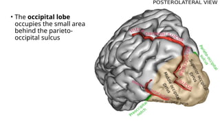
• The occipitallobe
occupies the small area
behind the parieto-
occipital sulcus
15.
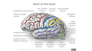
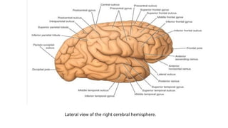
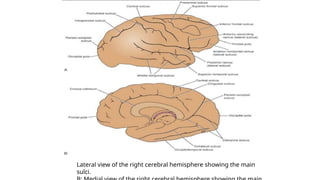
Lateral view ofthe right cerebral hemisphere showing the main
sulci.
16.
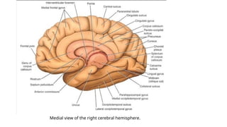
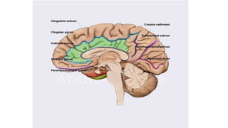
Medial and InferiorSurfaces of the
Hemisphere
• The cerebral hemisphere’s lobes are not clearly defined on
medial and inferior surfaces, but important areas include
the corpus callosum, cingulate gyrus, and superior
frontal gyrus. The cingulate gyrus is separated from the
corpus callosum and superior frontal gyrus by the cingulate
sulcus.
• The paracentral lobule is the cerebral cortex surrounding
the central sulcus indentation, extending from the
precentral gyrus on the superior lateral surface to the
postcentral gyrus.
17.
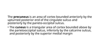
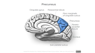
The precuneus isan area of cortex bounded anteriorly by the
upturned posterior end of the cingulate sulcus and
posteriorly by the parieto-occipital sulcus.
• The cuneus is a triangular area of cortex bounded above by
the parietooccipital sulcus, inferiorly by the calcarine sulcus,
and posteriorly by the superior medial margin
19.
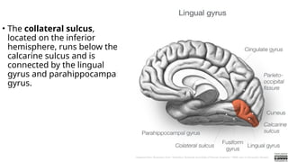
• The collateralsulcus,
located on the inferior
hemisphere, runs below the
calcarine sulcus and is
connected by the lingual
gyrus and parahippocampal
gyrus.
20.
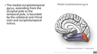
• The medialoccipitotemporal
gyrus, extending from the
occipital pole to the
temporal pole, is bounded
by the collateral and rhinal
sulci and occipitotemporal
sulcus.
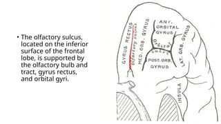
21.
• The olfactorysulcus,
located on the inferior
surface of the frontal
lobe, is supported by
the olfactory bulb and
tract, gyrus rectus,
and orbital gyri.
Frontal Lobe:
• (1)TheMotor area )area 4
• Site: occupies the precentral gyrus & extends to occupy the
ant part of the paracentral lobule on the medial Surface of
the cerebral hemisphere
• Function: it Contains the giant pyramidal cells of Betz which
give origin to 80%. the pyramidal tract which controls the
motor activity of the skeletal muscles of the opposite half of
the body (except the eye
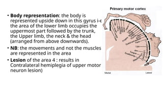
24.
• Body representation:the body is
represented upside down in this gyrus i-e
the area of the lower limb occupies the
uppermost part followed by the trunk,
the Upper limb, the neck & the head
(arranged from above downwards).
• NB: the movements and not the muscles
are represented in the area
• Lesion of the area 4 : results in
Contralateral hemiplegia of upper motor
neuron lesion)
25.
2) Premotor area(area 6
• Site: infront & parallel to the motor area. It is wide above
(scm)&narrow below (1 cm)
• Function: it is the main extrapyramidal area for the body
except the eye (which is found in the occipital lobe)
• (3) Frontal eye field (area 8):
• Site: infront of the premator area in the post part of the
middle frontal gyrus.
26.
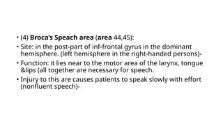
• (4) Broca’sSpeach area (area 44,45):
• Site: in the post-part of inf-frontal gyrus in the dominant
hemisphere. (left hemisphere in the right-handed persons)-
• Function: it lies near to the motor area of the larynx, tongue
&lips (all together are necessary for speech.
• Injury to this are causes patients to speak slowly with effort
(nonfluent speech)-
27.
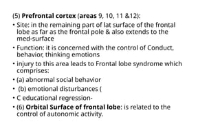
(5) Prefrontal cortex(areas 9, 10, 11 &12):
• Site: in the remaining part of lat surface of the frontal
lobe as far as the frontal pole & also extends to the
med-surface
• Function: it is concerned with the control of Conduct,
behavior, thinking emotions
• injury to this area leads to Frontal lobe syndrome which
comprises:
• (a) abnormal social behavior
• (b) emotional disturbances (
• C educational regression-
• (6) Orbital Surface of frontal lobe: is related to the
control of autonomic activity.
29.
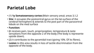
Parietal Lobe
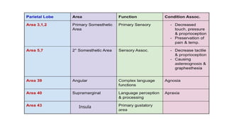
• (1)1y Somatosensory cortex (Main sensory area): areas 3,1,2
• Site: it occupies the postcentral gyrus on the lat-surface of the
cerebral hemisphere & extends to the post part of the paracentral
lobule on the med surface
• Function:
• (it receives pain, touch, proprioception, temperature & taste
sensations from the opposite 2 of the body (The body is represente
upside down).
• (2) it contributes to the pyramidal tract (giving 10%. Of its fibres).
• Lesion to this area results in loss of tactile discrimination from the
opposite of the body.
30.
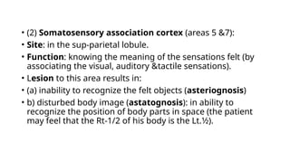
• (2) Somatosensoryassociation cortex (areas 5 &7):
• Site: in the sup-parietal lobule.
• Function: knowing the meaning of the sensations felt (by
associating the visual, auditory &tactile sensations).
• Lesion to this area results in:
• (a) inability to recognize the felt objects (asteriognosis)
• b) disturbed body image (astatognosis): in ability to
recognize the position of body parts in space (the patient
may feel that the Rt-1/2 of his body is the Lt.1⁄2).
31.
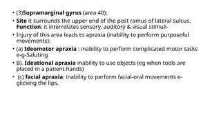
• (3)Supramarginal gyrus(area 40):
• Site it surrounds the upper end of the post ramus of lateral sulcus.
Function: it interrelates sensory, auditory & visual stimuli-
• Injury of this area leads to apraxia (inability to perform purposeful
movements):
• (a) Ideomotor apraxia : inability to perform complicated motor tasks
e-g-Saluting
• B). Ideational apraxia inability to use objects (eg when tools are
placed in a patient hands)
• (c) facial apraxia: inability to perform facial-oral movements e-
glicking the lips.
32.
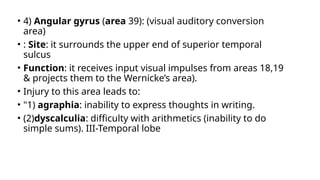
• 4) Angulargyrus (area 39): (visual auditory conversion
area)
• : Site: it surrounds the upper end of superior temporal
sulcus
• Function: it receives input visual impulses from areas 18,19
& projects them to the Wernicke’s area).
• Injury to this area leads to:
• "1) agraphia: inability to express thoughts in writing.
• (2)dyscalculia: difficulty with arithmetics (inability to do
simple sums). III-Temporal lobe
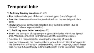
Temporal lobe
1) AuditorySensory area (area 41,42):
• Site: in the middle part of the sup.temporal gyrus (Heschl’s gyrus)
• Function: it receives the auditory radiation from the medial geniculate
body.
• Injury: unilateral destruction results in only partial deafness (due to
bilateral Cochlear representation).
• (2) Auditory association area (area 22)
• Site in the post-part of Sup-temporal gyrus-It includes Werniches Speech
area. Which is connected to Broca’s area by the arcuate fasciculus.
• Function: it is responsible for knowing the meaning of sounds
• Injury of this area in the dominant hemisphere leads to Wernicke’s aphasia:
the patient finds difficulty in understanding spoken language, speaks faster
than normal & has difficulty in Finding the right words to express himself:
35.
• (3) TheInsula
• Site: deep within the lateral sulcus
• Function: the gustatory cortex (area 43) in the parietal
operculum & para-. Insular Cortex It receives taste impulses
from the thalamus.
36.
• 1) VisualSensory area (area 17):
• Site: found mainly in the med-surface of occipital lobe in both banks of
Calcarine sulcus
• Function: it receives input visual sensation from the lat geniculate
body via the optic radiation
• Injury: results in visual field defects eg contralateral homonymous
hemianopia
• (2) Visual association area (areas 18,19):
• Site: in the cuneus & lingual gyrus, surrounding the visual sensory area
17.
• Function: it is responsible for knowing the meaning of the pictures
seen.
• Injury: may result in visual hallucinations.
The occipital lobe