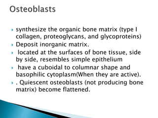
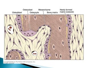
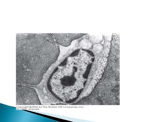
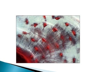
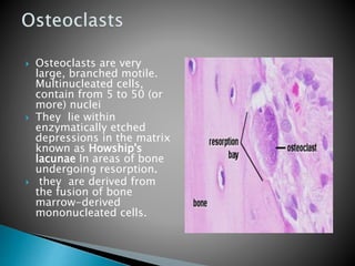
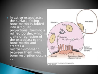
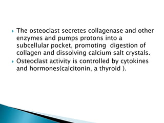
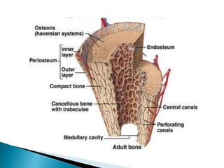
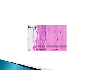
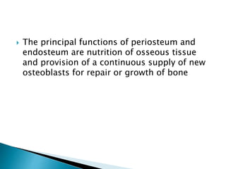
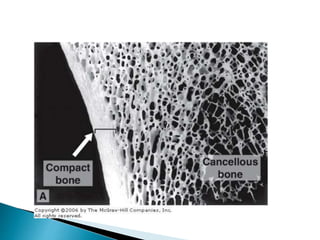
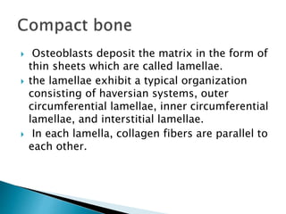
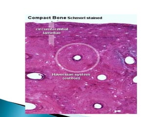
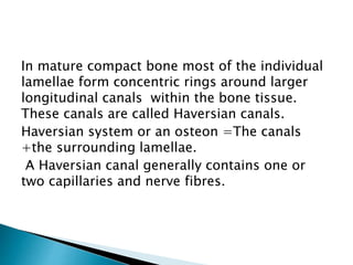
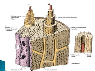
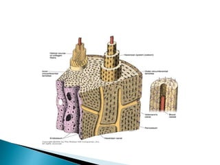
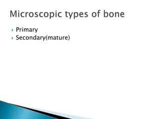
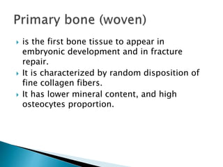
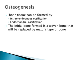
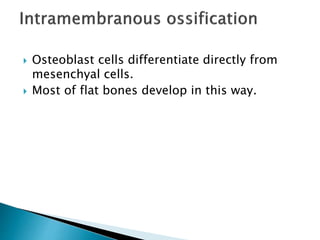
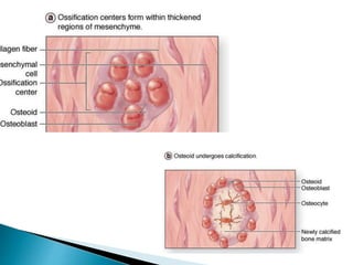
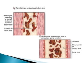
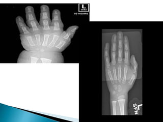
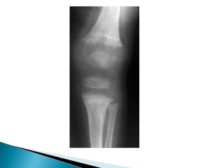
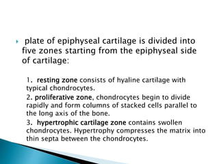
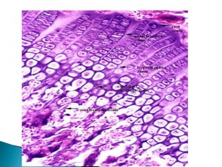
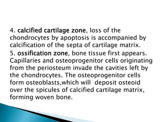
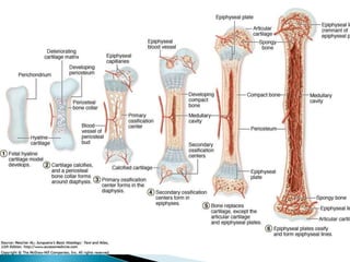
Dr. shiraz yousif
 Bone function
 Composition
 Periosteum
 Bone is the main component of the skeleton
in the adult human.
 It is a specialized form of connective tissue.
 Composed of intercellular calcified structures.
 Multiply and transform the force generated by
muscles into body movement
 supports the body against gravity.
 protects vital organs.
 harbors the bone marrow, where.
 metabolically serves as a reservoir of
calcium, phosphate, and other ions.
 Cells
 Matrix
 synthesize the organic bone matrix (type I
collagen, proteoglycans, and glycoproteins)
 Deposit inorganic matrix.
 located at the surfaces of bone tissue, side
by side, resembles simple epithelium
 have a cuboidal to columnar shape and
basophilic cytoplasm(When they are active).
 . Quiescent osteoblasts (not producing bone
matrix) become flattened.
 osteoblasts are polarized cells.
 bone apposition is a process when matrix
components are secreted at the osteoblast
cell surface, which is in contact with older
bone matrix, producing a layer of new
osteoid matrix ( not yet calcified).
 This process is completed by subsequent
deposition of calcium salts into the osteoid.
 Some osteoblasts are gradually surrounded
by newly formed matrix and become
osteocytes.
 derive from osteoblasts
 each osteocyte lie in the separate lacunae
situated between lamellae (leaf) of matrix
 Have canaliculi which thin, cylindrical matrix
house their cytoplasmic processes through
which they make contact with adjacent cells
via gap junctions, and exchange between
osteocytes and blood vessels also takes place
.
 This exchange can provide nourishment for a
chain of about 15 cells.
 They have flat shape, reduced rough
endoplasmic reticulum and Golgi complex
and condensed nuclear chromatin.
 They are actively involved in the maintenance
of the bony matrix, and their death is
followed by resorption of this matrix.
 Osteoclasts are very
large, branched motile.
Multinucleated cells,
contain from 5 to 50 (or
more) nuclei
 They lie within
enzymatically etched
depressions in the matrix
known as Howship's
lacunae In areas of bone
undergoing resorption.
 they are derived from
the fusion of bone
marrow-derived
mononucleated cells.
 In active osteoclasts,
the surface-facing
bone matrix is folded
into irregular
projections, forming a
ruffled border, which is
a site of adhesion of
the osteoclast to the
bone matrix and
creates a
microenvironment
between them which
bone resorption occurs
. .
 The osteoclast secretes collagenase and other
enzymes and pumps protons into a
subcellular pocket, promoting digestion of
collagen and dissolving calcium salt crystals.
 Osteoclast activity is controlled by cytokines
and hormones(calcitonin, a thyroid ).
 Organic component include type I collagen,
proteoglycans, and glycoproteins.
 Inorganic matter represents about 50% of the
dry weight of bone matrix. Mostly Calcium
and phosphate
 Inorganic matter
 represents about 50% of the dry weight of
bone matrix.
 Contains mainly calcium and phosphorus in
form of hydroxyapatite crystals
 IThe surface ions of hydroxyapatite are
hydrated, and a layer of water and ions forms
around the crystal. This layer, the hydration
shell, facilitates the exchange of ions
between the crystal and the body fluids
 The organic matter
 type I collagen and , proteoglycan aggregates and
glycoproteins.
 Bone glycoproteins may be responsible for
promoting calcification of bone matrix.
 The association of minerals with collagen fibers is
responsible for the hardness and resistance of bone
tissue.
 After a bone is decalcified, its shape is preserved,
but it becomes as flexible as a tendon.
 Removal of the organic part of the matrix leaves the
bone with its original shape but fragile, breaking
easily.
 layers of bone-forming cells and connective tissue
covering external and internal surfaces of bone .
 The periosteum:
◦ consists of two layers:outer layer of collagen fibers and
fibroblasts
◦ The inner, more cellular and composed of osteoprogenitor
cells, with the potential to divide by mitosis and
differentiate into osteoblasts.
◦ Sharpey's fibers.Bundles of periosteal collagen binding the
periosteum to bone.
 Endosteum:
 lines internal cavities within the bone and is
composed of a single layer of flattened
osteoprogenitor cells and a very small amount of
connective tissue.
 The principal functions of periosteum and
endosteum are nutrition of osseous tissue
and provision of a continuous supply of new
osteoblasts for repair or growth of bone.
 The principal functions of periosteum and
endosteum are nutrition of osseous tissue
and provision of a continuous supply of new
osteoblasts for repair or growth of bone
 Two types of bone macroscopically:
 Compact bone: dense outer sheet covering
flat bones and shaft of long bones
 Trabecular/cancellous/spongy bone: present
within the central marrow cavity comprised of
network of delicate bars and sheet of
trabecular bone which branch and intersect to
form a sponge-like network
 Osteoblasts deposit the matrix in the form of
thin sheets which are called lamellae.
 the lamellae exhibit a typical organization
consisting of haversian systems, outer
circumferential lamellae, inner circumferential
lamellae, and interstitial lamellae.
 In each lamella, collagen fibers are parallel to
each other.
In mature compact bone most of the individual
lamellae form concentric rings around larger
longitudinal canals within the bone tissue.
These canals are called Haversian canals.
Haversian system or an osteon =The canals
+the surrounding lamellae.
A Haversian canal generally contains one or
two capillaries and nerve fibres.
 A second system of canals, called Volkmann's
canals, penetrates the bone perpendicular to
its surface.
 These canals establish connections of the
Haversian canals with the inner and outer
surfaces of the bone
 the matrix of trabecular bone is also
deposited in the form of lamellae.
 In mature bones, trabecular bone will also be
lamellar bone. However, lamellae in
trabecular bone do not form Haversian
systems.
 Primary
 Secondary(mature)
 is the first bone tissue to appear in
embryonic development and in fracture
repair.
 It is characterized by random disposition of
fine collagen fibers.
 It has lower mineral content, and high
osteocytes proportion.
 Primary bone tissue is usually temporary and
is replaced in adults by secondary bone tissue
except in a very few places in the body, eg,
near the sutures of the calvaria, in tooth
sockets, and in the insertions of some
tendons.
 …………………………………………………………
…………………………………………………………
…………………………………………………
 bone tissue can be formed by
◦ Intramembranous ossification
◦ Endochondral ossification
 The initial bone formed is a woven bone that
will be replaced by mature type of bone
 Osteoblast cells differentiate directly from
mesenchyal cells.
 Most of flat bones develop in this way.
 ossification center appear in mesenchymal
membrane.
 The cells will differentiate into osteoblast.
 Osteoblasts will secret the ECM, and when they
are trapped within the osteon they will be
osteocytes.
 The collagen fibres of the osteoid form a woven
network without a preferred orientation.
 Further deposition and calcification of osteoid
at sites where compact bone is needed leads
to the formation of primitive compact bone.
 The portions of the connective tissue layer
that do not undergo ossification give rise to
the endosteum and the periosteum of the
new bone.
 ossification takes place within a piece of
hyaline cartilage whose shape resembles a
small version, or model, of the bone to be
formed.
 This type of ossification is principally
responsible for the formation of short and
long bones
 Osteoblast within the surrounding
perichondrium will produce the first bone
collar surrounding the diaphysis of the
cartilage model.
 this will decrease the diffusion of oxygen
and nutrients into the underlying cartilage,
leading to degenerative changes. The
chondrocytes will swell up (hypertrophy),
enlarging their lacunae. and the matrix will be
calcified.
 Blood vessels from the former perichondrium
now the periosteum penetrate through the
new bone formed, bringing osteoprogenitor
cells to the central region.
 osteoblasts adhere to the calcified cartilage
matrix and produce continuous layers of
primary bone that surround the cartilaginous
matrix
 Primary centers appear in the diaphysis
before birth
 Secondary centers appear later in life in the
epiphysis.
 In the secondary ossification centers,
cartilage remains in two regions:
◦ the articular cartilage
◦ the epiphyseal cartilage ( epiphyseal plate or
growth plate)

 The epiphyseal cartilage is responsible for the
growth in length of the bone and disappears
in adults.
 Disappearance(closure) of this plate occurs at
different time in different bone and this is
important is identifying bone age.
 plate of epiphyseal cartilage is divided into
five zones starting from the epiphyseal side
of cartilage:
1. resting zone consists of hyaline cartilage with
typical chondrocytes.
2. proliferative zone, chondrocytes begin to divide
rapidly and form columns of stacked cells parallel to
the long axis of the bone.
3. hypertrophic cartilage zone contains swollen
chondrocytes. Hypertrophy compresses the matrix into
thin septa between the chondrocytes.
4. calcified cartilage zone, loss of the
chondrocytes by apoptosis is accompanied by
calcification of the septa of cartilage matrix.
5. ossification zone, bone tissue first appears.
Capillaries and osteoprogenitor cells originating
from the periosteum invade the cavities left by
the chondrocytes. The osteoprogenitor cells
form osteoblasts,which will deposit osteoid
over the spicules of calcified cartilage matrix,
forming woven bone.
Basic Histology number 8 bone tissue .pptx

Basic Histology number 8 bone tissue .pptx

  • 1.
  • 2.
     Bone function Composition  Periosteum
  • 3.
     Bone isthe main component of the skeleton in the adult human.  It is a specialized form of connective tissue.  Composed of intercellular calcified structures.
  • 4.
     Multiply andtransform the force generated by muscles into body movement  supports the body against gravity.  protects vital organs.  harbors the bone marrow, where.  metabolically serves as a reservoir of calcium, phosphate, and other ions.
  • 5.
  • 7.
     synthesize theorganic bone matrix (type I collagen, proteoglycans, and glycoproteins)  Deposit inorganic matrix.  located at the surfaces of bone tissue, side by side, resembles simple epithelium  have a cuboidal to columnar shape and basophilic cytoplasm(When they are active).  . Quiescent osteoblasts (not producing bone matrix) become flattened.
  • 9.
     osteoblasts arepolarized cells.  bone apposition is a process when matrix components are secreted at the osteoblast cell surface, which is in contact with older bone matrix, producing a layer of new osteoid matrix ( not yet calcified).  This process is completed by subsequent deposition of calcium salts into the osteoid.  Some osteoblasts are gradually surrounded by newly formed matrix and become osteocytes.
  • 10.
     derive fromosteoblasts  each osteocyte lie in the separate lacunae situated between lamellae (leaf) of matrix  Have canaliculi which thin, cylindrical matrix house their cytoplasmic processes through which they make contact with adjacent cells via gap junctions, and exchange between osteocytes and blood vessels also takes place .  This exchange can provide nourishment for a chain of about 15 cells.
  • 13.
     They haveflat shape, reduced rough endoplasmic reticulum and Golgi complex and condensed nuclear chromatin.  They are actively involved in the maintenance of the bony matrix, and their death is followed by resorption of this matrix.
  • 14.
     Osteoclasts arevery large, branched motile. Multinucleated cells, contain from 5 to 50 (or more) nuclei  They lie within enzymatically etched depressions in the matrix known as Howship's lacunae In areas of bone undergoing resorption.  they are derived from the fusion of bone marrow-derived mononucleated cells.
  • 15.
     In activeosteoclasts, the surface-facing bone matrix is folded into irregular projections, forming a ruffled border, which is a site of adhesion of the osteoclast to the bone matrix and creates a microenvironment between them which bone resorption occurs . .
  • 16.
     The osteoclastsecretes collagenase and other enzymes and pumps protons into a subcellular pocket, promoting digestion of collagen and dissolving calcium salt crystals.  Osteoclast activity is controlled by cytokines and hormones(calcitonin, a thyroid ).
  • 17.
     Organic componentinclude type I collagen, proteoglycans, and glycoproteins.  Inorganic matter represents about 50% of the dry weight of bone matrix. Mostly Calcium and phosphate
  • 18.
     Inorganic matter represents about 50% of the dry weight of bone matrix.  Contains mainly calcium and phosphorus in form of hydroxyapatite crystals  IThe surface ions of hydroxyapatite are hydrated, and a layer of water and ions forms around the crystal. This layer, the hydration shell, facilitates the exchange of ions between the crystal and the body fluids
  • 19.
     The organicmatter  type I collagen and , proteoglycan aggregates and glycoproteins.  Bone glycoproteins may be responsible for promoting calcification of bone matrix.  The association of minerals with collagen fibers is responsible for the hardness and resistance of bone tissue.  After a bone is decalcified, its shape is preserved, but it becomes as flexible as a tendon.  Removal of the organic part of the matrix leaves the bone with its original shape but fragile, breaking easily.
  • 21.
     layers ofbone-forming cells and connective tissue covering external and internal surfaces of bone .  The periosteum: ◦ consists of two layers:outer layer of collagen fibers and fibroblasts ◦ The inner, more cellular and composed of osteoprogenitor cells, with the potential to divide by mitosis and differentiate into osteoblasts. ◦ Sharpey's fibers.Bundles of periosteal collagen binding the periosteum to bone.  Endosteum:  lines internal cavities within the bone and is composed of a single layer of flattened osteoprogenitor cells and a very small amount of connective tissue.
  • 24.
     The principalfunctions of periosteum and endosteum are nutrition of osseous tissue and provision of a continuous supply of new osteoblasts for repair or growth of bone.
  • 25.
     The principalfunctions of periosteum and endosteum are nutrition of osseous tissue and provision of a continuous supply of new osteoblasts for repair or growth of bone
  • 26.
     Two typesof bone macroscopically:  Compact bone: dense outer sheet covering flat bones and shaft of long bones  Trabecular/cancellous/spongy bone: present within the central marrow cavity comprised of network of delicate bars and sheet of trabecular bone which branch and intersect to form a sponge-like network
  • 28.
     Osteoblasts depositthe matrix in the form of thin sheets which are called lamellae.  the lamellae exhibit a typical organization consisting of haversian systems, outer circumferential lamellae, inner circumferential lamellae, and interstitial lamellae.  In each lamella, collagen fibers are parallel to each other.
  • 30.
    In mature compactbone most of the individual lamellae form concentric rings around larger longitudinal canals within the bone tissue. These canals are called Haversian canals. Haversian system or an osteon =The canals +the surrounding lamellae. A Haversian canal generally contains one or two capillaries and nerve fibres.
  • 32.
     A secondsystem of canals, called Volkmann's canals, penetrates the bone perpendicular to its surface.  These canals establish connections of the Haversian canals with the inner and outer surfaces of the bone
  • 34.
     the matrixof trabecular bone is also deposited in the form of lamellae.  In mature bones, trabecular bone will also be lamellar bone. However, lamellae in trabecular bone do not form Haversian systems.
  • 38.
  • 39.
     is thefirst bone tissue to appear in embryonic development and in fracture repair.  It is characterized by random disposition of fine collagen fibers.  It has lower mineral content, and high osteocytes proportion.
  • 40.
     Primary bonetissue is usually temporary and is replaced in adults by secondary bone tissue except in a very few places in the body, eg, near the sutures of the calvaria, in tooth sockets, and in the insertions of some tendons.
  • 41.
  • 42.
     bone tissuecan be formed by ◦ Intramembranous ossification ◦ Endochondral ossification  The initial bone formed is a woven bone that will be replaced by mature type of bone
  • 43.
     Osteoblast cellsdifferentiate directly from mesenchyal cells.  Most of flat bones develop in this way.
  • 44.
     ossification centerappear in mesenchymal membrane.  The cells will differentiate into osteoblast.  Osteoblasts will secret the ECM, and when they are trapped within the osteon they will be osteocytes.  The collagen fibres of the osteoid form a woven network without a preferred orientation.
  • 48.
     Further depositionand calcification of osteoid at sites where compact bone is needed leads to the formation of primitive compact bone.  The portions of the connective tissue layer that do not undergo ossification give rise to the endosteum and the periosteum of the new bone.
  • 49.
     ossification takesplace within a piece of hyaline cartilage whose shape resembles a small version, or model, of the bone to be formed.  This type of ossification is principally responsible for the formation of short and long bones
  • 50.
     Osteoblast withinthe surrounding perichondrium will produce the first bone collar surrounding the diaphysis of the cartilage model.  this will decrease the diffusion of oxygen and nutrients into the underlying cartilage, leading to degenerative changes. The chondrocytes will swell up (hypertrophy), enlarging their lacunae. and the matrix will be calcified.
  • 51.
     Blood vesselsfrom the former perichondrium now the periosteum penetrate through the new bone formed, bringing osteoprogenitor cells to the central region.  osteoblasts adhere to the calcified cartilage matrix and produce continuous layers of primary bone that surround the cartilaginous matrix
  • 53.
     Primary centersappear in the diaphysis before birth  Secondary centers appear later in life in the epiphysis.  In the secondary ossification centers, cartilage remains in two regions: ◦ the articular cartilage ◦ the epiphyseal cartilage ( epiphyseal plate or growth plate) 
  • 54.
     The epiphysealcartilage is responsible for the growth in length of the bone and disappears in adults.  Disappearance(closure) of this plate occurs at different time in different bone and this is important is identifying bone age.
  • 57.
     plate ofepiphyseal cartilage is divided into five zones starting from the epiphyseal side of cartilage: 1. resting zone consists of hyaline cartilage with typical chondrocytes. 2. proliferative zone, chondrocytes begin to divide rapidly and form columns of stacked cells parallel to the long axis of the bone. 3. hypertrophic cartilage zone contains swollen chondrocytes. Hypertrophy compresses the matrix into thin septa between the chondrocytes.
  • 59.
    4. calcified cartilagezone, loss of the chondrocytes by apoptosis is accompanied by calcification of the septa of cartilage matrix. 5. ossification zone, bone tissue first appears. Capillaries and osteoprogenitor cells originating from the periosteum invade the cavities left by the chondrocytes. The osteoprogenitor cells form osteoblasts,which will deposit osteoid over the spicules of calcified cartilage matrix, forming woven bone.

Editor's Notes

  • #5 that can be released or stored in a controlled fashion to maintain constant concentrations of these important ions in body fluids.
  • #8 When their synthesizing activity declines, they flatten, and cytoplasmic basophilia declines
  • #16 Surrounding the ruffled border is a cytoplasmic .the clear zone that is devoid of organelles and rich in actin filaments
  • #17 cytokines (small signaling proteins that act as local mediator
  • #20 Other tissues containing type I collagen are not normally calcified and do not contain these glycoproteins. Because of its high collagen content, decalcified bone matrix intensely binds stains for collagen fiber
  • #22 Osteoprogenitor cells play a prominent role in bone growth and repair.